
HISTOLOGIC DIFFICULTIES IN MOHS MICROGRAPHIC SURGERY Dr Pablo
HISTOLOGIC DIFFICULTIES IN MOHS MICROGRAPHIC SURGERY Dr. Pablo Umbert, Dr. Mònica Quintana Instituto P. Umbert. Clínica Corachán Hospital Universitario Sagrado Corazón UB
• We present our experience of clinical, histological and educational findings • 1981 -2016: 6102 procedures • Exceptionally: immunohistochemistry in challenging cases using paraffin debulking tissue
TECHNICAL DETAILS - Debulking should be limited to the tumour mass - Palpation and light epiluminescence - This facilitates the flattening of the tissue when sectioning with the cryostat, giving a 3 D image
PREVIOUS STEPS • Immediate histological examination from the debulking tissue. Perpendicular cut under the cryostat and staining in order to confirm the diagnosis and the histological invasion pattern • 40% of basal cell carcinomas present a mixed pattern • Clinical photography is made before surgery. • Anaesthesia precautions. The inclusion of adrenaline and serum is important: it extends surgery time and reduces toxicity • All patients referred for MMS must be previously seen by the surgeon
SURGERY OF THE OBLIQUEHORIZONTAL CUT The technician should be present at the moment of the excision for correct orientation and mapping
• Histologic examination of frozen material from the cryostat to transfer the findings on the map • It does not have the quality of paraffin embedding tissue and therefore is more difficult to interpret • It requires learning with a dermatopathologist • Ultimate responsibility rests with the dermatopathologist
HISTOLOGICAL INTERPRETATION • There are histological simulators in basal cell carcinoma and in squamous cell carcinoma • Examining sequential images (+/- 4 sections) resolves most doubts • In case of doubt, do a further stage • Embed in paraffin and do immunohistochemistry of the suspicious sections later
BASAL CELL CARCINOMA SIMULATORS • • Dense inflammatory infiltrates Basaloid proliferations Hair follicles Adnexal structures Endothelial cells Nevus cells Squamous cell carcinoma islet
Dense inflammation masks adnexal structures, residual primary basal cell carcinoma and perineural invasion
Differentiation of Folliculocentric Basaloid Proliferation From Basal Cell Carcinoma on Frozen Sections • Folliculocentric basaloid proliferation shows histologic overlap with other benign basaloid proliferations including trichoepithelioma, trichoblastoma, and follicular basaloid hamartoma. Follicule Baseloid proliferation
HISTOLOGICAL CLUES - Axial and folliculocentric configuration Radial pinwheel or clover-like image Superficial location Normal dermis
BASAL CELL CARCINOMA SIMULATORS • • • Basaloid proliferations Hair follicles Adnexal structures Endothelial cells Nevus cells Squamous cell carcinoma islets
BASAL CELL CARCINOMA SIMULATORS • • • Basaloid proliferations Hair follicles Adnexal structures Endothelial cells Nevus cells Squamous cell carcinoma islets
BASAL CELL CARCINOMA SIMULATORS • • • Basaloid proliferations Hair follicles Adnexal structures Endothelial cells Nevi cells Squamous cell carcinoma islets
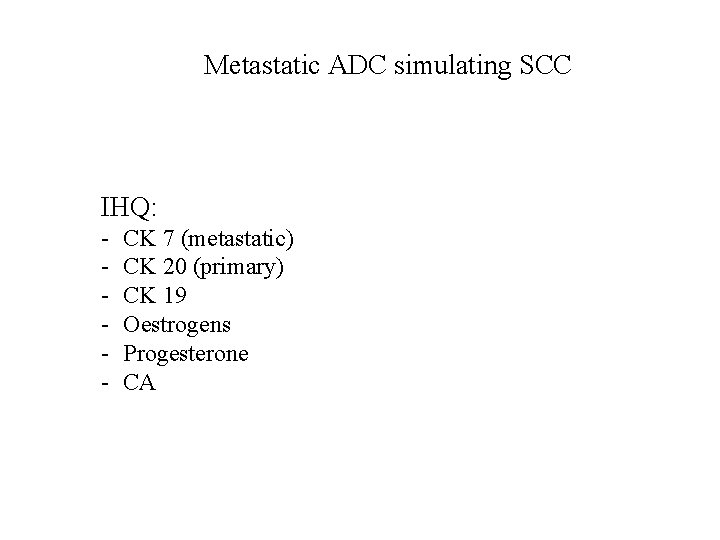
Metastatic ADC simulating SCC IHQ: - CK 7 (metastatic) CK 20 (primary) CK 19 Oestrogens Progesterone CA
BASAL CELL CARCINOMA SIMULATORS • • Trabecular Merkel cell carcinoma Metastatic adenocarcinoma Syringoid carcinoma Metatypical carcinoma Trichoblastoma Desmoplastic trichoepithelioma Lymphadenoma Lympho-epithelioma-like carcinoma
MERKEL Deeply-basophilic highly-cellular tumor with small to medium sized cells, with dense round nuclei Sparse cytoplasm Apoptotic tumor cells with mitosis Patterns: diffuse, trabecular or nested CK 20, Pan cytokeratin is diagnostic S 100 and LCA are negative
BASAL CELL CARCINOMA SIMULATORS • • Trabecular Merkel cell carcinoma Adenocarcinoma metastasis Syringoid carcinoma Metatypical carcinomas Trichoblastomas Desmoplastic trichoepithelioma Lymphadenoma Lymphoepithelioma-like carcinoma
BASAL CELL CARCINOMA SIMULATORS • • Trabecular Merkel cell carcinoma Adenocarcinoma metastasis Syringoid carcinoma Metatypical carcinomas Trichoblastomas Desmoplastic trichoepithelioma Lymphoadenoma Lymphoepithelioma-like carcinoma
Desmoplastic trichoepithelioma • Tumor cells growing as cellular sheets within a sclerotic dermis
Lymphadenoma- Trichoblastoma TRICOEPITELIOMA spectrum
BASAL CELL CARCINOMA SIMULATORS • • Trabecular Merkel cell carcinoma Adenocarcinoma metastasis Syringoid carcinoma Metatypical carcinomas Trichoblastomas Desmoplastic trichoepithelioma Lymphadenoma Lympho-epithelioma-like carcinoma
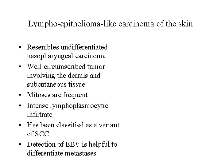
Lympho-epithelioma-like carcinoma of the skin • Resembles undifferentiated nasopharyngeal carcinoma • Well-circumscribed tumor involving the dermis and subcutaneous tissue • Mitoses are frequent • Intense lymphoplasmocytic infiltrate • Has been classified as a variant of SCC • Detection of EBV is helpful to differentiate metastases
SQUAMOUS CELL CARCINOMA SIMULATORS • • • Inverted follicular keratosis Pseudo-epitheliomatous hyperplasia Glandular sialometaplasia Scars Keratin granulomas Nasal muco-epidermoid carcinoma Sebaceous carcinoma Desmoplastic melanoma Nodular fasciitis
Inverted follicular keratosis • Proliferative downgrowths of mature squamous epithelium with infundibular keratinization
SQUAMOUS CELL CARCINOMA SIMULATORS • • • Inverted follicular keratosis Pseudo-epitheliomatous hyperplasia Glandular sialometaplasia Scars Keratin granulomas Nasal muco-epidermoid carcinoma Sebaceous carcinoma Desmoplastic melanoma Nodular fasciitis
Pseudoepitheliomatous hyperplasia • Occurs in the setting of chronic skin ulcers, abscesses, burns and infections • In challenging cases, close clinical correlation and follow-up are required; rare cases need repeated biopsy for evaluation
Pseudo-epitheliomatous hyperplasia • A – Reactive: it could be due to a banal epiphenomenon – – Border of a venous leg ulcer Chronic inflammation of dermal tumors Insect bite Infection: leishmaniasis, verrucous TB, blastomycosis, etc – Toxicodermia: iododerma and bromoderma
• Neutrophilic microabscesses are typically seen in halogenodermas, infections like chromomycosis, blastomycosis, inguinal granuloma, leishmaniasis.
Pseudo-epitheliomatous hyperplasia • B – Immunologic lesions – Pemphigus vegetans – Pyoderma gangrenosum • C – Lymphomatous – Anaplastic large cell lymphoma – Lymphomatoid papulosis CD 30 + “Multiple eruptive keratoacanthomas” (personal observation)
SQUAMOUS CELL CARCINOMA SIMULATORS • • • Inverted follicular keratosis Pseudo-epitheliomatous hyperplasia Glandular sialometaplasia Scars Keratin granulomas Nasal muco-epidermoid carcinoma Sebaceous carcinoma Desmoplastic melanoma Nodular fasciitis
Ecrine squamous syringometaplasia • Is well described as an adverse drug reaction, mostly to chemotherapy • Necrotic sialometaplasia occurs following biopsy or surgical treatment of buccal mucosa (salivary glands)
SQUAMOUS CELL CARCINOMA SIMULATORS • • • Inverted follicular keratosis Pseudo-epitheliomatous hyperplasia Glandular sialometaplasia Scars Keratin granulomas Nasal muco-epidermoid carcinoma Sebaceous carcinoma Desmoplastic melanoma Nodular fasciitis
SQUAMOUS CELL CARCINOMA SIMULATORS • • Inverted follicular keratosis Pseudo-epitheliomatous hyperplasia Glandular sialometaplasia Scars Keratin granulomas Desmoplastic melanoma Nodular fasciitis
Desmoplastic melanoma • It is a variant of melanoma with fibroblast-like spindled melanocytes surrounded by abundant collagen fibres • Immunostaining: – HMB 45 negative – S 100 positive – SOX 10 positive
SQUAMOUS CELL CARCINOMA SIMULATORS • • • Inverted follicular keratosis Pseudo-epitheliomatous hyperplasia Glandular sialometaplasia Scars Keratin granulomas Nasal muco-epidermoid carcinoma Sebaceous carcinoma Desmoplastic melanoma Nodular fasciitis
Nodular fasciitis • 51 -year-old female • 8 -month history of a nodular lesion with progressive growth on the cheek • Size: 3 cm, adhered to skin but not to bone • Skin biopsy malignant mesenchymal tumour • MOHS MICROGRAPHIC SURGERY
Nodular fasciitis – It’s important to recognise this reactive pattern to avoid misdiagnosing a sarcoma and to perform unnecessary radical surgical treatment
DERMATOFIBROSARCOMA PROTUBERANS
Dermatofibrosarcoma protuberans • Histopathologic findings: – Dermal and hypodermal tumour – Irregular infiltration in extension and depth between the different dermal planes and subcutaneous tissue – Patterns: cartwheel, honeycomb, in bands, storiform – Mitosis: < 5 (x 10 field) – CD 34 +
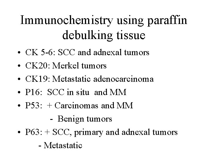
Immunochemistry using paraffin debulking tissue • • • CK 5 -6: SCC and adnexal tumors CK 20: Merkel tumors CK 19: Metastatic adenocarcinoma P 16: SCC in situ and MM P 53: + Carcinomas and MM - Benign tumors • P 63: + SCC, primary and adnexal tumors - Metastatic
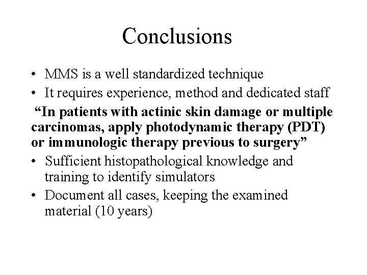
Conclusions • MMS is a well standardized technique • It requires experience, method and dedicated staff “In patients with actinic skin damage or multiple carcinomas, apply photodynamic therapy (PDT) or immunologic therapy previous to surgery” • Sufficient histopathological knowledge and training to identify simulators • Document all cases, keeping the examined material (10 years)
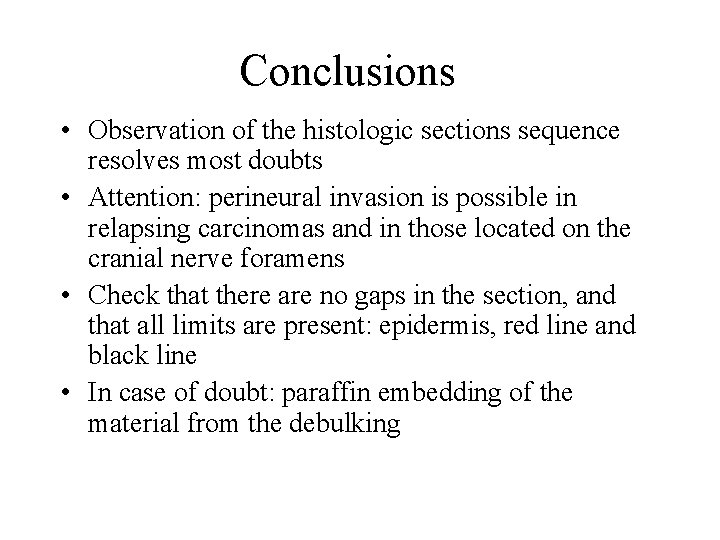
Conclusions • Observation of the histologic sections sequence resolves most doubts • Attention: perineural invasion is possible in relapsing carcinomas and in those located on the cranial nerve foramens • Check that there are no gaps in the section, and that all limits are present: epidermis, red line and black line • In case of doubt: paraffin embedding of the material from the debulking

END
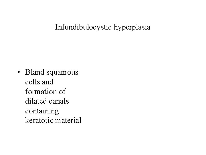
Infundibulocystic hyperplasia • Bland squamous cells and formation of dilated canals containing keratotic material
Dermatofibrosarcoma protuberans • Histopathologic findings: – Aggressiveness signs: > mitotic activity, pleomorphism, necrosis – Variants: • • Granular Bednar tumour (pigmented) Fibrosarcoma – DFSP Myxoid
Nasal muco-epidermoid carcinoma The lining of the nasal cavity, it is continuous with the skin in the vestibule of the nose and with the mucosa of the nasopharynx, the paranasal sinuses Sinonasal papillomas are benign epithelial neoplasms arising from Schneiderian mucosa. The three subtypes, exophytic, oncocytic, and inverted (inverted papilloma [IP]), should be distinguished from one another histopathologically. 5% of inverted SCC Schneiderian papillomas are benign neoplasms that are associated with three key characteristics: tendency to recur, capacity for local destruction
Tratamiento DFSP • Excisión de 5 cms de margen , llegando a fascia 20% de recidivas • Tratamiento de elección: CM Mohs • Exéresis ganglionar si se palpan adenopatías (forma sarcomatosa del DFSP). Metástasis 5% • Tratamiento de los DFSP no rescables, recidivantes o metastizantes : IMATINIB : Inhibidores plaquetarios de los receptores de factores de crecimiento
False relapses • Chance of transporting affected or potentially affected skin (AK) with a flap or graft • Growth of a new tumour after more than 5 years (debatable) or of a different lineage • Previously irradiated area • Collision tumours
Histologic criteria to distinguish false relapses • New primary tumour: It appears on the skin of the graft or flap, and DOES NOT EXTEND inside the scar tissue (fibrosis) • Relapsing tumour: It extends inside the scar tissue (fibrosis)
Risk situations • Carcinomas located on the cranial nerves foramens Perineural invasion • Scars. They should be considered, especially if there is a previous flap • Multiple carcinomatosis and actinic skin damage: false relapses and appearance of sequential carcinomas
Nasal muco-epidermoid carcinoma • 56 -year-old male. Rapid growth of an erythematous, infiltrated plaque on the naso-ocular left area for one month *Zhao G. et al. Cutaneous involvement by nasal mucoepidermoid carcinoma: the tip of the iceberg phenomenon. J Cut Pathol. 2017; 44(2): 113 -17
Nasal muco-epidermoid carcinoma • “The tip of the iceberg phenomenon” • Schneiderian mucosa: The Schneider or sinus membrane is a thin layer of cells. Respiratory polystratified epithelium.
- Slides: 53