HIP JOINT By Dr Mujahid Khan Articulation The

HIP JOINT By: Dr. Mujahid Khan

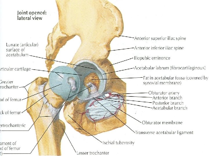
Articulation Ø The hip joint is the articulation between the hemispherical head of femur and the cup shaped acetabulum of the hip bone Ø The articular surface of the acetabulum is horseshoe shaped and is deficient inferiorly at the acetabular notch
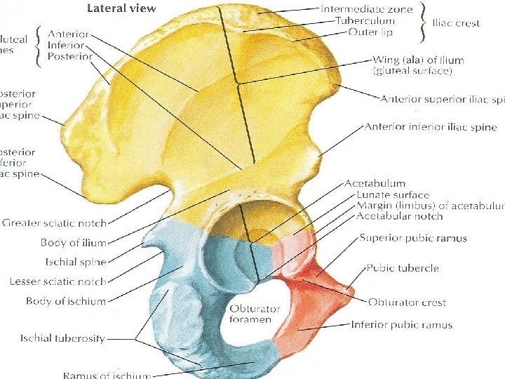

Articulation Ø The cavity of acetabulum is deepened by the presence of a fibrocartilaginous rim called acetabular labrum Ø The labrum bridges across the acetabular notch and is here called the transverse acetabular ligament Ø The articular surfaces are covered with hyaline cartilage

Type & Capsule Ø It is a synovial ball and socket joint Ø The capsule encloses the joint and is attached to the acetabular labrum medially Ø Laterally it is attached to the intertrochanteric line of the femur in front and along the posterior aspect of the neck of the bone behind

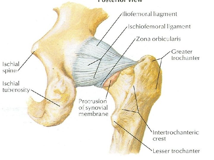
Iliofemoral Ligaments Ø It is a strong, inverted Y-shaped ligament Ø Its base is attached to the anterior inferior iliac spine above Ø Below the two limbs of Y are attached to the upper and lower parts of the intertrochanteric line of the femur Ø The strong ligament prevents overextension during standing
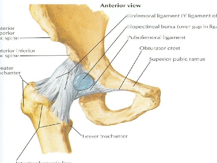

Pubofemoral Ligament Ø It is a triangular ligament Ø The base of the ligament is attached to the superior ramus of the pubis Ø The apex is attached below to the lower part of the intertrochanteric line Ø This ligament limits extension and abduction

Ischiofemoral Ligament Ø It is a spiral shaped ligament Ø Attached to the body of the ischium near the acetabular margin Ø Fibers pass upward and laterally and attached to the greater trochanter Ø This ligament limits the extension


Transverse Acetabular Ligament Ø It is formed by the acetabular labrum as it bridges the acetabular notch Ø It converts the notch into a tunnel through which blood vessels and nerves enter the joint


Ligament of Head of Femur Ø It is flat and triangular ligament Ø It is attached by its apex to the pit on the head of the femur (fovea capitis) Ø Attached by its base to the transverse ligament and the margins of the acetabular notch Ø It lies within the joint and is ensheathed by synovial membrane

Synovial Membrane Ø The synovial membrane lines the capsule Ø It is attached to the margins of the articular surfaces Ø It covers the portion of the neck of the femur that lies within the joint capsule Ø It ensheathes the ligament of the head of the femur


Synovial Membrane Ø It covers the pad of fat contained in the acetabular fossa Ø A pouch of synovial membrane frequently protrudes through a gap in the anterior wall of the capsule Ø Forms the psoas bursa beneath the psoas tendon

Nerve Supply Ø Femoral nerve Ø Obturator nerve Ø Sciatic nerve Ø Nerve to the quadratus femoris

Movements Ø The hip joint has a wide range of movement but less so than the shoulder joint Ø Some of the movement has been sacrificed to provide strength and stability Ø The strength of the joint depends largely on the shape of the bones taking part in the articulation and on strong ligaments

Movements Ø When the knee is flexed, flexion is limited by the anterior surface of the thigh coming in contact with the anterior abdominal wall Ø When the knee is extended, flexion is limited by the tension of the hamstring muscles Ø Abduction is limited by the tension of the pubofemoral ligament

Movements Ø Adduction is limited by contact with the opposite limb and by the tension of the ligament of the head of the femur Ø Lateral rotation is limited by the tension in the iliofemoral and pubofemoral ligaments Ø Medial rotation is limited by the ischiofemoral ligament

Movements Ø Flexion: It is performed by the iliopsoas, rectus femoris, sartorius, also by adductor muscles Ø Extension: it is performed by the gluteus maximus and the hamstring muscles Ø Abduction: It is performed by the gluteus medius and minimus, assisted by sartorius, tensor fasciae latae, and piriformis

Movements Ø Adduction: It is performed by the adductor longus and brevis and the adductor fibers of the adductor magnus Ø Lateral rotation: It is performed by the piriformis, obturator internus and externus, superior and inferior gamelli Ø Medial rotation: It is performed by the anterior fibers of gluteus medius and gluteus minimus and the tensor fasciae latae Ø Circumduction: It is a combination of the previous movements

Movements Ø The extensor group of muscles is more powerful than the flexor group Ø The lateral rotators are more powerful than the medial rotators

Relations Ø Anteriorly: Iliopsoas, pectineus, and rectus femoris Ø Posteriorly: The obturator internus, the gamelli, and the quadratus femoris muscle separate the joint from sciatic nerve Ø Superiorly: Piriformis and gluteus minimus Ø Inferiorly: Obturator externus tendon
- Slides: 26