HighResolution Chest CT Practical Clinical Applications Paul L


 • High spatial frequency reconstruction • Windows")


 • Desquamative interstitial pneumonitis • Non-specific interstitial pneumonitis")


 • Langerhans Cell Histiocytosis (EG) • End-stage (honeycomb) lung")


- Slides: 76
High-Resolution Chest CT: Practical Clinical Applications Paul L. Molina, M. D. Department of Radiology University of North Carolina at Chapel Hill
Disclosures None
Objectives • Identify current clinical indications for the use of HRCT • Review proper technique for performance of HRCT • Summarize the characteristic patterns of abnormality seen on HRCT and the most common diseases resulting in their formation
HRCT - Indication • Evaluation of patients with suspected infiltrative lung disease but normal or nonspecific findings on CXR
HRCT - Indication • Further characterization of known or suspected diffuse lung disease
HRCT - Indication • Evaluation of patients in whom radiographic findings are not in keeping with clinical findings or pulmonary function tests
HRCT - Indication • Delineation of disease prior to lung biopsy as a guide to the optimal type and site of biopsy
HRCT Technique • Thin collimation (1 mm) • High spatial frequency reconstruction • Windows -700/1000 -1500 HU • • Prone scans – differentiate atelectasis Expiratory scans – air trapping
HRCT Findings • Septal thickening • Reticular densities • Nodules • Increased lung opacity • Decreased lung opacity
Septal Thickening • Pulmonary edema • Lymphangitic carcinomatosis • Sarcoidosis • Asbestosis • Idiopathic pulmonary fibrosis
Reticular Densities • Idiopathic pulmonary fibrosis • Collagen vascular disease • Asbestosis • Chronic hypersensitivity pneumonitis • Sarcoidosis
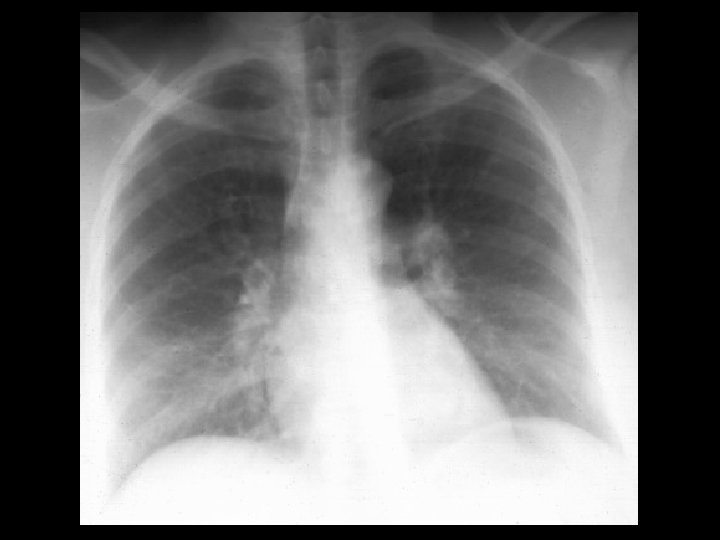
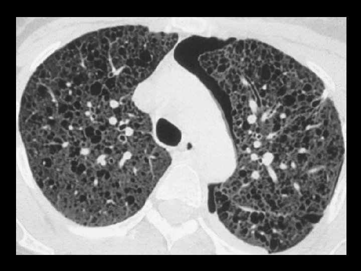
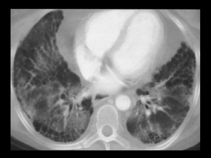
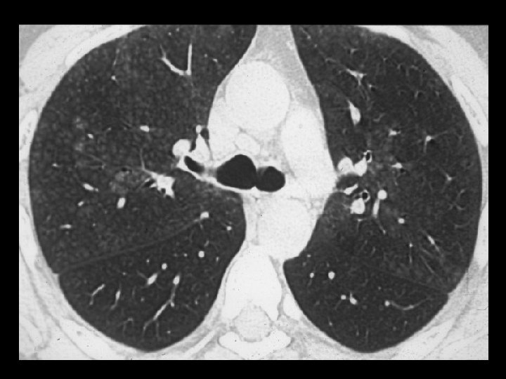
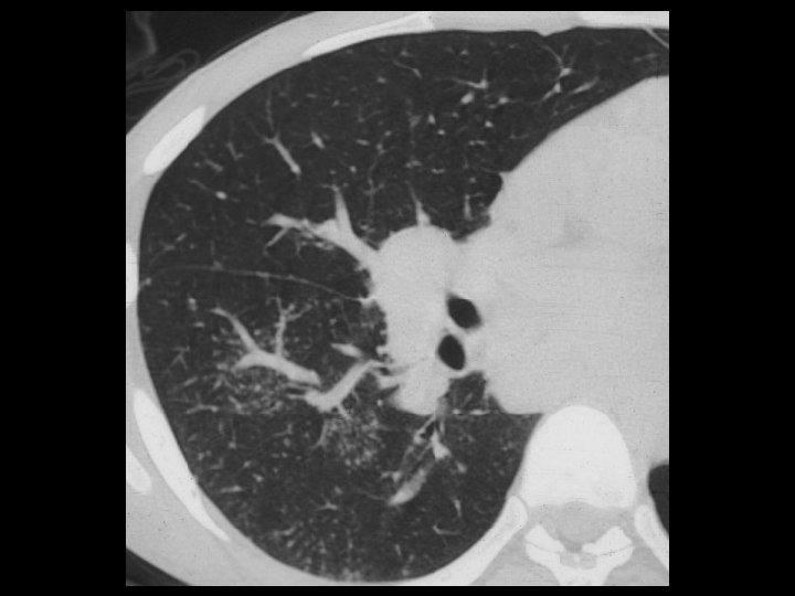
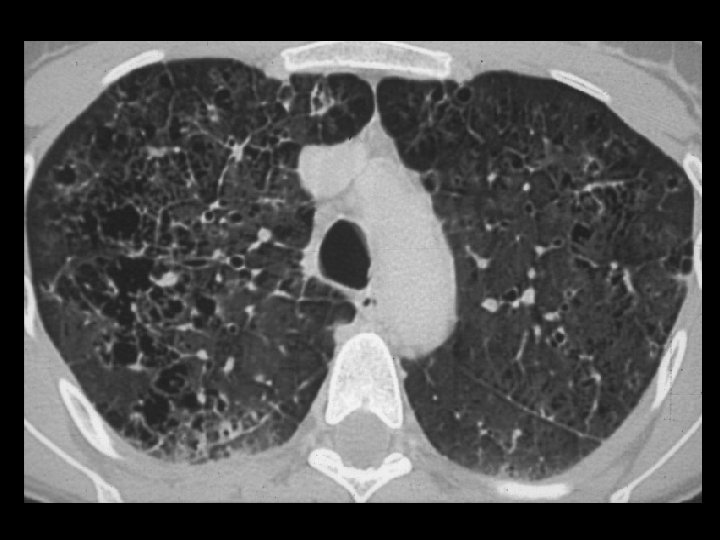
UIP
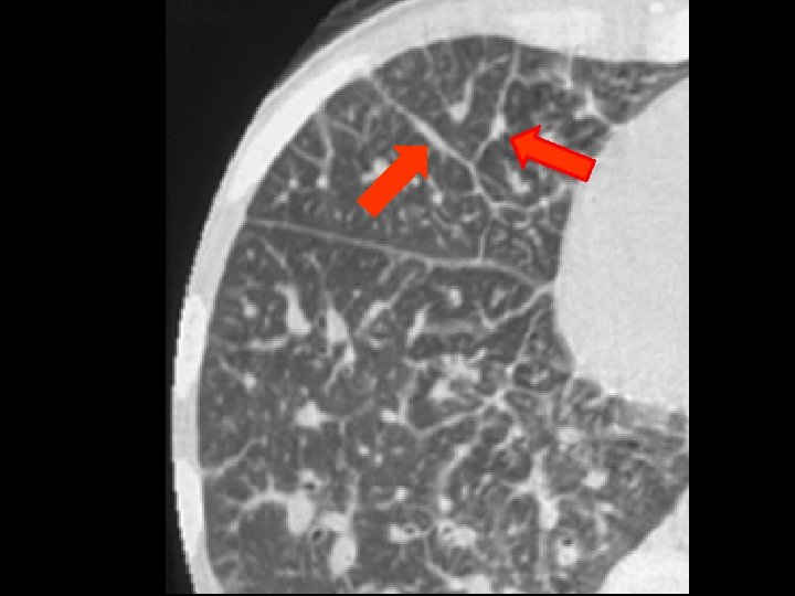
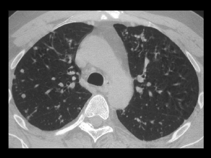
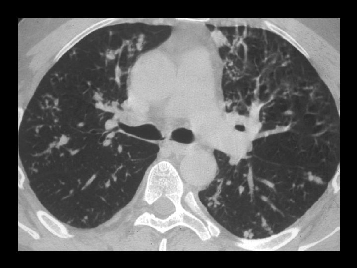
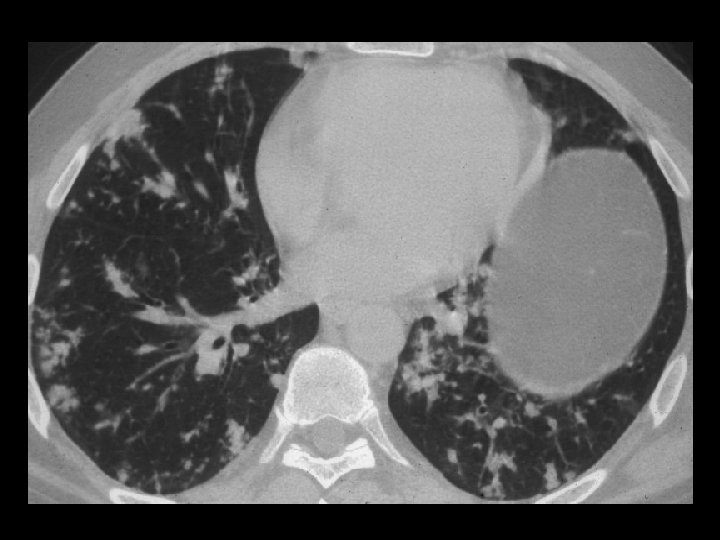
Nodular Opacities • Sarcoidosis • Silicosis • Coal worker’s pneumoconiosis • Hypersensitivity pneumonitis • Tuberculosis • Metastatic disease
Nodular Opacities • Perilymphatic nodules • Random distribution • Centrilobular nodules
Perilymphatic Nodules • Sarcoidosis • Silicosis • Lymphangitic Ca
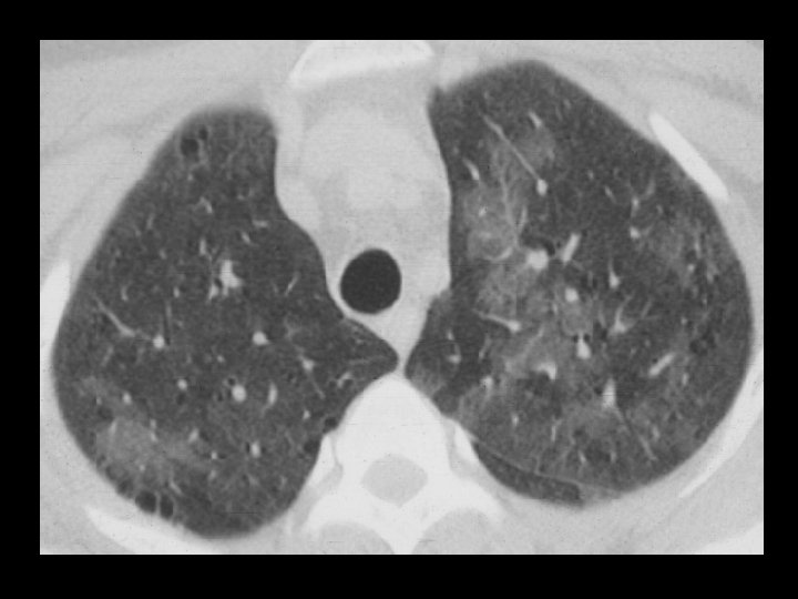
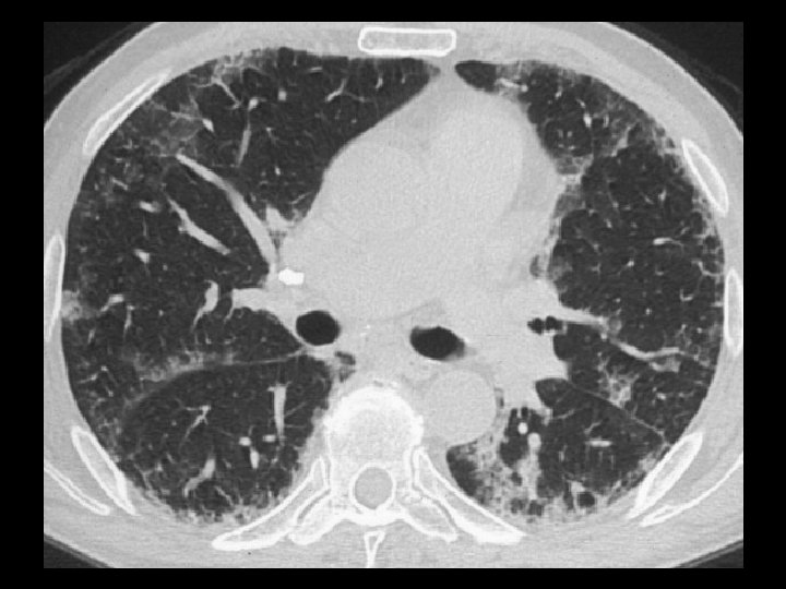
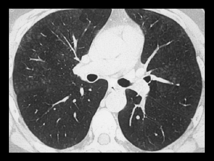
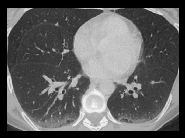
Silicosis
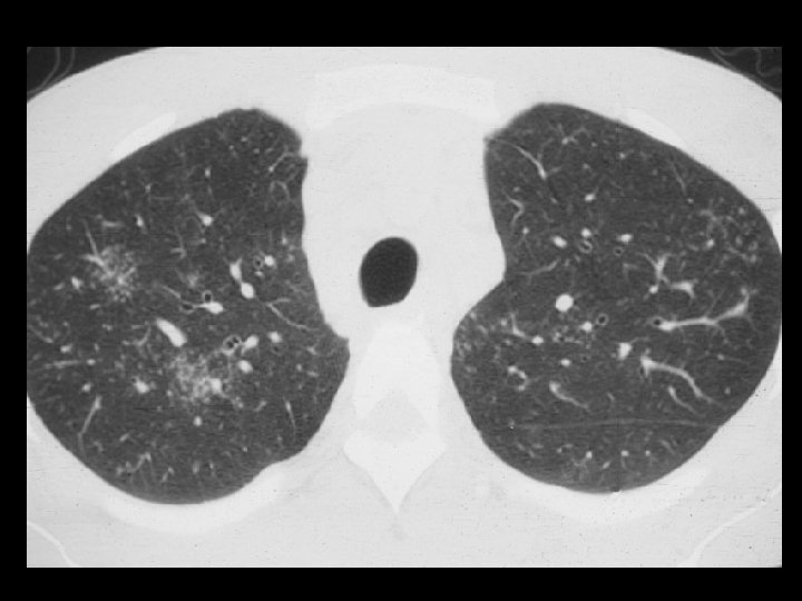
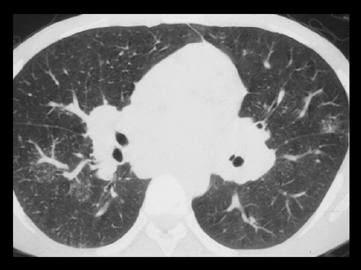
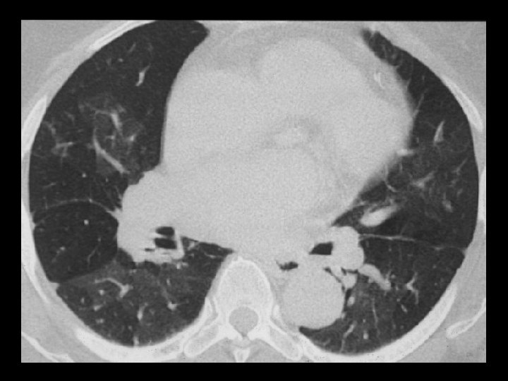
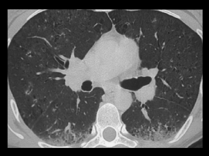
Random Nodules • Miliary TB • Hematogenous mets m
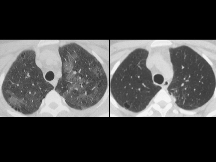
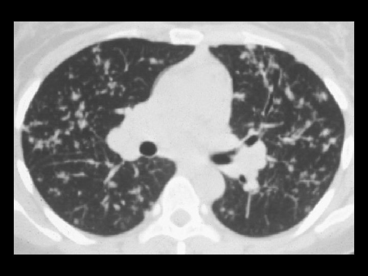
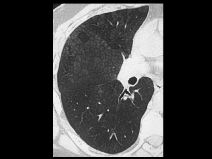
Metastatic adenoca
Centrilobular Nodules • Endobronchial spread of TB or other infection • Hypersensitivity pneumonitis • Endobronchial tumor spread
Nodular Opacities • Perilymphatic nodules • Random distribution • Centrilobular nodules
Increased Lung Opacity • Ground-glass opacity • Air-space consolidation
Ground-glass Opacity • Hypersensitivity pneumonitis (subacute) • Desquamative interstitial pneumonitis • Non-specific interstitial pneumonitis • Sarcoidosis • Alveolar proteinosis
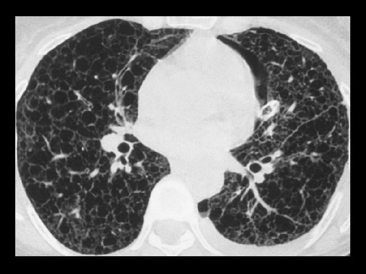
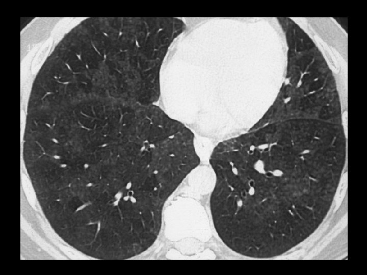
DIP
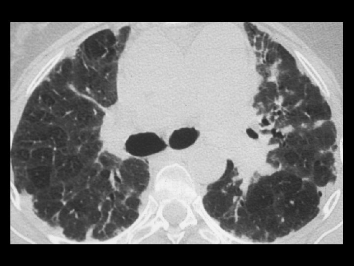
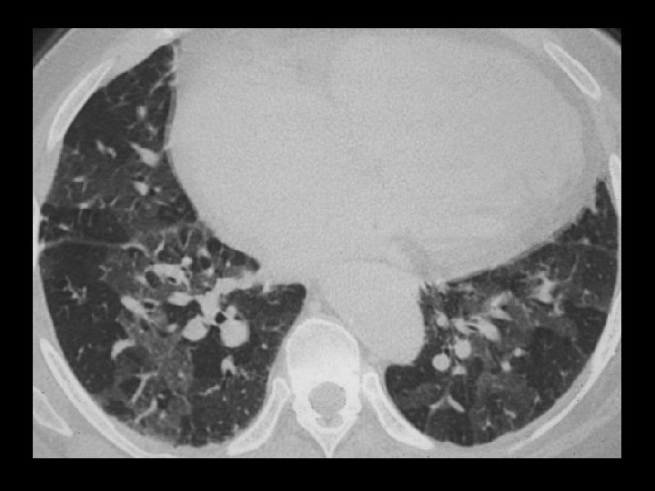
Non-specific Interstitial Pneumonitis
Crazy-Paving Alveolar Proteinosis
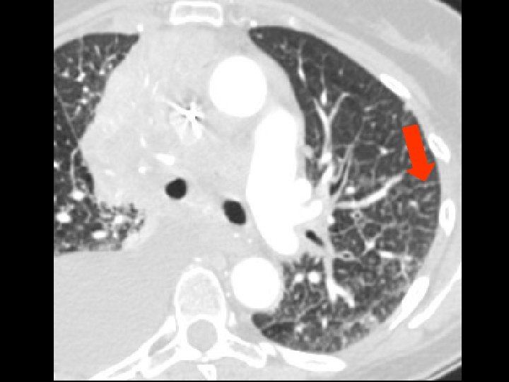
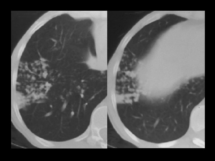
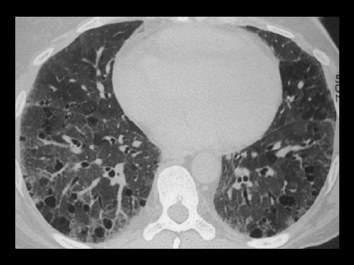
Mosaic Pefusion
Consolidation • Obscures underlying vessels • Solid, opaque • Air bronchograms
Consolidation • Chronic eosinophilic pneumonia • BOOP / COP • Bronchoalveolar cell carcinoma • Lymphoma
Chronic Eosinophilic Pneumonia
BOOP / COP
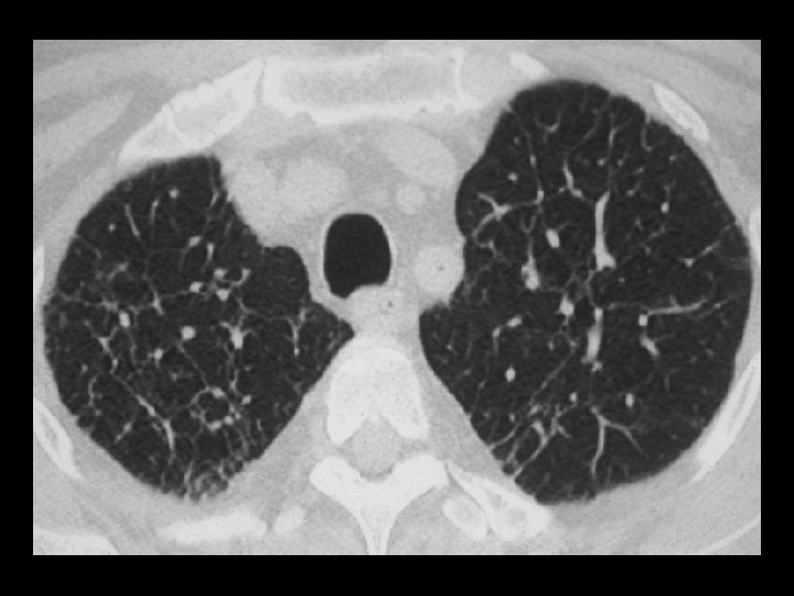
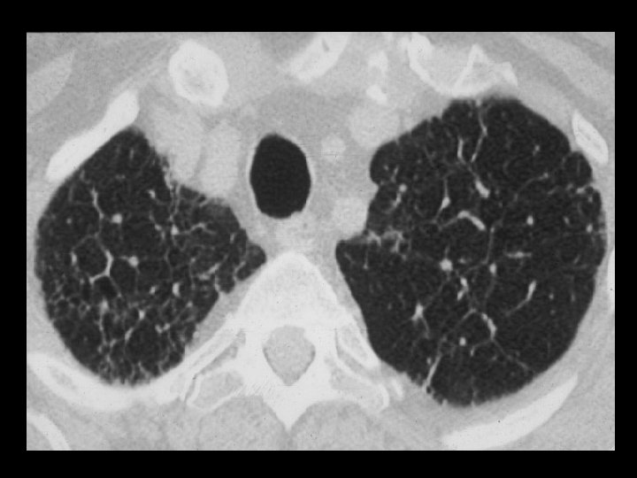
Decreased Lung Opacity • Emphysema • Cystic airspaces • Mosaic perfusion
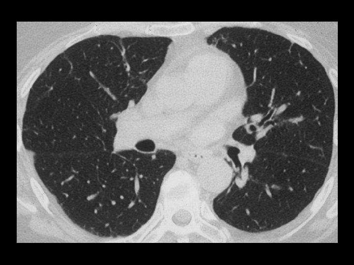
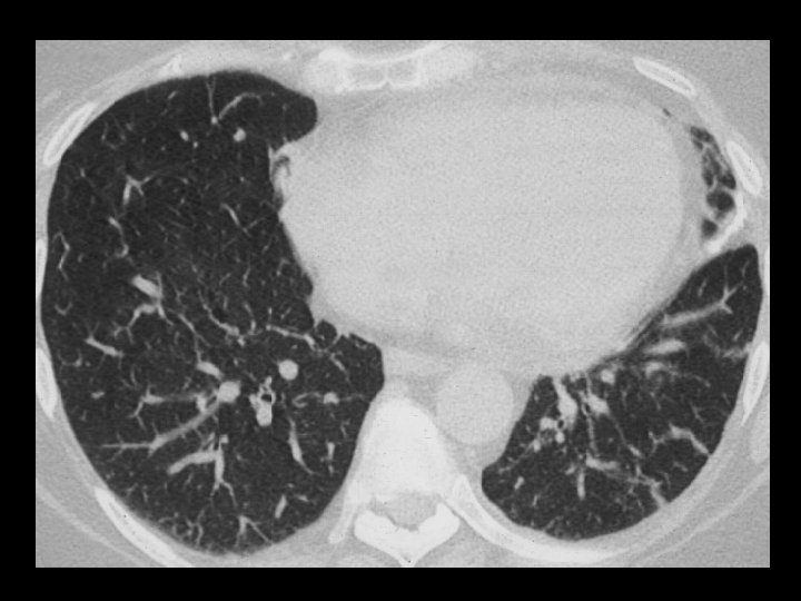
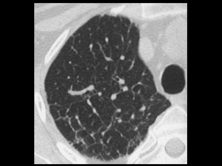
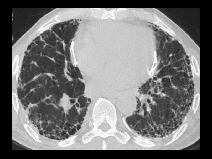
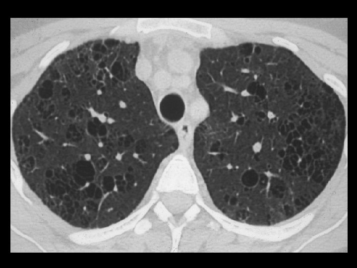
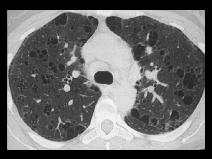
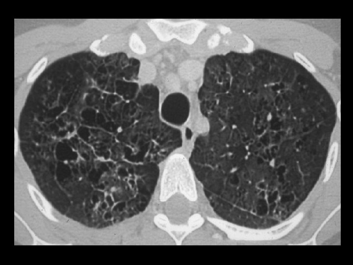
Cystic Airspaces • Lymphangioleiomyomatosis (LAM) • Langerhans Cell Histiocytosis (EG) • End-stage (honeycomb) lung
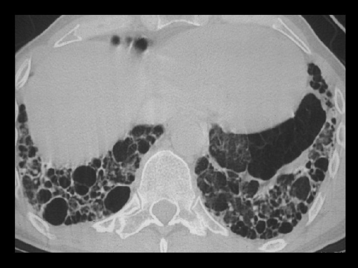
LAM
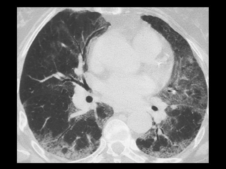
EG
HRCT - Indications • Suspected infiltrative disease but normal or nonspecific CXR • Further characterize diffuse disease • CXR findings not in keeping with clinical findings or PFT’s • Guide type and site of biopsy
HRCT Findings • Septal thickening • Reticular opacities • Nodular opacities • Increased lung opacity • Decreased lung opacity