
Higher Human Biology Unit 2 Physiology and Health
Higher Human Biology Unit 2 – Physiology and Health Section 13 – Structure and Function of Arteries, Capillaries and Veins
What Can You Remember? • Name the four chambers of the heart • What is the main artery in the heart? • Describe the difference in structure between an artery and a vein. • Explain the process of gas exchange at the alveoli • What protein transports oxygen around the body • What type of protein is mentioned in the previous question?
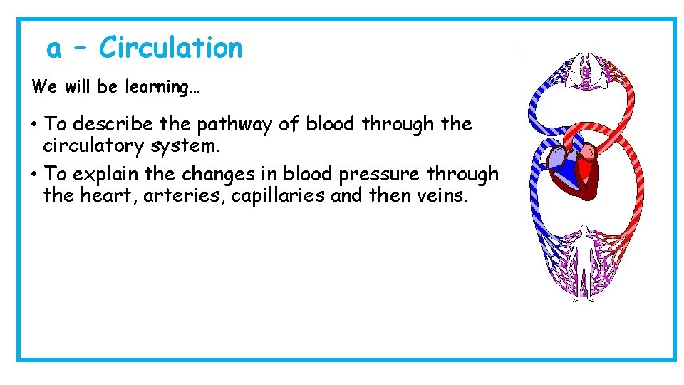
a – Circulation We will be learning… • To describe the pathway of blood through the circulatory system. • To explain the changes in blood pressure through the heart, arteries, capillaries and then veins.
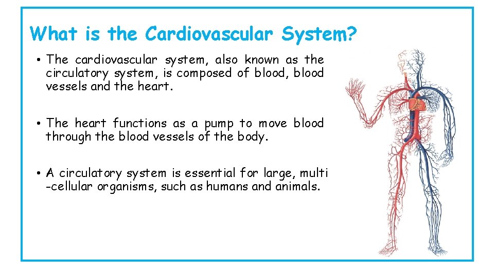
What is the Cardiovascular System? • The cardiovascular system, also known as the circulatory system, is composed of blood, blood vessels and the heart. • The heart functions as a pump to move blood through the blood vessels of the body. • A circulatory system is essential for large, multi -cellular organisms, such as humans and animals.
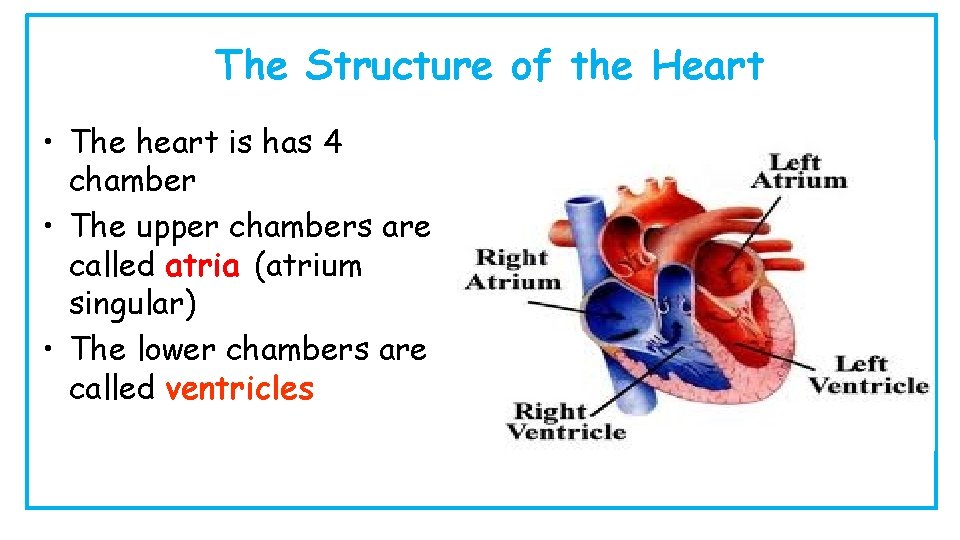
The Structure of the Heart • The heart is has 4 chamber • The upper chambers are called atria (atrium singular) • The lower chambers are called ventricles
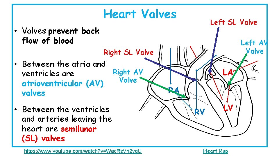
Heart Valves Left SL Valve • Valves prevent back flow of blood Left AV Valve Right SL Valve • Between the atria and Right AV ventricles are Valve atrioventricular (AV) valves • Between the ventricles and arteries leaving the heart are semilunar (SL) valves https: //www. youtube. com/watch? v=Wac. Rs. Vn 2 yg. U LA RA RV LV Heart Rap
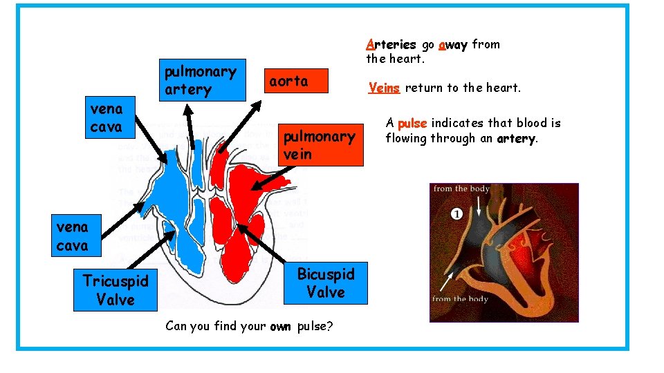
vena cava pulmonary artery Arteries go away from the heart. aorta pulmonary vein vena cava Tricuspid Valve Bicuspid Valve Can you find your own pulse? Veins return to the heart. A pulse indicates that blood is flowing through an artery.
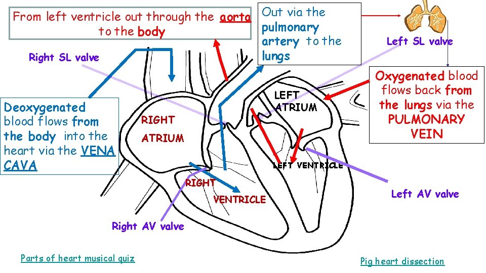
From left ventricle out through the aorta Out via the pulmonary to the body artery to the lungs Right SL valve Deoxygenated blood flows from the body into the heart via the VENA CAVA LEFT ATRIUM RIGHT ATRIUM Left SL valve Oxygenated blood flows back from the lungs via the PULMONARY VEIN LEFT VENTRICLE RIGHT VENTRICLE Left AV valve Right AV valve Parts of heart musical quiz Pig heart dissection
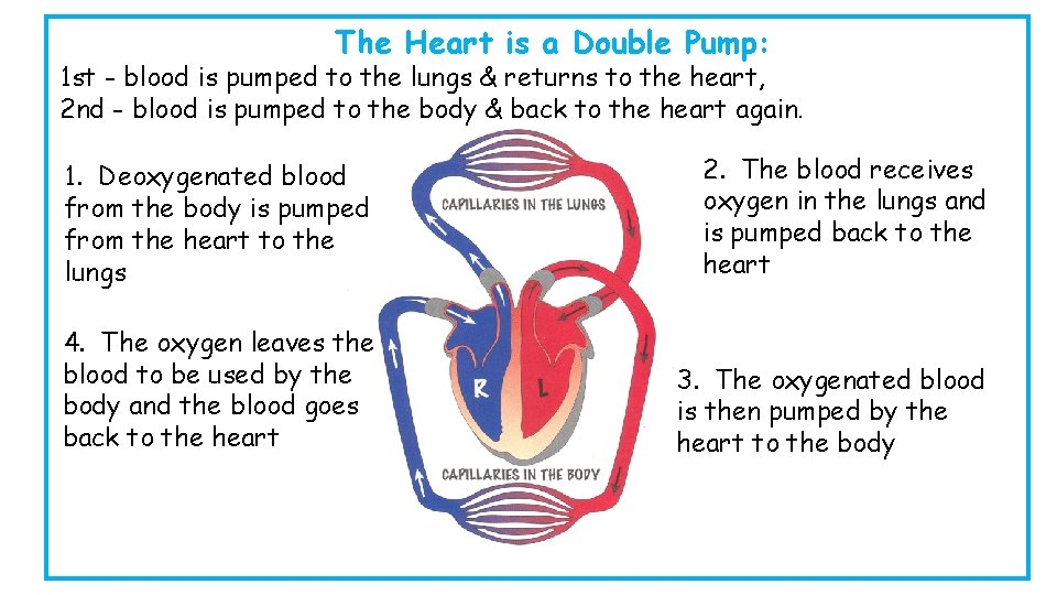
The Heart is a Double Pump: 1 st - blood is pumped to the lungs & returns to the heart, 2 nd - blood is pumped to the body & back to the heart again. 1. Deoxygenated blood from the body is pumped from the heart to the lungs 2. The blood receives oxygen in the lungs and is pumped back to the heart 4. The oxygen leaves the blood to be used by the body and the blood goes back to the heart 3. The oxygenated blood is then pumped by the heart to the body
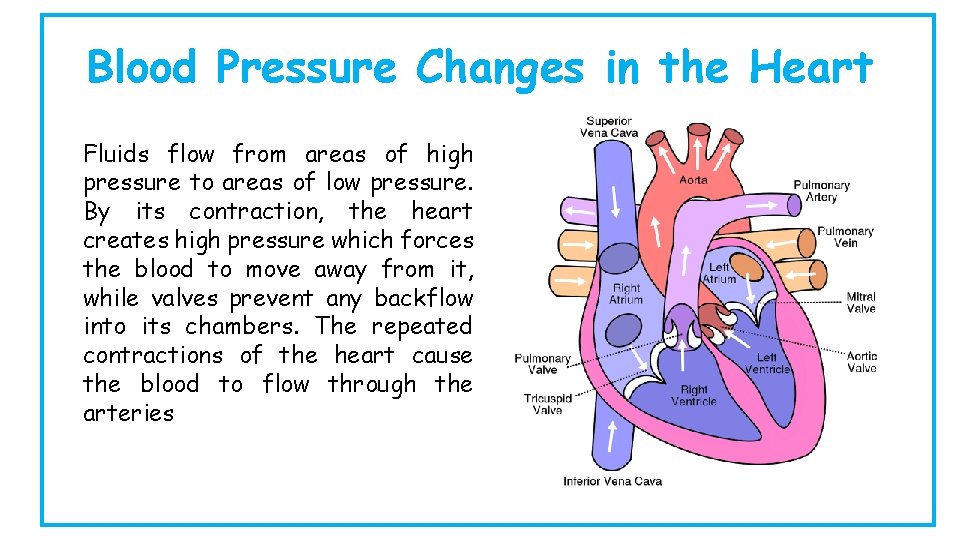
Blood Pressure Changes in the Heart Fluids flow from areas of high pressure to areas of low pressure. By its contraction, the heart creates high pressure which forces the blood to move away from it, while valves prevent any backflow into its chambers. The repeated contractions of the heart cause the blood to flow through the arteries
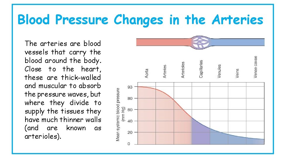
Blood Pressure Changes in the Arteries The arteries are blood vessels that carry the blood around the body. Close to the heart, these are thick-walled and muscular to absorb the pressure waves, but where they divide to supply the tissues they have much thinner walls (and are known as arterioles).
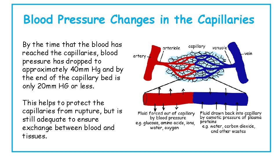
Blood Pressure Changes in the Capillaries By the time that the blood has reached the capillaries, blood pressure has dropped to approximately 40 mm Hg and by the end of the capillary bed is only 20 mm HG or less. This helps to protect the capillaries from rupture, but is still adequate to ensure exchange between blood and tissues.
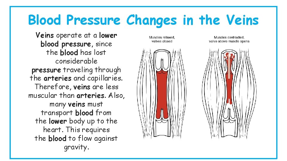
Blood Pressure Changes in the Veins operate at a lower blood pressure, since the blood has lost considerable pressure traveling through the arteries and capillaries. Therefore, veins are less muscular than arteries. Also, many veins must transport blood from the lower body up to the heart. This requires the blood to flow against gravity.
b – Structure and Function of Arteries, Capillaries and Veins We will be learning… • To be able to describe the structure and function of arteries, capillaries and veins. • To be able to describe the structure and function of the endothelium, central lumen, connective tissue, elastic fibres, smooth muscle and valves. • To explain the role of vasoconstriction • To state that capillaries allow exchange of substances with tissues through their thin walls • To explain the role of vasodilation
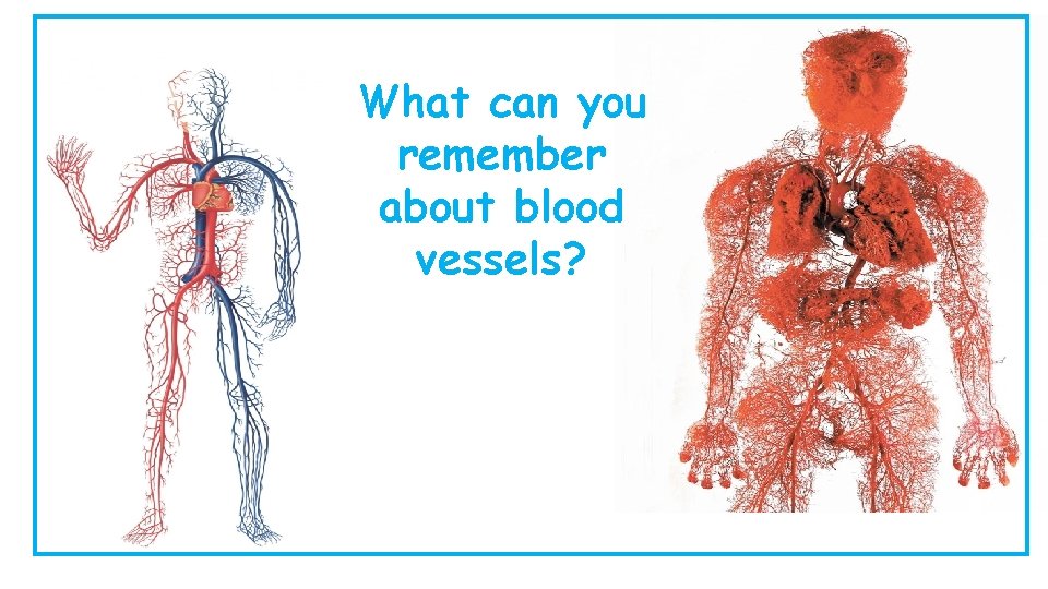
What can you remember about blood vessels?
Who Am I? 1. I leave the right ventricle and go to the lungs Pulmonary Artery 2. I take deoxygenated blood to the heart Vena Cava 3. I prevent backflow of blood into the left atrium Left AV Valve 4. I come from the lungs and go back to the heart Pulmonary Vein 5. I prevent backflow of blood into the right ventricle Right SL Valve 6. I leave the left ventricle of the heart Aorta
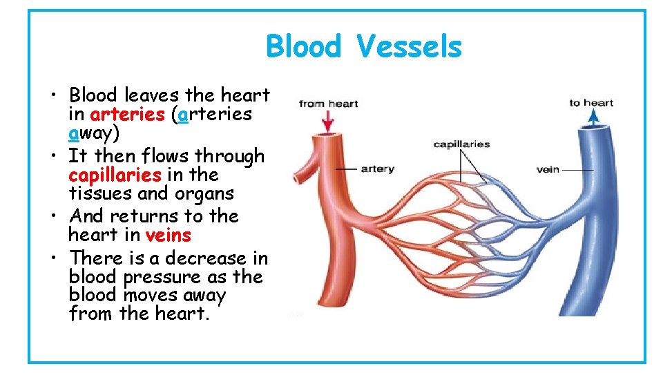
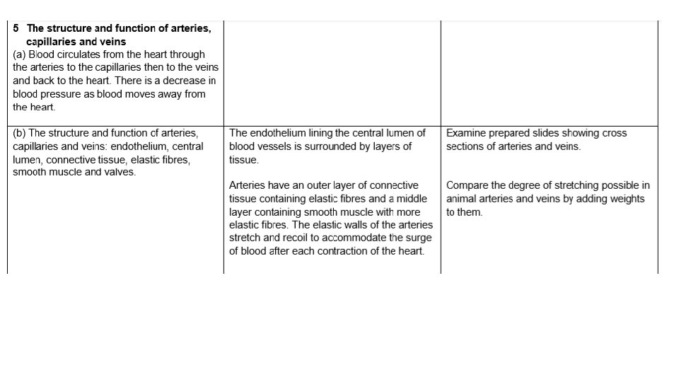
Blood Vessels • Blood leaves the heart in arteries (arteries away) • It then flows through capillaries in the tissues and organs • And returns to the heart in veins • There is a decrease in blood pressure as the blood moves away from the heart.
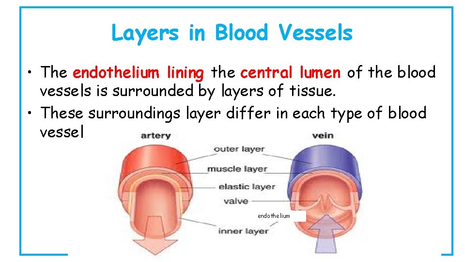
Layers in Blood Vessels • The endothelium lining the central lumen of the blood vessels is surrounded by layers of tissue. • These surroundings layer differ in each type of blood vessel endothelium
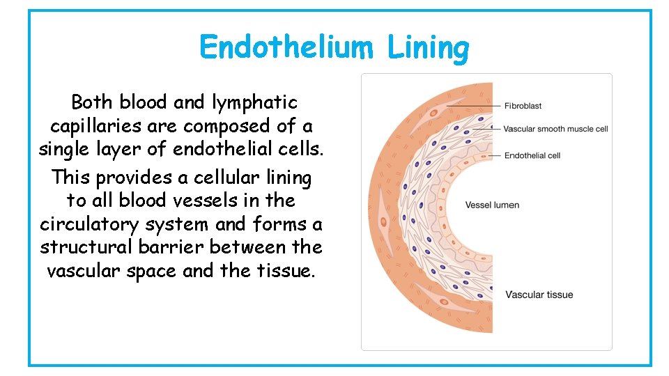
Endothelium Lining Both blood and lymphatic capillaries are composed of a single layer of endothelial cells. This provides a cellular lining to all blood vessels in the circulatory system and forms a structural barrier between the vascular space and the tissue.
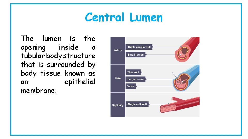
Central Lumen The lumen is the opening inside a tubular body structure that is surrounded by body tissue known as an epithelial membrane.
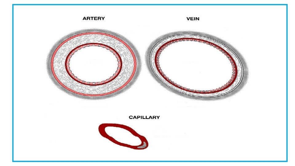
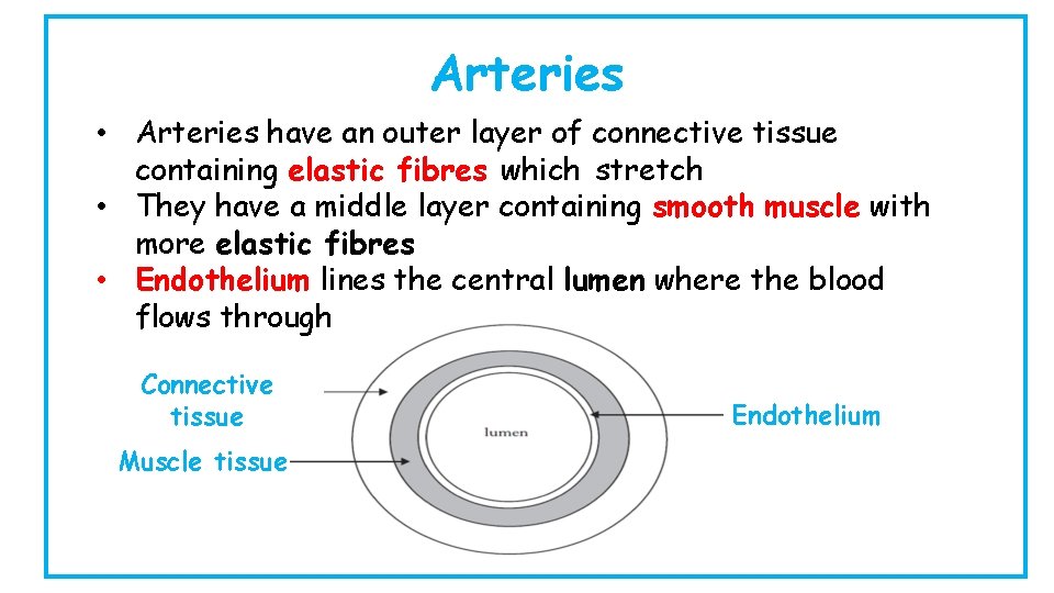
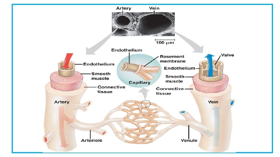
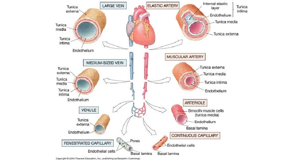
Arteries • Arteries have an outer layer of connective tissue containing elastic fibres which stretch • They have a middle layer containing smooth muscle with more elastic fibres • Endothelium lines the central lumen where the blood flows through Connective tissue Muscle tissue Endothelium
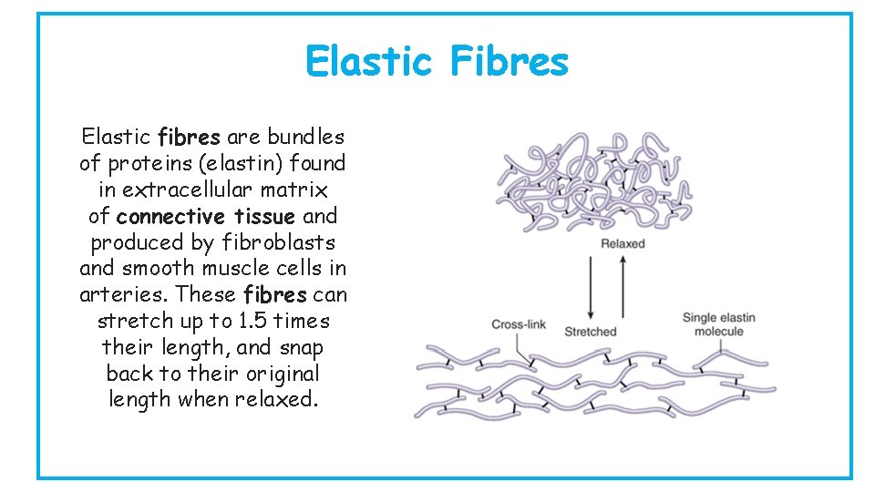
Elastic Fibres Elastic fibres are bundles of proteins (elastin) found in extracellular matrix of connective tissue and produced by fibroblasts and smooth muscle cells in arteries. These fibres can stretch up to 1. 5 times their length, and snap back to their original length when relaxed.
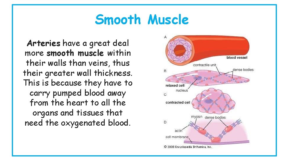
Smooth Muscle Arteries have a great deal more smooth muscle within their walls than veins, thus their greater wall thickness. This is because they have to carry pumped blood away from the heart to all the organs and tissues that need the oxygenated blood.
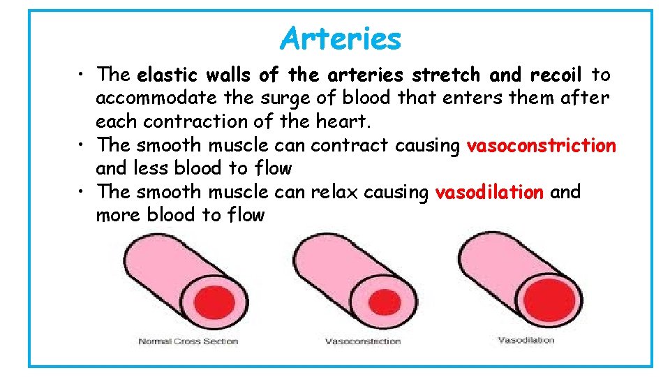
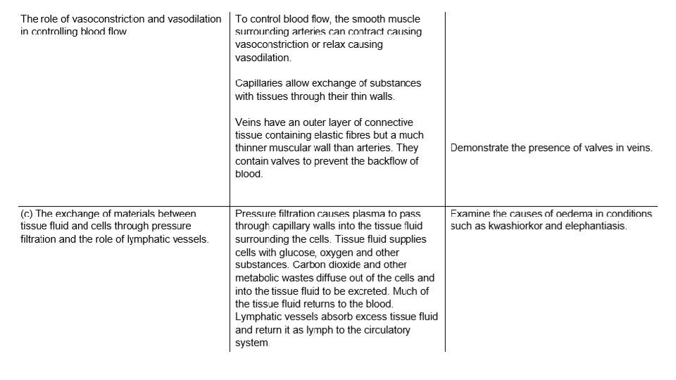
Arteries • The elastic walls of the arteries stretch and recoil to accommodate the surge of blood that enters them after each contraction of the heart. • The smooth muscle can contract causing vasoconstriction and less blood to flow • The smooth muscle can relax causing vasodilation and more blood to flow
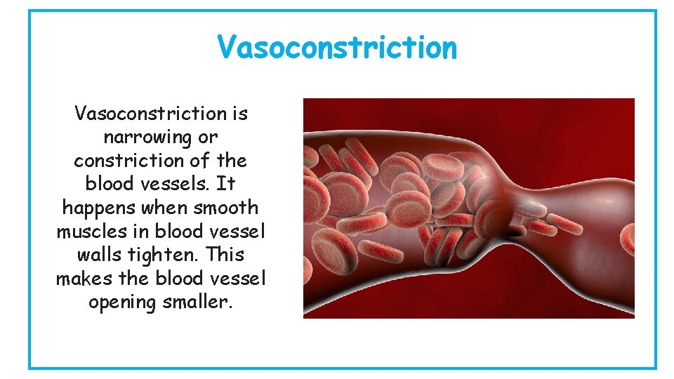
Vasoconstriction is narrowing or constriction of the blood vessels. It happens when smooth muscles in blood vessel walls tighten. This makes the blood vessel opening smaller.
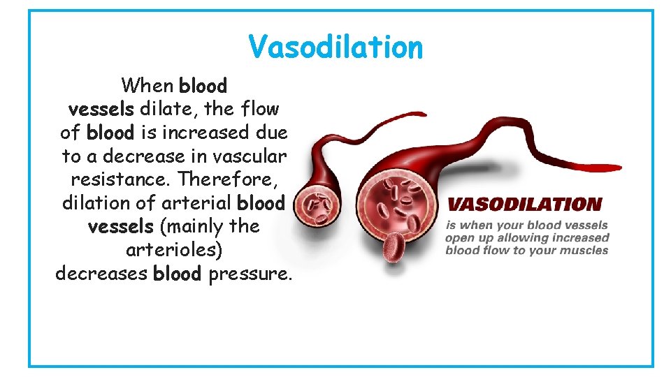
Vasodilation When blood vessels dilate, the flow of blood is increased due to a decrease in vascular resistance. Therefore, dilation of arterial blood vessels (mainly the arterioles) decreases blood pressure.
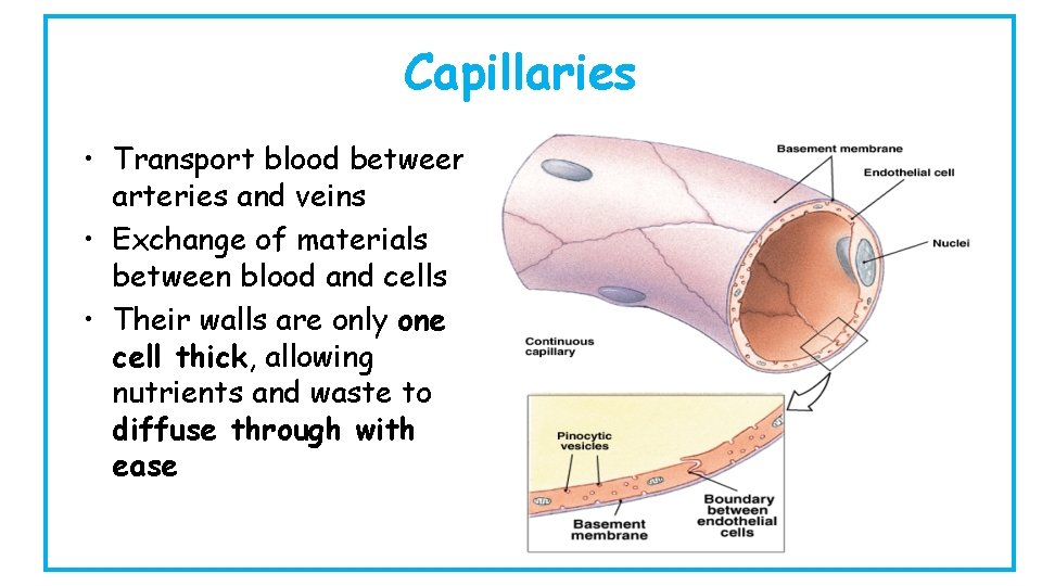
Capillaries • Transport blood between arteries and veins • Exchange of materials between blood and cells • Their walls are only one cell thick, allowing nutrients and waste to diffuse through with ease
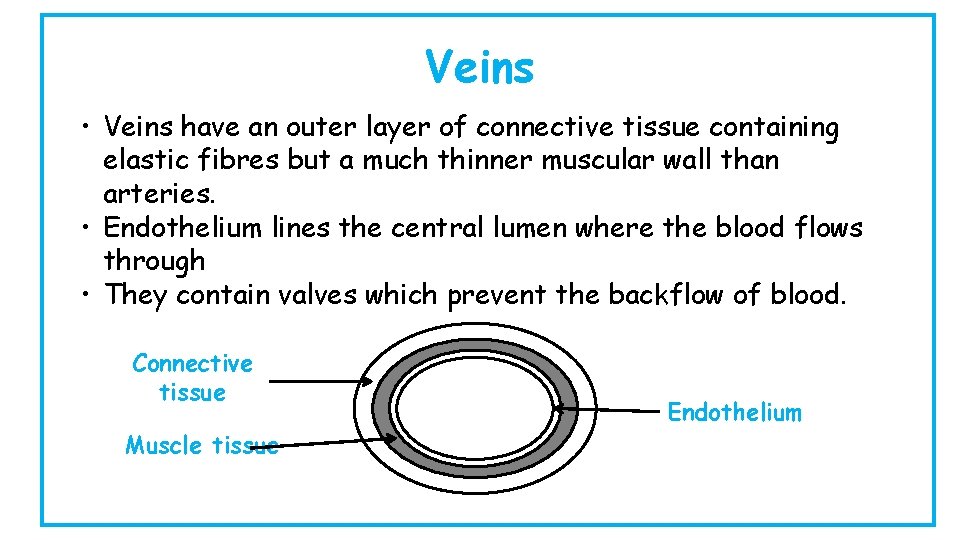
Veins • Veins have an outer layer of connective tissue containing elastic fibres but a much thinner muscular wall than arteries. • Endothelium lines the central lumen where the blood flows through • They contain valves which prevent the backflow of blood. Connective tissue Muscle tissue Endothelium
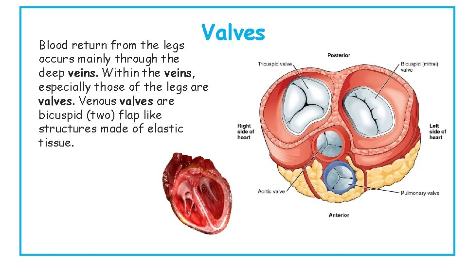
Valves Blood return from the legs occurs mainly through the deep veins. Within the veins, especially those of the legs are valves. Venous valves are bicuspid (two) flap like structures made of elastic tissue.
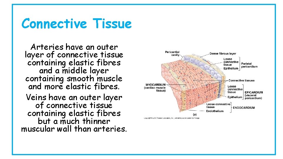
Connective Tissue Arteries have an outer layer of connective tissue containing elastic fibres and a middle layer containing smooth muscle and more elastic fibres. Veins have an outer layer of connective tissue containing elastic fibres but a much thinner muscular wall than arteries.
c – Material Exchange between Tissue Fluid and Cells We will be learning… • To explain the process of material exchange between tissue fluid and cells through pressure filtration • State what materials move from the tissue fluid into cells • State what materials move from cells to the tissue fluid • To describe the role of the lymphatic vessels • Explain the causes of oedema in conditions such as kwashiorkor and elephantiatis
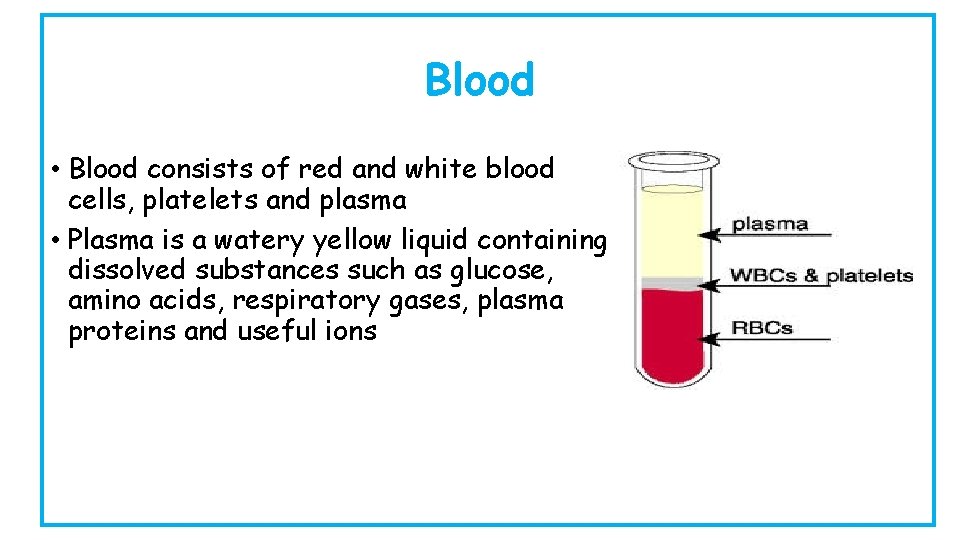
Blood • Blood consists of red and white blood cells, platelets and plasma • Plasma is a watery yellow liquid containing dissolved substances such as glucose, amino acids, respiratory gases, plasma proteins and useful ions
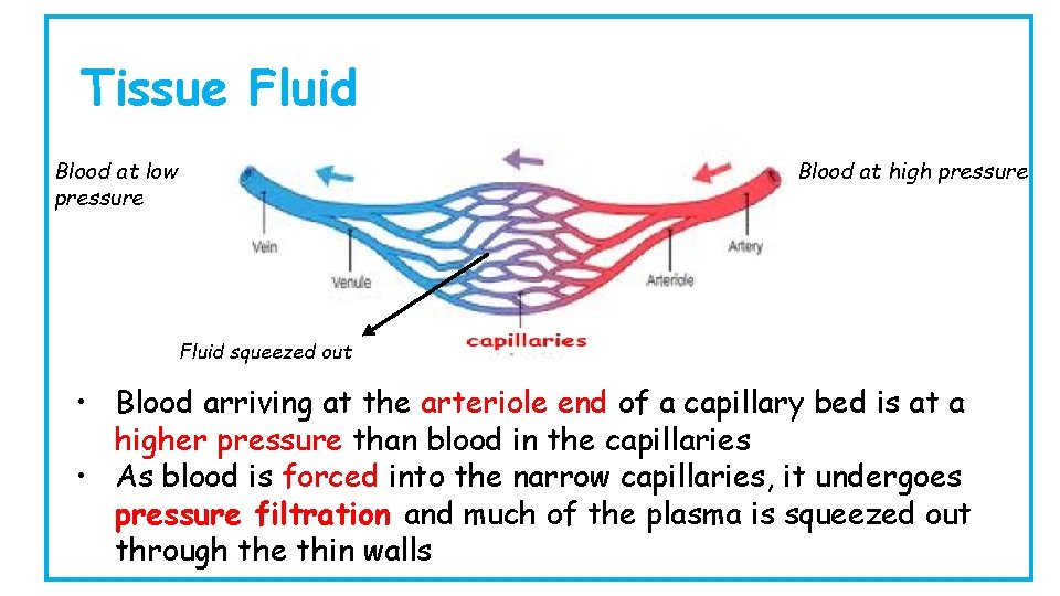
Tissue Fluid Blood at low pressure Blood at high pressure Fluid squeezed out • Blood arriving at the arteriole end of a capillary bed is at a higher pressure than blood in the capillaries • As blood is forced into the narrow capillaries, it undergoes pressure filtration and much of the plasma is squeezed out through the thin walls
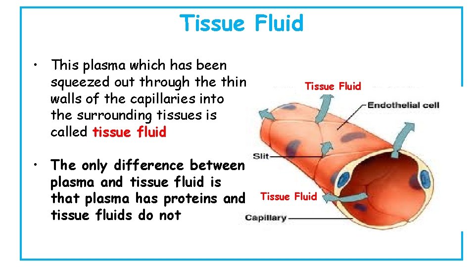
Tissue Fluid • This plasma which has been squeezed out through the thin walls of the capillaries into the surrounding tissues is called tissue fluid • The only difference between plasma and tissue fluid is that plasma has proteins and tissue fluids do not Tissue Fluid
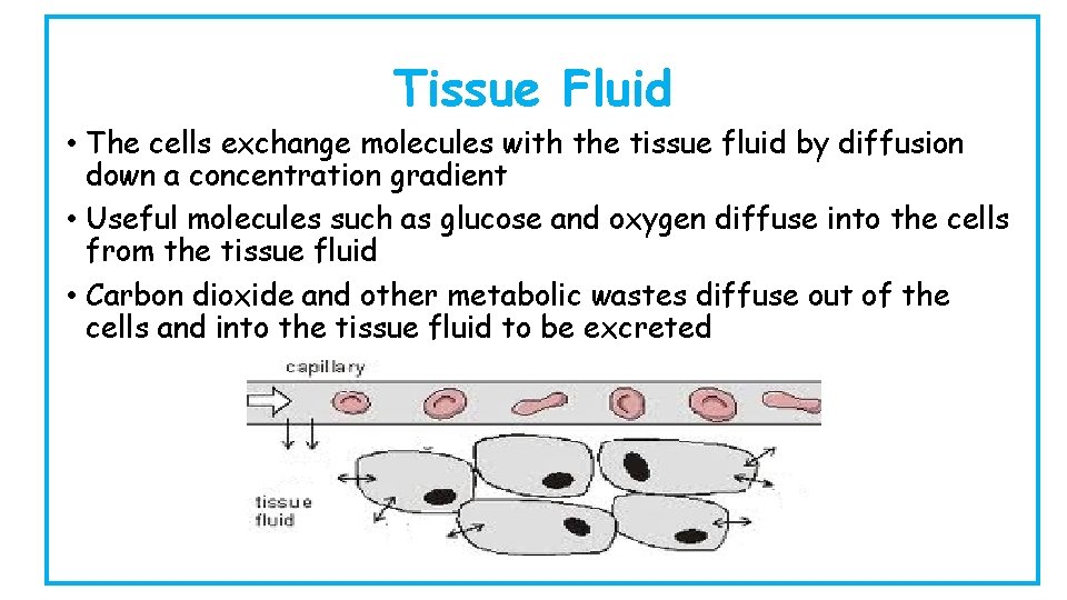
Tissue Fluid • The cells exchange molecules with the tissue fluid by diffusion down a concentration gradient • Useful molecules such as glucose and oxygen diffuse into the cells from the tissue fluid • Carbon dioxide and other metabolic wastes diffuse out of the cells and into the tissue fluid to be excreted
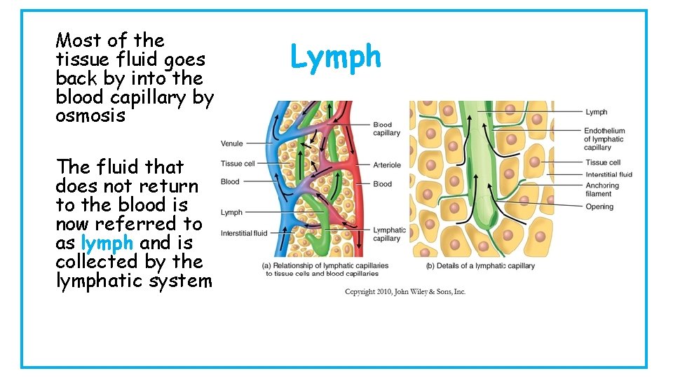
Most of the tissue fluid goes back by into the blood capillary by osmosis The fluid that does not return to the blood is now referred to as lymph and is collected by the lymphatic system Lymph
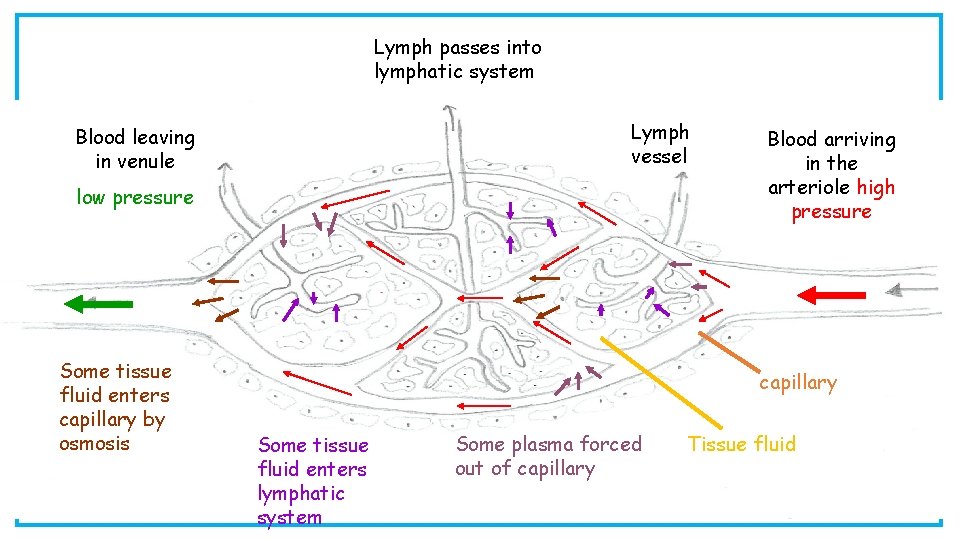
Lymph passes into lymphatic system Lymph vessel Blood leaving in venule low pressure Some tissue fluid enters capillary by osmosis Blood arriving in the arteriole high pressure capillary Some tissue fluid enters lymphatic system Some plasma forced out of capillary Tissue fluid
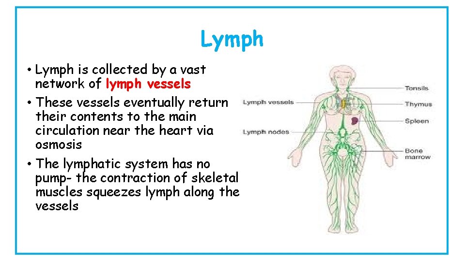
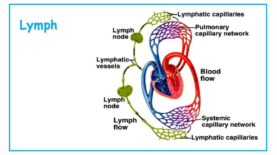
Lymph • Lymph is collected by a vast network of lymph vessels • These vessels eventually return their contents to the main circulation near the heart via osmosis • The lymphatic system has no pump- the contraction of skeletal muscles squeezes lymph along the vessels
Lymph
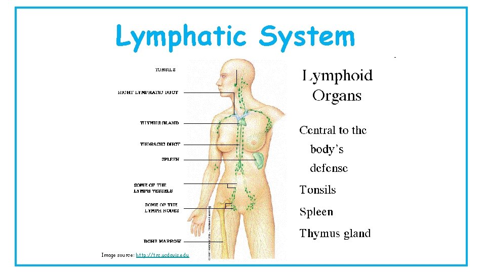
Lymphatic System Image source: http: //trc. ucdavis. edu
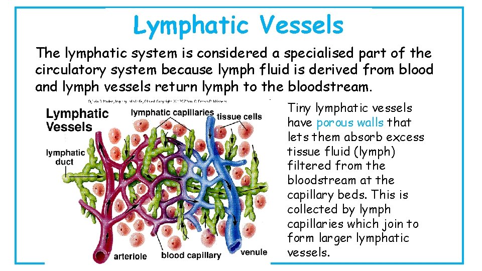
Lymphatic Vessels The lymphatic system is considered a specialised part of the circulatory system because lymph fluid is derived from blood and lymph vessels return lymph to the bloodstream. Tiny lymphatic vessels have porous walls that lets them absorb excess tissue fluid (lymph) filtered from the bloodstream at the capillary beds. This is collected by lymph capillaries which join to form larger lymphatic vessels.
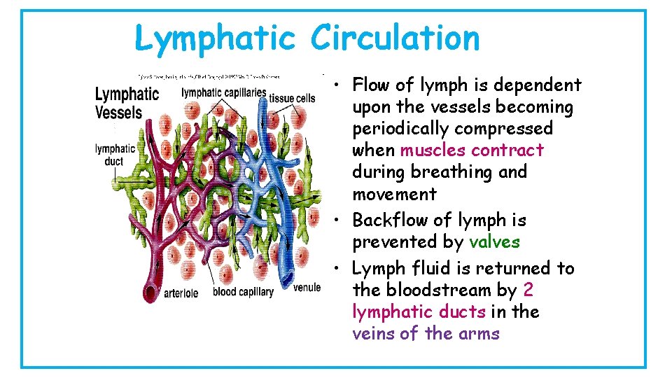
Lymphatic Circulation • Flow of lymph is dependent upon the vessels becoming periodically compressed when muscles contract during breathing and movement • Backflow of lymph is prevented by valves • Lymph fluid is returned to the bloodstream by 2 lymphatic ducts in the veins of the arms
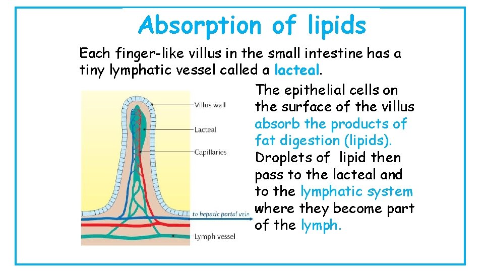
Absorption of lipids Each finger-like villus in the small intestine has a tiny lymphatic vessel called a lacteal. The epithelial cells on the surface of the villus absorb the products of fat digestion (lipids). Droplets of lipid then pass to the lacteal and to the lymphatic system where they become part of the lymph.
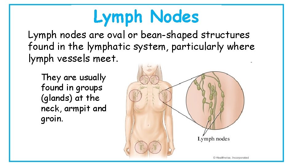
Lymph Nodes Lymph nodes are oval or bean-shaped structures found in the lymphatic system, particularly where lymph vessels meet. They are usually found in groups (glands) at the neck, armpit and groin.
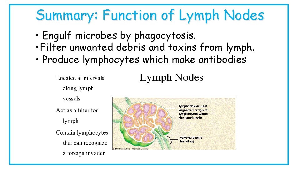
Summary: Function of Lymph Nodes • Engulf microbes by phagocytosis. • Filter unwanted debris and toxins from lymph. • Produce lymphocytes which make antibodies
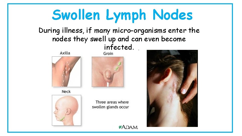
Swollen Lymph Nodes During illness, if many micro-organisms enter the nodes they swell up and can even become infected.
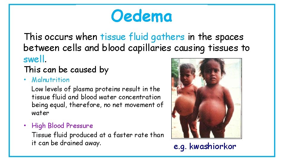
Oedema This occurs when tissue fluid gathers in the spaces between cells and blood capillaries causing tissues to swell. This can be caused by • Malnutrition Low levels of plasma proteins result in the tissue fluid and blood water concentration being equal, therefore, no net movement of water • High Blood Pressure Tissue fluid produced at a faster rate than it can be drained away. e. g. kwashiorkor
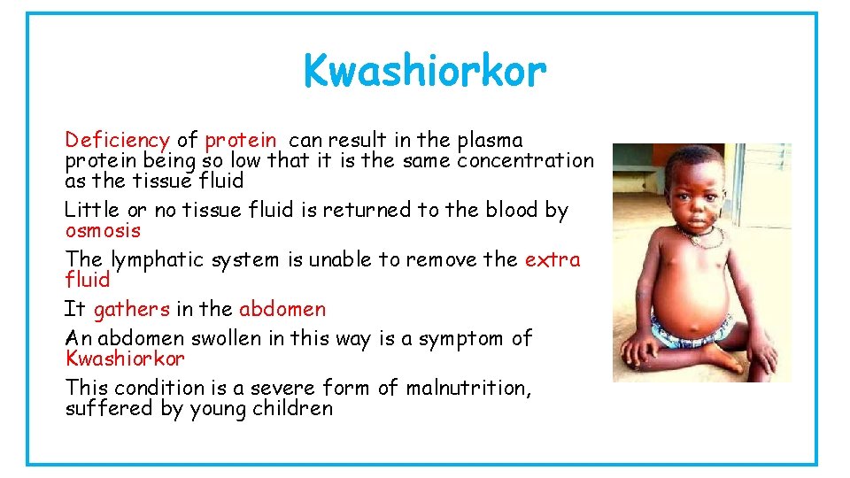
Kwashiorkor Deficiency of protein can result in the plasma protein being so low that it is the same concentration as the tissue fluid Little or no tissue fluid is returned to the blood by osmosis The lymphatic system is unable to remove the extra fluid It gathers in the abdomen An abdomen swollen in this way is a symptom of Kwashiorkor This condition is a severe form of malnutrition, suffered by young children
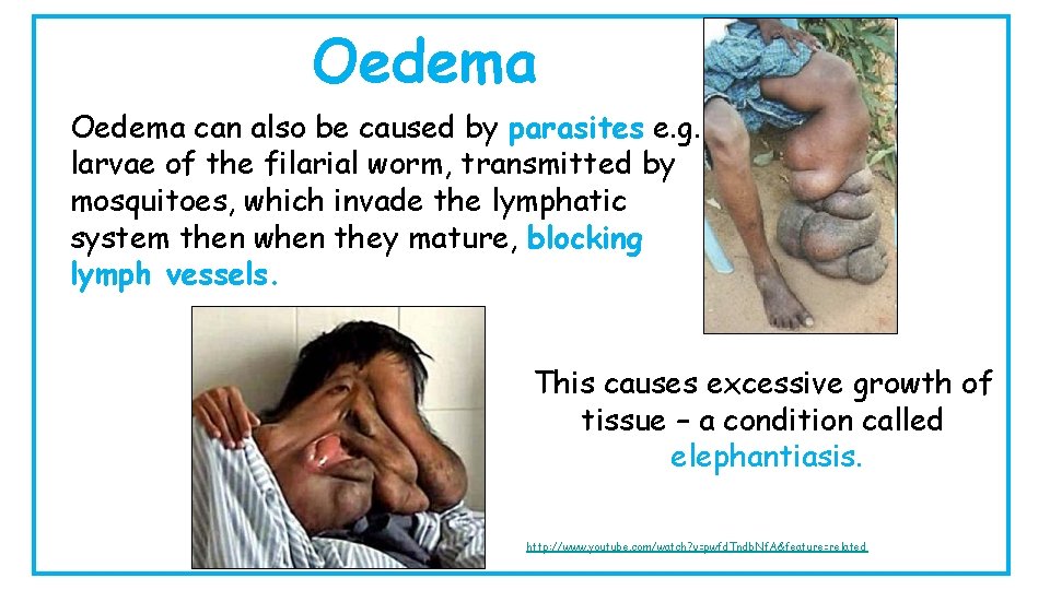
Oedema can also be caused by parasites e. g. larvae of the filarial worm, transmitted by mosquitoes, which invade the lymphatic system then when they mature, blocking lymph vessels. This causes excessive growth of tissue – a condition called elephantiasis. http: //www. youtube. com/watch? v=pwfd. Tndb. Nf. A&feature=related
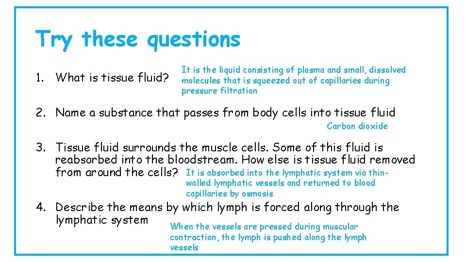
Try these questions 1. What is tissue fluid? It is the liquid consisting of plasma and small, dissolved molecules that is squeezed out of capillaries during pressure filtration 2. Name a substance that passes from body cells into tissue fluid Carbon dioxide 3. Tissue fluid surrounds the muscle cells. Some of this fluid is reabsorbed into the bloodstream. How else is tissue fluid removed from around the cells? It is absorbed into the lymphatic system via thinwalled lymphatic vessels and returned to blood capillaries by osmosis 4. Describe the means by which lymph is forced along through the lymphatic system When the vessels are pressed during muscular contraction, the lymph is pushed along the lymph vessels
a – Circulation Now I can…. . • Describe the pathway of blood through the circulatory system. • Explain the changes in blood pressure through the heart, arteries, capillaries and then veins.
b – Structure and Function of Arteries, Capillaries and Veins Now I can…. . • Describe the structure and function of arteries, capillaries and veins. • Describe the structure and function of the endothelium, central lumen, connective tissue, elastic fibres, smooth muscle and valves. • Explain the role of vasoconstriction • State that capillaries allow exchange of substances with tissues through their thin walls • Explain the role of vasodilation
c – Material Exchange between Tissue Fluid and Cells Now I can…. . • Explain the process of material exchange between tissue fluid and cells through pressure filtration • State what materials move from the tissue fluid into cells • State what materials move from cells to the tissue fluid • Describe the role of the lymphatic vessels • Explain the causes of oedema in conditions such as kwashiorkor and elephantiasis
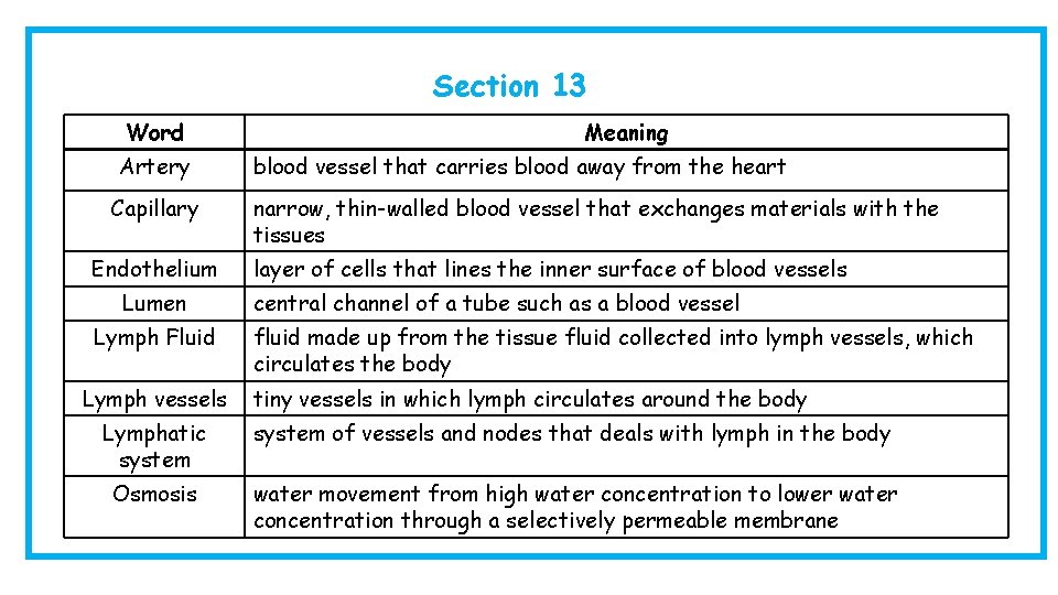
Section 13 Word Artery Capillary Endothelium Lumen Lymph Fluid Lymph vessels Meaning blood vessel that carries blood away from the heart narrow, thin-walled blood vessel that exchanges materials with the tissues layer of cells that lines the inner surface of blood vessels central channel of a tube such as a blood vessel fluid made up from the tissue fluid collected into lymph vessels, which circulates the body tiny vessels in which lymph circulates around the body Lymphatic system of vessels and nodes that deals with lymph in the body Osmosis water movement from high water concentration to lower water concentration through a selectively permeable membrane
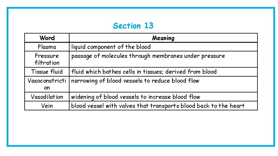
Section 13 Word Plasma Pressure filtration Tissue fluid Meaning liquid component of the blood passage of molecules through membranes under pressure fluid which bathes cells in tissues; derived from blood Vasoconstricti narrowing of blood vessels to reduce blood flow on Vasodilation Vein widening of blood vessels to increase blood flow blood vessel with valves that transports blood back to the heart
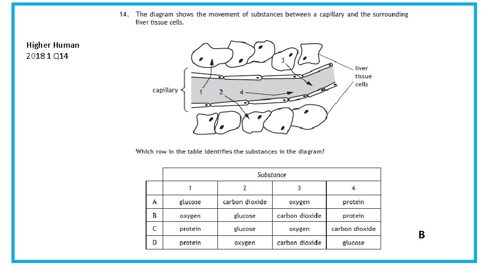
Higher Human 2018 1 Q 14 B
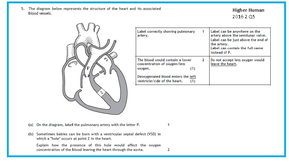
Higher Human 2016 2 Q 5 P
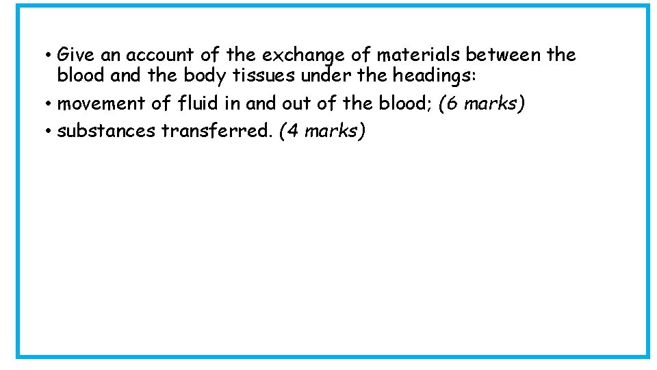
• Give an account of the exchange of materials between the blood and the body tissues under the headings: • movement of fluid in and out of the blood; (6 marks) • substances transferred. (4 marks)
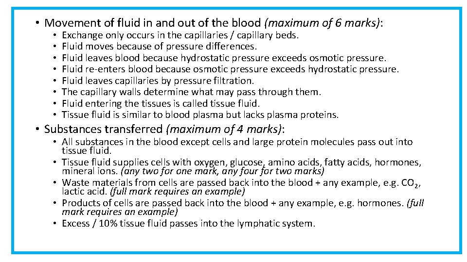
• Movement of fluid in and out of the blood (maximum of 6 marks): • • Exchange only occurs in the capillaries / capillary beds. Fluid moves because of pressure differences. Fluid leaves blood because hydrostatic pressure exceeds osmotic pressure. Fluid re-enters blood because osmotic pressure exceeds hydrostatic pressure. Fluid leaves capillaries by pressure filtration. The capillary walls determine what may pass through them. Fluid entering the tissues is called tissue fluid. Tissue fluid is similar to blood plasma but lacks plasma proteins. • Substances transferred (maximum of 4 marks): • All substances in the blood except cells and large protein molecules pass out into tissue fluid. • Tissue fluid supplies cells with oxygen, glucose, amino acids, fatty acids, hormones, mineral ions. (any two for one mark, any four for two marks) • Waste materials from cells are passed back into the blood + any example, e. g. CO 2, lactic acid. (full mark requires an example) • Products of cells are passed back into the blood + any example, e. g. hormones. (full mark requires an example) • Excess / 10% tissue fluid passes into the lymphatic system.
Suggested activities • Heart model and human torso • Heart dissection • Circulation diagram
- Slides: 65