Hernia of the anterolateral abdominal wall Definition w

Hernia of the anterolateral abdominal wall

Definition w Progressive protrusion through the abdominal wall of the peritoneum, with tendency to progress, together with an abdominal viscus w SO – An abdominal viscus will HAVE to leave the abdominal cavity – There must be a peritoneal covering

NOT real hernias by this definition w Embrionic or fetal hernia where there is an anomaly in development w Protrusions of the organs of the retroperitoneum without peritoneal cover.

Common manifestations of hernia

HERNIA? Pathological aspects

Hernia development – HERNIATION POINTw First step in develeopment w The protrusion of serosa begins like a small bulge through a small PARIETAL DEFECT w CLINICAL SIGNS: – Pain of variable intensity – Digital examination may be inconclusive, except for a large defect

Hernia development – Interstitial herniaw Peritoneal diverticulum increases in size w Protrusion within the muscular-fascial structures of the abdominal wall w Peritoneal serosa becomes thick and becomes a herniation sac w CLINICALLY: – Pain through compression on viscera or traction on mesentery. Possible pain through interstitial compression – All signs of a hernia can be identified

Hernia development – COMPLETE HERNIAw Herniation sac = completely passed through the wall w Clinical signs are complete both in uncomplicated and complicated form

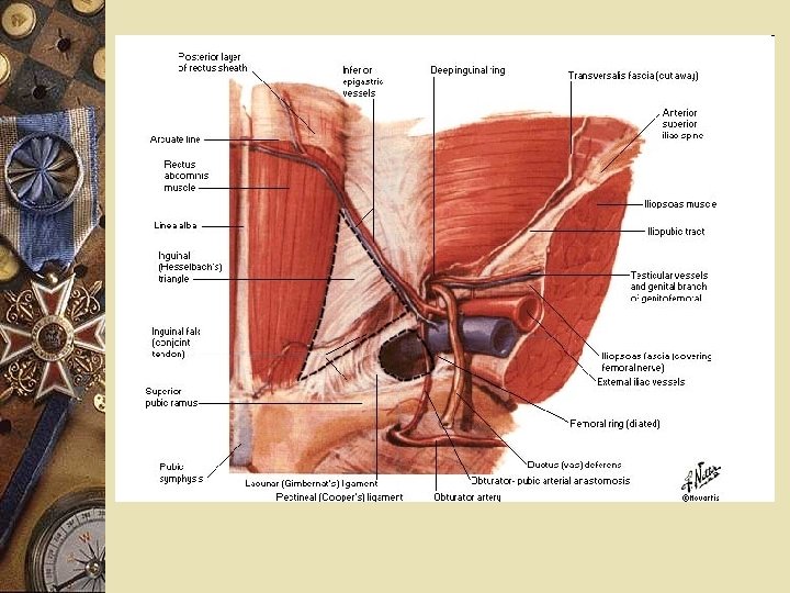
PATHOLOGIC CHANGES w Wall defect – the abnormality in the abdominal wall – – Fibrous (umbilical hernia) Fibro-muscular (epigastric hernia) Fibro-osseous (obturator hernia) True channel (inghuinal hernia) w Hernia wall or coverings w Hernia content

Complete hernia – structures of the wall w Skin and subcutaneous fat w Sac (peritoneum which is stretched + fat and structures migrating from under the peritoneum) – Fundus area – Neck area

Causes w Conflict: pressure inside the abdominal cavity and possibility of the abdominal wall to content that pressure w Fragile balance – if imbalance appears a herniation point and a hernia will develop

Causes w Congenital: the sac preexists at birth or defect of development w Acquired hernia : in areas of minimal resistence of the abdominal wall
 or chronic")
Causes -high intraabdominal pressurew An increase in abdominal pressure acute (muscular rupture) or chronic (long term increase in stress over the abdominal wall) may increase the risk of hernia development – Increase respiratory effort: chronic respiratory diseases associated with cough; jobs that require increase expiratory effort. – Tumors or peritoneal effusion in large quantity (pregnancy, ascites, peritoneal dialyses) – Straining or effort with closed epiglotis – Functional disorders with chronic effort (prostate adenoma, chronic constipation) – Pathologic causes – colonic tumor!!!!!

Causes -wall defectsw Abdominal structure is not homogenous WEAK POINTS – Natural communications between abdominal cavity and other cavities – Passing of nerves or vessels towards superficial structures – Scars (posttraumatic, postoperative) – Intersection of fascial structures

Causes -wall defects w Other factors essential in hernia develoment – Loss of tissue elasticity and resistence – usually associated with agging – Genetic factors – hernias predominant in some families: defects in synthesis and structure of colagen fibers – Trauma – tissue distruction + scars. Infection is a major contributor in incisional hernia – Metabolic abnormalities

Hernia formation w Hernia with preexisting sac: development abnormalities when the peritoneal diverticula is preexistent. There is no wall defect. w Pushing hernia: association of high intraabdominal hernia and weak point w Sliding henria: similar but organs attached to peritoneum slide in the sac. w Hernia with abnormally distended sac – peritoneum fixed at the level of the neck is blown up and loses its characteristics (umbilical hernia)

Clinical signs in uncomplicated hernia w Pseudo-tumoral bulge with variable medical history that is apparent to the patient w Discomfort; difficulties in dressing +/- skin lesion through friction; the patient notices that it can be reduced and may need an orthopedic support. w Pain: traction or compression on nerves or mesentery. Usually it is bothersome but not major. Small hernia with small defects will be more painful. w Incomplete obstruction – when bowel is present in large hernia w Esthetic problem

Clinical examination -uncomplicated herniaw Positio of the patient : – Standing up : COMPULSORY as an initial assesment – Laying down - compare the size and dynamic of tumor when intraabdominal pressure changes – ALL WEAK ABDOMINAL POINTS should be examined, as more hernias can be present w Protect the patient’s sensibility

Clinical examination -uncomplicated herniaw Inspection: – Tumor, bulging, in an area known as weak area of the abdominal wall – “Tumor” is changing volume according to changes in abdominal pressure (standin/laying down, coughing, straining) – Skin covering is normal – Volume increases while coughing – Progression of hernia follows a trajectory which is the herniation channel

Clinical examination -uncomplicated herniaw Superficial palpation – Check the sensibility – Tumor has elastic consistency – Pear-like shape with a neck that continues in the abdominal cavity!!! (very important) – Content: diferentiate between bowel and non digestive structures – Reduce the hernia content in the abdominal cavity REDUCTIBLE HERNIA – Hernia forms back after reduction: COERCIBILE VS NONCOERCIBLE

Clinical examination -uncomplicated herniaw Palpation of the abdominal wall after reduction of the content – Evaluation of the well defect (dimension, structure, position) – The “tumor” follows the finger to progress during a coughing effort, following the direction of your finger EXPANSSION – The “tumor” knocks your finger during a coughing effort PULSATE WITH COUGH

Clinical examination -uncomplicated herniaw Percussion – Tympanic – presence of air = bowel – Dull = omentum or retroperitoneal fat, but bowel can also be present but does not contain air.

Clinical examination -uncomplicated herniaw Auscultation – NOT significant but you may hear hydro-aeric sounds characteristic for bowel content

POSITIV DIAGNOSTIC IN UNCOMPLICATED HERNIA w w w “Tumor” or bulge + in a weak point Normal skin Volume changes with postural changes Pedicle inside the abdominal cavity Communication through a defect in the abdominal wall - palpable w Reducible + expansion during cough w Pulsation during cough

Lab exploration w Barium enema-colon in hernia + colonic tumors w Small bowel followup w Ultrasound scan content w Laparoscopy – “gold standard” for small hernia

Natural history w Hernia of the adult never heal spontaneously!!! w Hernia with a large defect are well tolerated but represent a handicap w Rigid defect: can produce a strangulation at any time w COMPLICATIONS – given enough time all hernias will complicate

Complications w Irreducible w Incarceration w To large to be adapted in the peritoneal cavity “no right to stay in the abdomen” w Strangulation w Incomplete intestinal obstruction peritonitis in the sac w Complications due to compression (testicular atrophie, changes in urinary habits, respiratory disfunction) w Trauma to the hernia w Tumors in the hernia w Foreign body in the hernia

Strangulation w The most serious complication: transforms a benign pathology in one potentially lethal w CAUSES that favor strangulation: – Inextensible parietal defect (orifice) – Narrow or sclerotic neck of hernia sac – Adhesions in the sac

Pathogenesis of strangulation w Effort with sudden increase in intra-abdominal pressure w A larger volume of bowel/viscus is pushed in the hernia w Increases the pressure inside hernia sac – Much more so at the level of the inextensible hernia orificeor neck of hernia w Impediment in the venous retur with consecutive edema. w Further increase in intra-sacular pressure and of hernia volume w Pressure inside the hernia becomes bigger then arterial pressure = ischemia SPEED OF PROGRESSION towards ireversible lesions is greater in tight strangulation.

Lesions w Sac: same changes edematous – eritematous – liquid initially serous+/- bloody the puss or fecal w Intestinal loop: 3 stages 1. Congestion (venous stasis): congesitve loop, cyanosis, visible strangulation ridge. REVERSIBLE LESION w 2. Intermediate bowel becomes purple – black, more rapidly at the strangulation area, the loop wall is destroyed and reduced to serosa 3. Necrosis and perforation the lopp becomes green (necrotic) like a dead leaf. Partial or total rupture of the wall + contamination of the peritoneum of the sac.

Pathology w Mesentery in strangulated area – Edematous, friable with distended veins and trombosis w Omentum – Similar as above, can progress towards necrosis
 a clinical manifestation of complete obstruction")
Intestinal obstruction w Strangulation = (with few exception) a clinical manifestation of complete obstruction – Loops above hernia are dilated, with active peristalsis – Loops below hernia are emtpy w After perforation – peritonitis (either localized in the hernia sac or generalized peritonitis
 – Strangulation of a segment of circumference on")
Unusual forms w Lateral pinch (Richter) – Strangulation of a segment of circumference on the anti-mesenteric border – Incomplete clinical manifestations of intestinal obstruction (lumen is free) – Manual reduction of hernia is possible but ischemic lesion of the loop may progress in the abdomen – when the necrotic tissue is delimitated and falls of = PERITONITIS – More frequent in femoral hernia

Unusual forms w Retrograde strangulation “In W” – A large loop is in the hernia but strangulation involves a segment of loop situated in the abdominal cavity with a part of mesentery in the hernia – Greatest risk – during the surgical cure in the emergency settings – the intraabdominal loop may not be noticed - PERITONITIS

Clinical signs in strangulation w SHARP PAIN at the level of hernia, continuous – SIGNAL - viability of the loop is threatened w INTESTINAL OBSTRUCTION w Colicky abdominal pain (obstruction) w Nausea, vomiting (at first food, the bile, then fecal aspect) w No intestinal transit but diarrhea is posible

General signs w Very good at first w Tachycardia w Anxiety

Clinica examination w Patient is known to have a hernia BUT not always (strangulation as a first symptom) w Hernia is large and painful (in particular at the level of the neck) w DISAPPEAR impulsion and expansion with cough w Henria becomes irreducible: TAXISUL (forceful reduction) is very dangerous – and more so after one hour from onset – En bloc reduction together with peritoneum – Non vital loop being reduced in the peritoneum

Clinical examination w Abdomen: classic appearance of intestinal obstruction – Meteorism – Hyper-peristaltic loops – Borborism w Peritonitis; it is a “normal” evolution of clinical aspect a strangulated hernia neglected for too long w Abscess formation – may open spontaneously producing a digestive fistula

Positive diagnosis w Hernia can not be reduced ANY MORE w NO impulsion NO expansion w Hernia becomes painful - continuous pain w Intestinal obstruction w Peritonitis

Treatment of strangulation w URGENT: operated as soon as possible to save the loop w Hemo-dynamic control w Gastric aspiration (naso-gastric tube) w Surgical treatment using any type of anesthesia

Hernia SAC w Open but isolate as it may be contaminated Laparotomy – if abdominal contamination is probable! – – Treat content Resection of sac Close peritoneum Drain the contaminated area (+/-)

Content w Incise the neck and decompress the strangulation area w Evaluate viability of bowel loop – If viable – reintroduce in the peritoneal cavity – Not viable – resect – In doubt: warm saline + infiltrations in the mesenter; wait and see

Orifice w Close the orifice and repair the defect w Exception: – Massive contamination. Repair can be put in danger by septic complications
 – nothing")
Peritoneal cavity w Non-contaminated (infection limited at the level of the neck) – nothing special but need to be checked intraoperatively w Contaminated – LAPAROTOMY (LAPAROSCOPY) irrigate and drain – Intestinal resection

Irreducible hernia w Henria content can not be reduce anymore – does not affect viability of the loop – In general it is progressive. The hernia is more and more difficult to be reduced. BUT sudden henriation of a larger volume can induce this complication. – Intra-sacular adherences w Old hernia with step by step development of irreducibility w Differential diagnosis – strangulation: all strangulated hernias are ireducible
 w Rare complication")
NO right to stay anymore in the abdomen (not welcome anymore) w Rare complication of very large hernia that recur immediately after reduction w Large volume outside the abdominal cavity for a long time = abdomen is reshaped on a smaller content w Reduction immediately increases the abdominal pressure and the whole volume can not be reduce or recurs immediately
 w Consequences :")
NO right to stay anymore in the abdomen (not welcome anymore) w Consequences : – hernia is incoercible – Forceful reduction and contention is accompanied by respiratory distress – Treatment is very problematic • Need to increase the abdominal volume in time • Organ resections to reduce the pressure • Large synthetic meshes

Trauma to the hernia w Organs in the hernia are exposed. Much so if traumatized they do not have the liberty to retract in the abdomen. Entraped. w Diagnostic problems – Lesions that can progress in 2 steps – Intra-sacular peritonitis is non specific and few symptoms may be present. May develop generalized peritonitis.

Peritonitis in the hernia w Unusual complication w Secondary to infectious complications of intra-sacular organs (appendicitis, diverticulitis, etc) w Clinical signs: increase in volume, becomes painful, ireducible, local signs of inflamation
- Slides: 50