HERNIA Koray Topgl MD Prof General Surgery Dept


 inguinal ring")


 • Direct Inguinal")


 2. Occupation ( nature ? ?")
 Tender? Skin changes? Palpable edges Genitalia Rectal")


 or conjoint")


- Slides: 38
HERNIA Koray Topgül, MD, Prof. General Surgery Dept.
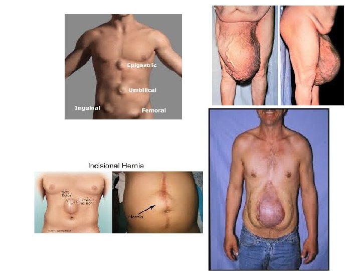
Definition A hernia is a protrusion of a viscus or part of a viscus through an abnormal opening in the walls of its containing cavity.
Common Hernia • Umbilical • Incisional • Inguinal – Direct and indirect • Femoral
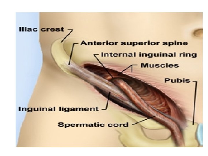
Anatomy The inguinal canal : - The inguinal canal is approximately 4 cm long and is 4 cm long directed obliquely inferomedially through the inferomedially inferior part of the anterolateral abdominal wall. The canal lies parallel and 2 -4 cm superior to the medial half of the inguinal ligament. This ligament extends from the anterior superior iliac spine to the pubic tubercle.
The inguinal canal has openings at either end : The deep (internal) inguinal ring is the entrance to The deep (internal) inguinal ring the inguinal canal. It is thesite of an outpouching of the transversalis fascia. This is approximately 1. 25 cm superior to the middle of the inguinal ligament The superficial, or external inguinal ring is the exit The superficial, or external inguinal from the inguinal canal. It is a slitlke opening between the diagonal fibres of the aponeurosis of the external oblique
Inguinal canal Walls of the inguinal canal : The anterior wall is formed mainly by the aponeurosis of the external Oblique The posterior wall is formed mainly by transversalis fascia The roof is formed by the arching fibres of the internal oblique and transverse abdominal muscles. transverse abdominal The floor is formed by the inguinal ligament, which forms a shallow trough. It is reinforced in its most medial part by the lacunar ligament.
Groin Anatomy--Anterior • • Inguinal ligament Layers External ring Internal ring Spermatic cord Inferior epigastrics Hesselbach’s triangle • Femoral vessels
Groin Anatomy--Posterior
Transperitoneal Anatomy
Content : 1. Spermatic cord ( round ligament of the uterus in female ) Spermatic cord The Cord Itself. —The contents of the spermatic cord are (a) the ductus (vas) deferens and its artery. (b) the testicular artery and venous (pampiniform) plexus. (c) the genital branch of the genitofemoral nerve. (d) lymphatic vessels and sympathetic nerve fibers. (e) fat and connective tissue surrounding the cord and its coverings in various amounts 2. Ilioinguinal nerve 3. Ilioinguinal lymph node
Femoral Canal Contains, from lateral to medial, the femoral artery, femoral vein, and femoral canal. Other features of the femoral triangle include the femoral nerve, which lies lateral to the sheath, Wall of The Femoral canal Anterior is the inguinal ligament Posterior is the iliopsoas, pectineal, and long adductor muscles (floor). Medial is lacunar ligament Lateral is femoral vessle
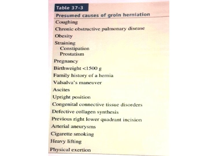
Predisposing: All hernias occur at the site of WEAKNESS OF THE ABDOMINAL WALL which are acted on by repeated INCREASE in abdominal pressure
• Indirect Inguinal Hernia through the inguinal canal (Lateral Hernia) • Direct Inguinal Hernia The sac passes through a weakness or defect of the transversalis fascia in the posterior wall of the inguinal canal-Hasselbach triangle. . (Medial Hernia) • Femoral Hernia medial to femoral vessels under inguinal ligament • Umbilical Hernia through the umbilical ring
Paraumbilical Hernia A protrusion through the linea alba just above or sometimes just below the umbilicus Epigastric Hernia Protrusion of extraperitoneal fat through the linea alba anywhere between the xiphoid process and the umbilicus Incisional Hernia through an incisional site an incisional Lumber Hernia occur through the inferior lumber triangle of Petit
Repeated INCREASE in abdominal pressure is usually due to Chronic cough Straining Bladder neck or urethral obstruction Pregnancy Vomiting Sever muscular effort Ascetic fluid
Inguinal hernia History: 1. Age (young vs. old) 2. Occupation ( nature ? ? ) 3. Local symptoms: Swelling, discomfort and pain 4. Systemic symptoms: if there is obstruction or strangulation 5. Precipitating factors
Pertinent Exam • • Location Reducible? (incarcerated=irreductible) Tender? Skin changes? Palpable edges Genitalia Rectal
Inguinal hernia Examination: 1. Inspection for site, size, shape and color. 2. Palpation for surface, temp, tenderness, composition and reducibility. 3. Expansible cough impulse. 4. General exam: for common causes of increase intra abdominal pressure
Indirect Versus Direct inguinal hernias Indirect is the most common form of hernia and its usually congenital due to congenital patent processus vaginalis Direct usually acquired occur in old men with weak abdominal muscles. weak abdominal
Indirect Versus Direct inguinal hernias Indirect Inguinal Hernia Direct Inguinal Hernia Pass through inguinal canal. Bulge from the posterior wall of the inguinal canal Can descend into the scrotum. Cannot descent into the scrotum. Lateral to inferior epigastric vessels. Medial to inferior epigastric vessels. Reduced: upward, then laterally and backward. Reduced: upward, then straight backward. Controlled: after reduction by pressure over the internal (deep) inguinal ring. Not controlled: after reduction by pressure over the internal (deep) inguinal ring. The defect is not palpable (it is behind the The defect may be felt in the abdominal wall fibers of the external oblique muscle). above the pubic tubercle. After reduction: the bulge appears in the middle of inguinal region and then flows medially before turning down to the scrotum. After reduction: the bulge reappears exactly where it was before. Common in children and young adults. Common in old age.
Femoral hernia Small femoral hernia may be unnoticed by the patient or disregarded for years perhaps until the day it strangulates. Adherence of the greater omentum sometimes causes a dragging pain. Rarely a large sac is present.
Femoral hernia History Age ; uncommon in children , most common in old age female. Sex; women > men (but still commonest hernia in women the inguinal hernia ) The patient came with local symptoms 1 - discomfort and pain 2 - swelling in the groin General ; femoral hernia is more likely to be strangulated than the inguinal hernia Multiplicity ; often bilateral
Femoral hernia versus inguinal hernia Inguinal hernia Femoral hernia 1 - more common in male 1 - more common in females 2 - pass through the inguinal canal 2 - pass through the femoral canal 3 - less common to be strangulated 4 - more common to be strangulated 4 - can be treated without surgery 5 - must be treated surgically
Umbilical Hernia through the umbilical scar , so it is a true umbilical hernia. Not common and is usually secondary to increase intra abdominal pressure. The most common causes 1 - pregnancy 2 - ascitis 3 - ovarian cyst 4 - fibrosis 5 - bowel distention
Incision hernia Signs and symptoms Previous operation or accidental trauma Age ; all ages , but more common in old age. Symptom ; lump , pain , intestinal obstruction ( distention , colic, vomiting , constipation , sever pain in the lump ) Examination 1 - reducible lump 2 - expansile cough impulse 3 - if the lump dose not reduse and dose not have cough impulse , than it may be not a hernia Ddx Tumor Chronic abscess Hematoma Foreign body granuloma
Etiology of Incisional Hernia Patient-related factors—DM, Obesity, Smoke, Chronic Patient-related factors obstructive pulmonary disease, multiple surgery, cancer, chemotherapy, radiotherapy, malnutrition, jaundice, CRF. . Technical factors---bad surgical technique, wrong echnical factors surgical materials Mechanical factors---Mechanical factors increase the Mechanical factors intra-abdominal pressure after an operation and causes the hernia. Common causes include-
Chronic cough- Coughing and vomiting are associated with a brief but significant increase in intra-abdominal pressure. This, again, leads to too much tension on the incision and possible breakdown of the incision. Lifting heavy weights- Obviously, if a patient lifts something too heavy immediately after the operation, he or she can also tear the incision and cause a hernia. Postoperative ileus- This occurs when the abdomen becomes distended because the intestines are not working properly after an operation. The swelling of the intestines increases the pressure in the abdomen and places tension on the incision. The tension then leads to defective healing. Constipation-Straining to move bowels after surgery
Management and Repair
Reduction Uncomplicated: Manual Gentle pressure over hernia Gentle traction over the mass sedation Complicated (strangulated): no attempt!! should be made to reduce the hernia because of potential reduction of gangrenous segment of bowel with the hernial sac.
Surgical Treatment 1. choice of anesthetic: Elective open repair : Local/Spinal anesthesia is preferred Laproscopic hernia repair: more commonly under GA.
Inguinal Floor Reconstruction 3. Inguinal Floor Reconstruction Some method of reconstruction of the inguinal floor is necessary in all adult hernia repairs to prevent recurrence. Primary tissue repair Open tension free repair Laproscopic & preperitoneal repairs
1. Primary tissue repair Bassini repair: inferior arch of transversalis fascia (TF) or conjoint tendon is approximated to shelving portion of inguinal ligament. Mc. Vay: TF is sutured to cooper ligament. Shouldice: TF is incised and reapproximated.
Shouldice Repair • Imbricated, running repair
2. Open tension free repair Lichtenstein repair &Patch and Plug technique: Mesh is used to reconstruct inguinal floor Mesh plug technique : place mesh in the hernial defect
Mesh repair • Tension free • Less painful • Foreign body