HEMOSTASIS Stages of Blood Clotting 1 Vascular Spasms
")
HEMOSTASIS (Stages of Blood Clotting)

1. Vascular Spasms • In response to damage of an artery or arteriole the rings of smooth muscle in the walls of the vessel begin to spasm immediately • This reduces blood loss and gives time for other mechanisms to begin • The spasms are caused by chemicals that are released by the smooth muscle, platelets, and pain receptors in the area
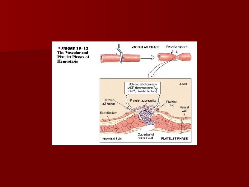

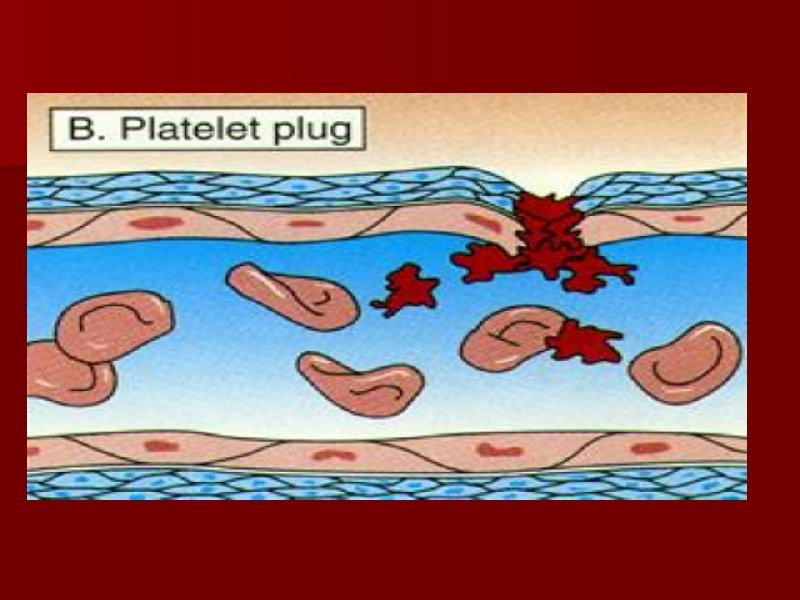
2. Platelet Plug Formation • Platelets come into contact with the damaged vessel and stick to the parts of it that are torn (called platelet adhesion) • The adhesion activates the platelets and they change • Each platelet extends projections that allow them to touch one anther and they begin to empty the contents of their vesicles (called platelet release reaction)

2. Cont. • ADP and thromboxane A 2 are released to activate nearby platelets • Serotonin and thromboxane A 2 cause vasoconstriction of the blood vessel which in turn reduces blood flow from the damaged vessel • The ADP makes the platelets nearby sticky and they migrate and join the first responder platelets (platelet aggregation) • This will lead to the formation of a solid mass called the platelet plug.

Platelet Plug Formation Process Diagrams Step-by-Step Copyright © 2007 by John Wiley & Sons, Inc.

Red blood cell Platelet Collagen fibers and damaged endothelium 1 1 Platelet adhesion

Red blood cell Platelet Collagen fibers and damaged endothelium 1 1 Platelet adhesion Liberated ADP, serotonin, and thromboxane A 2 2 2 Platelet release reaction

Red blood cell Platelet Collagen fibers and damaged endothelium 1 1 Platelet adhesion Liberated ADP, serotonin, and thromboxane A 2 2 2 Platelet release reaction Platelet plug 3 3 Platelet aggregation
 • Takes advantage of the fact that blood only")
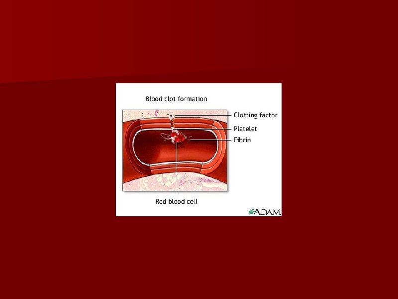
3. Blood Clotting (Coagulation Events) • Takes advantage of the fact that blood only remains a liquid when contained in vessels • Dependent on injured tissues releasing tissue factor (TF) which interacts with PF 3 on platelets • The first reactions form the molecule prothrombin (all controlled by thromboplastin) • Thromboplastin converts prothrombin into an enzyme called thrombin via reaction with Ca 2+ ions • Thrombin converts soluble fibrinogen into insoluble fibrin which forms the threads of the clot

After Affects • Once the clot is formed it will slowly begin to retract and pull the damaged ends of the vessel together • During this phase some blood serum can escape but the actual blood cells cannot • Eventually fibroblasts will begin to form new tissues in the area and new endothelial cells will repair the damaged lining • In the end the vessel will return back to normal. • Calcium ions (Ca 2+) are the main ion involved in the clotting process


Blood Clotting Process Diagrams Step-by-Step Copyright © 2007 by John Wiley & Sons, Inc.
 Extrinsic pathway Tissue trauma (b) Intrinsic pathway Blood trauma Damaged endothelial cells expose")
(a) Extrinsic pathway Tissue trauma (b) Intrinsic pathway Blood trauma Damaged endothelial cells expose collagen fibers Tissue factor (TF) Damaged platelets Activated XII Activated platelets Ca 2+ Platelet phospholipids Activated X V 1 V Ca 2+ PROTHROMBINASE
 Extrinsic pathway Tissue trauma (b) Intrinsic pathway Blood trauma Damaged endothelial cells expose")
(a) Extrinsic pathway Tissue trauma (b) Intrinsic pathway Blood trauma Damaged endothelial cells expose collagen fibers Tissue factor (TF) Damaged platelets Activated XII Activated platelets Ca 2+ + Platelet phospholipids Activated X V 1 V Ca 2+ + PROTHROMBINASE (c) Common pathway Ca 2+ Prothrombin (II) THROMBIN 2
 Extrinsic pathway Tissue trauma (b) Intrinsic pathway Blood trauma Damaged endothelial cells expose")
(a) Extrinsic pathway Tissue trauma (b) Intrinsic pathway Blood trauma Damaged endothelial cells expose collagen fibers Tissue factor (TF) Damaged platelets Activated XII Activated platelets Ca 2+ + Platelet phospholipids Activated X V 1 V Ca 2+ + PROTHROMBINASE (c) Common pathway Ca 2+ Prothrombin (II) THROMBIN Ca 2+ Fibrinogen (I) Loose fibrin threads 2 XIII Activated XIII STRENGTHENED FIBRIN THREADS 3
- Slides: 18