Hemostasis Hemostasis depends on the integrity of Blood

Hemostasis • Hemostasis depends on the integrity of – Blood vessels – Platelets – Coagulation factors – Anticoagulation factors

Initial Laboratory Tests For Bleeding Abnormalities • Platelet count. • Bleeding time: prolongation is seen in platelet diseases or in patients receiving drugs interfering with platelet function. • Partial thromboplastin time (PTT). – Prolongation is seen in hemophiliacs and in patients with lupus anti-coagulant. • Prothrombin time (PT). – Prolongation is seen in factor VII deficiency. – Factors V, X, prothrombin and fibrinogen affect APTT and PT. • Thrombin time.

Causes of Abnormal Bleeding • • Vascular disorders. Thrombocytopenia. Platelet function defects. Defective coagulation.

Coagulation disorders

Inherited Deficiencies of Coagulation Factors Deficiency Incidence in Gene on Mode of Population Chromosome Inheritance Fibrinogen 1: 1 million 4 AR Prothrombin 1: 2 million 11 AR Factor V 1: 1 million 1 AR Factor VII 1: 500, 000 13 AR Factor VIII 1: 10, 000 X XLR Factor IX 1: 600, 000 X XLR Factor X 1: 1 million 13 AR Factor XI 1: 1 million 4 AR Factor XIII 1: 2 million 6&1 AR

Nomenclature of Factor VIII Protein lacking or aberrant in hemophilia A Factor VIIIc Functional property of factor VIII missing in hemophilia A, measured by coagulation assays Factor VIIIAg Antigenic property of factor VIII, measured by immunoassays
. • Sex-linked. • 30% of")
Hemophilia A • Incidence: 1 in 10, 000 (common). • Sex-linked. • 30% of the cases have no family history (recent mutation or generations of silent carriers). • Caused by absolute reduction of factor VIII or normal amount but defective factor VIII (FVIII: C). • Severity of disease depends on factor VIII level – Normal level 100 U/dl – Severe cases level <2 U/dl (<1%) – Moderate cases level 2 -5 U/dl (2 -5%) – Mild cases level 5 -25 U/dl (5 -30%)

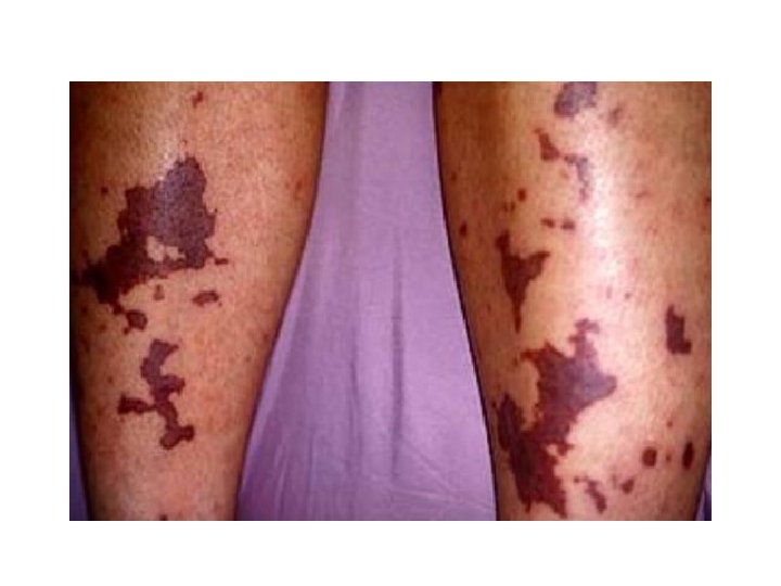
Hemophilia A • Affects male • Less commonly excessive bleeding occurs in heterozygous females…preferential inactivation of the X chromosome carrying the normal factor VIII gene ( unfavorable lyonization) • C/O: -Easy bruising and massive hemorrhage after trauma or operative procedure. -Petechia are characteristically absent.

Hemophilia A • Sites of bleeding: – – Large joints and soft tissue Urinary tract and GI tract Brain Nose • Laboratory tests: – Prolonged PTT (corrected by mixing study). Normal PT and TT. – Low factor VIII assay. • Treatment: replacement therapy (factor VIII concentrate or recombinant VIII). • Inhibitor antibodies develop in 18 -50% of patients
 and the FVIII Gene The gene is located at the tip")
Factor VIII (FVIII) and the FVIII Gene The gene is located at the tip of the X chromosome. Huge gene composed of 26 exons. FVIII m. RNA is ~9 kb, and encodes a 300 k. D protein. FVIII is a cofactor for F IX in the proteolytic activation of F X. • Bound to v. WF in plasma. • Markedly unstable in the absence of v. WF. • •

Hemophilia in Females Exceedingly rare, seen in: – Mating between a carrier mother and affected father – Carriers with abnormalities of X-chromosome • Extreme lyonization • X mosaicism or deletion • Newly mutant gene

Hemophilia B • Incidence: 1 in 60, 000. • Sex-linked. • Severity of disease depends on factor IX level – Normal level 100 U/dl – Severe cases level <2 U/dl – Moderate cases level 2 -5 U/dl – Mild cases level 5 -25 U/dl • Bleeding sites: similar to hemophilia A. • Laboratory tests: – Prolonged PTT. Normal PT and TT. – normal factor VIII assay.
 • Coded by a gene located on the short")
von Willebrand Factor (v. WF) • Coded by a gene located on the short arm of chromosome 12 • The primary protein product is a 250 KDa dimer. • Transformed by multiple disulfide bonds to form a series of multimers ranging in weight from 600 -20, 000 KDa

v. WF • Adhesive protein, bridges collagen to platelets receptor GPIb • Carrier protein to factor VIII • Ristocetin induces platelets agglutination in the presence of v. WF (RIPA) • Stored in Weibel-Palade bodies of endothelial cells and a granules of platelets
. •")
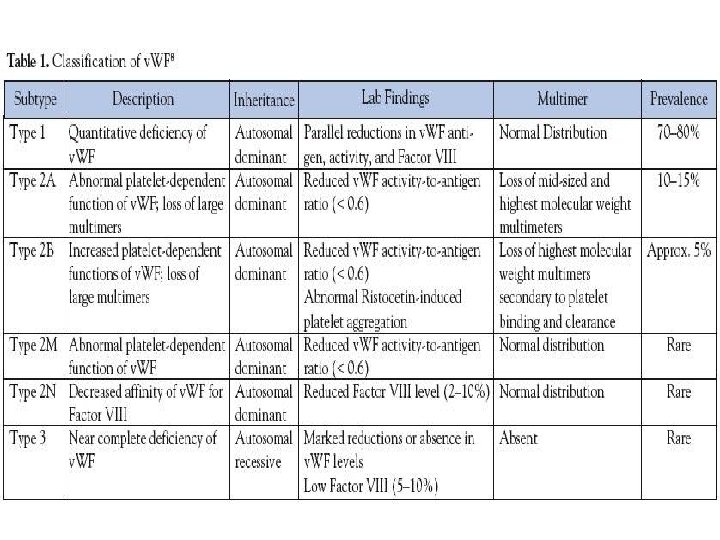
von Willebrand Disease *The most common hereditary bleeding disorder (prevalence 0. 1 -1%). • Mild bleeding problems – Mucous membrane bleeding – Easy bruising – menorrhagia – Post-operative bleeding • Most cases are inherited as Autosomal dominant disorder. • ? When to suspect: *Both sexes are affected, and presented with prolonged bleeding times (BT) despite normal platelet counts. ( early historical observation)

von Willebrand Disease *v. WD differs from classic Hemophilia A in 3 cardinal manifestations: 1. 2. 3. Autosomal inheritance rather than sex linked Consistently prolonged bleeding time (BT) Mucocutaneous bleeding rather than hemarthroses and deep muscle hemorrhage.

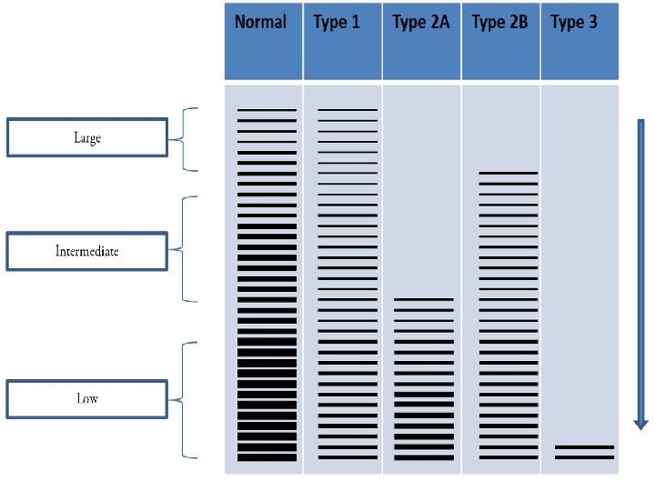
von Willebrand Disease *v. WD : quantitative or qualitative defects of plasma v. WF. • Either Dominant or Recessive. • Divided into 3 main type: 1, 2, and 3. • Type 2 v. WD is further subdivided into 4 subtypes, designated as 2 A, 2 B, 2 M, and 2 N. • Additional disease related to platelet v. WF receptor, platelet type/pseudo-v. WD (similar presentation of v. WD, but doesn’t involve a mutation of the v. WF gene).

von Willebrand Disease *v. WD : Severe forms generally present with A normal PT A prolonged PTT that corrects with mixing study Normal platelet count An abnormal BT *Lab. Assessment should include assays for: F VIII: C activity, v. WF: Ag, RIPA, and v. WF multimeric analysis.
 Accelerated platelet destruction in combination with coagulation factor consumption • Acute")
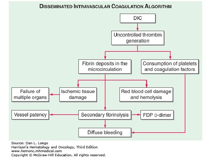
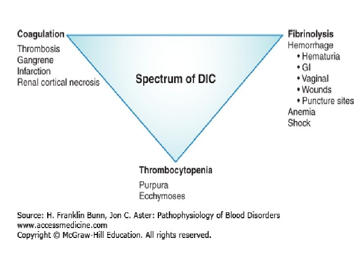
DIC (Consumptive Coagulopathy) Accelerated platelet destruction in combination with coagulation factor consumption • Acute presentation: Anemia and thrombocytopenia • Chronic: Thrombosis Imbalance between the action of Thrombin and the action of Plasmin Triggering mechanisms: • Activation of extrinsic, intrinsic pathways or direct activation of factor X/II
")
DIC • THROMBOSIS AND FIBRINOLYSIS • TRIGGERS: 1. Release of Thromboplastin (adenocarcinoma, leukemia, inflammation) 2. Widespread endothelial injury (release of TF and exposure of VWF)

DIC • TWO COMPONENTS: 1. Microangiopathic hemolytic anemia 2. Fibrinolysis/FDPs: a. b. Inhibit platelet coagulation Anti-thrombotic activity
 • Thrombosis, fibrinolysis, bleeding. • Obstetric complications, infections, cancers (panc, lung,")
DIC (Consumptive Coagulopathy) • Thrombosis, fibrinolysis, bleeding. • Obstetric complications, infections, cancers (panc, lung, prost, stomach, APML), massive tissue injury (trauma, burn), others (shock, liver, heat stroke, vasculitis, intravascular hemolysis). • Lab: Low plat, . High fibrinogen, PTT and FDP.

DIC

Platelet disorders

Relationship Between Platelet Count and Bleeding • Normal range 150 -450 X 103 per µl. • Levels above 60 x 103/µl will not cause bleeding under normal conditions. • Levels below 20 x 103/µl will cause: Petechiae, mucosal bleeding. Post-operative bleeding, CNS bleeding. • Levels around 5 x 10³/µl can lead to fatal CNS or GI hemorrhage. • Levels between 20 and 60 x 103/µl may cause bleeding (depending on platelets functional status).
 2 -Increased")
Classification of Thrombocytopenia 1 -Failure of production (aplastic anemia, radiation, chemo. Rx) 2 -Increased platelet destruction (ITP) 3 -Abnormal distribution (Splenic sequestration)

Thrombocytopenia Due to Increased Platelets Destruction • ITP • Secondary immune thrombocytopenia as in SLE • Drug related immune thrombocytopenia as in quinidine and heparin. • Post transfusion thrombocytopenia • Neonatal thrombocytopenia either due to autoantibodies or alloantibodies • DIC and microangiopathic hemolytic anemia
 or secondary • Acute (self limiting) or chronic.")
ITP • Primary (idiopathic) or secondary • Acute (self limiting) or chronic.
 • • Affects children. Develops acutely with 1 -2 week duration.")
Acute ITP (Idiopathic/Childhood) • • Affects children. Develops acutely with 1 -2 week duration. Bruising and petechia Preceded by infection or vaccination in 75% of cases. Initial Plt. count <20, 000 Self limited, Spontaneous remission in >90% of cases. Severe cases benefit from steroids or IV immunoglobulins.
 • High incidence in women of child bearing age")
Chronic Immune Thrombocytopenic Purpura (ITP) • High incidence in women of child bearing age (20 -50). • NO recent history of drug or recent infection. • Mostly idiopathic, secondary causes include SLE, HIV, CLL, Hodgkin’s disease, drugs (uncommon). • Autoantibodies against GP IIb/IIIa, or Ib/IX (30% of cases). • Platelets lifespan reduced to hours. • Megakaryocytes increased. • Petechial bleeding, easy bruising, menorrhagia.
. Hb. and WBCs are normal.")
ITP Diagnosis Decreased platelet count (10 -50 x 109/l). Hb. and WBCs are normal. PB: large platelet. BM: Increased Megakaryocytes numbers. BT: Mild prolongation; not as prolonged as the BT in other diseases ass/w same decrease in Plt. Count. • Antiplatelet antibodies. • • •
. High dose IV immunoglobulins.")
ITP Treatment • • Steroids. Splenectomy (long term Rx. ). High dose IV immunoglobulins. Immunosuppressive therapy.

Microangiopathic Thrombocytopenia TTP/HUS
 Thrombocytopenia and Microangiopathic hemolytic anemia characterize this disorder")
Thrombotic thrombocytopenic Purpura (TTP) Thrombocytopenia and Microangiopathic hemolytic anemia characterize this disorder
 • Usually affects adult • Pathogenic mechanism: Inherited and Sporadic:")
Thrombotic thrombocytopenic Purpura (TTP) • Usually affects adult • Pathogenic mechanism: Inherited and Sporadic: • Deficiency of metalloprotease (ADAMTS 13) needed for cleaving very HMW_v. WF (multimers). • More common, non-familial acquired: autoantibody against ADAMTS 13 • Platelet micro-aggregate (Hyaline microthrombi) formation. • Acute Thrombocytopenia, fever, microangiopathic hemolytic anemia, neurologic abnormalities, renal dysfunction.
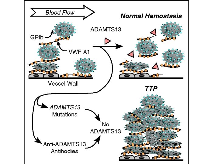

* Diagnosis should be suspected in any patient who presents acutely with thrombocytopenia. * Female > males * 3 rd-4 th decade * Hyaline (platelet rich) microthrombi are the characterstic pathologic feature (capillary of skin and gingiva). * D. D: DIC.

Laboratory Results: Microangiopathic hemolytic anemia picture; Schistocytes Reticulocytosis NRBC’s in PB Increase Mega in BM. Signs of hemolysis: Increase LDH Increase indirect bilirubin Decrease Haptoglobin Normal PT, PTT, D-Dimer but elevated BT. Rx: Plasma exchange.
 • • NON-IMMUNE THROMBOCYTOPENIA More commonly seen in pediatric population.")
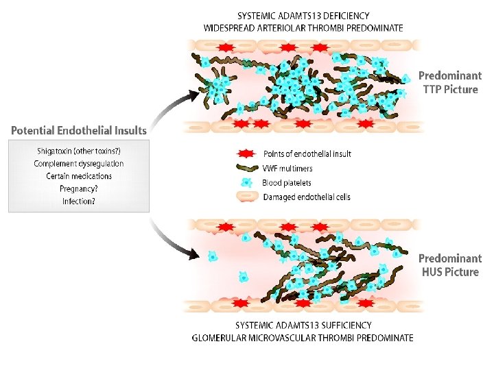
Hemolytic Uremic Syndrome (HUS) • • NON-IMMUNE THROMBOCYTOPENIA More commonly seen in pediatric population. E. coli O 157: H 7 (toxin induced endothelial damage) Bloody diarrhea followed by acute renal failure. Platelet microaggregate (Hyaline microthrombi) formation, usually limited to the glomerular capillaries. Acute Thrombocytopenia, Microangiopathic hemolytic anemia, Renal failure. Normal PT, PTT, D-Dimer but elevated BT. Rx: Conservative (dialysis, antihypertensive, …).
 * Resemble TTP but: - More seen in pediatric population")
Hemolytic Uremic Syndrome (HUS) * Resemble TTP but: - More seen in pediatric population - After viral/bacterial infection - Pathologic thrombi almost always limited to glomerular capillaries
 • DIC • TTP • HUS • DIC: activation of")
MAHA (MICROANGIOPATHIC HEMOLYTIC ANEMIA) • DIC • TTP • HUS • DIC: activation of coagulation pathway (prolonged PT, PTT) • TTP and HUS: Platelet activation (coagulation pathway not activated)
- Slides: 47