
Hemorrhage control III year BDS 29220 Hemostasis Defintion
Hemorrhage control III year BDS 29/2/20
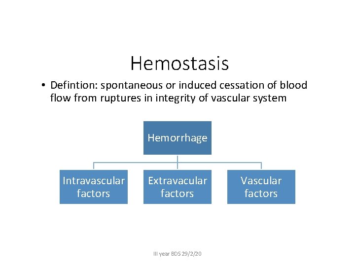
Hemostasis • Defintion: spontaneous or induced cessation of blood flow from ruptures in integrity of vascular system Hemorrhage Intravascular factors Extravacular factors III year BDS 29/2/20 Vascular factors
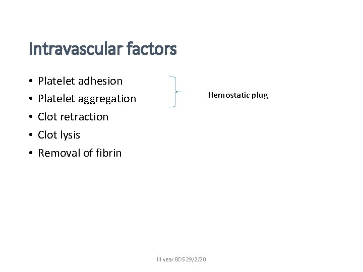
Intravascular factors • • • Platelet adhesion Platelet aggregation Clot retraction Clot lysis Removal of fibrin Hemostatic plug III year BDS 29/2/20
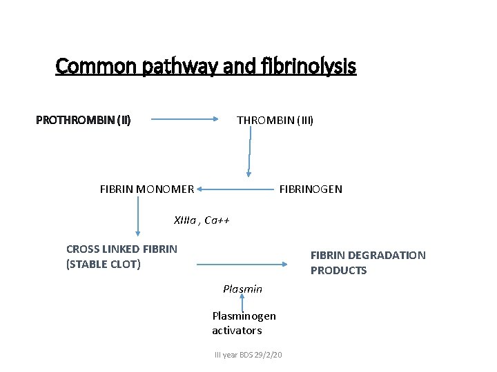
Common pathway and fibrinolysis PROTHROMBIN (II) THROMBIN (III) FIBRIN MONOMER FIBRINOGEN XIIIa , Ca++ CROSS LINKED FIBRIN (STABLE CLOT) FIBRIN DEGRADATION PRODUCTS Plasminogen activators III year BDS 29/2/20
• First stage of coagulation • Second stage • Final stage III year BDS 29/2/20
Extravascular factors • State of health • Mucosa, submucosa, cutaneous, subcutaneous , muscular and pther tissues that support yhe vessels • Example • Disorders that lead to atrophy of subcut (purpura senilis) • Fragility of skin (Cushings syndrome) III year BDS 29/2/20
Vascular factors • Disorders of vessels themselves • capillaries, arteries or veins • Vit C deficiency, III year BDS 29/2/20
Laboratory tests • • • Prothrombin time (PT/INR) Clotting time Partial thromboplastin time (a. PTT) Bleeding time Platelet count III year BDS 29/2/20
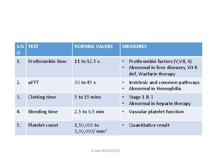
S. N TEST O NORMAL VALUES MEASURES 1. Prothrombin time 11 to 12. 5 s • Prothrombin factors (V, VII, X) • Abnormal in liver diseases, Vit K def, Warfarin therapy 2. a. PTT 30 to 45 s • Instrinsic and common pathways • Abnormal in Hemophilia 3. Clotting time 5 to 15 mins • Stage 1 & 3 • Abnormal in heparin therapy 4. Bleeding time 2. 5 to 9. 5 min • Vascular platelet function 5. Platelet count 1, 50, 000 to 3, 000/ mm 3 • Quantitative result III year BDS 29/2/20
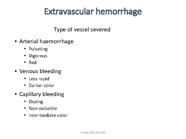
Extravascular hemorrhage Type of vessel severed • Arterial haemorrhage • Pulsating • Vigorous • Red • Venous bleeding • Less rapid • Darker color • Capillary bleeding • Oozing • Non-pulsatile • Intermediate color III year BDS 29/2/20
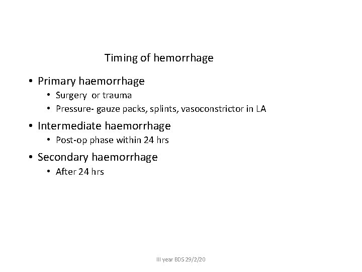
Timing of hemorrhage • Primary haemorrhage • Surgery or trauma • Pressure- gauze packs, splints, vasoconstrictor in LA • Intermediate haemorrhage • Post-op phase within 24 hrs • Secondary haemorrhage • After 24 hrs III year BDS 29/2/20
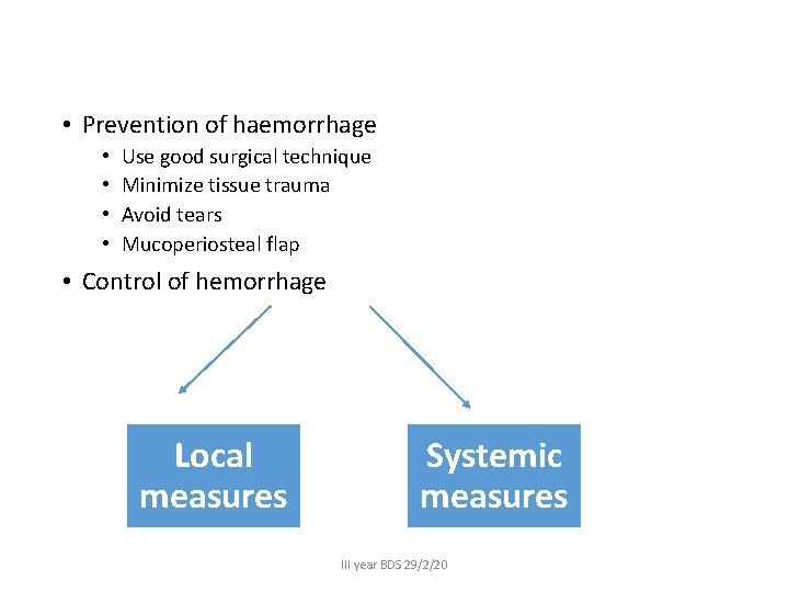
• Prevention of haemorrhage • • Use good surgical technique Minimize tissue trauma Avoid tears Mucoperiosteal flap • Control of hemorrhage Local measures Systemic measures III year BDS 29/2/20
Local measures • Direct pressure • Gauze/bandages • Digital pressure • ECA- Bt angle and SCM • Facial artery- premasseteric notch • Sup temporal artery- Bt tragus and root of zygoma • Direct occlusion of vessel • Mosquito hemostats • Ties (silk) • Coagulation of small capillaries III year BDS 29/2/20
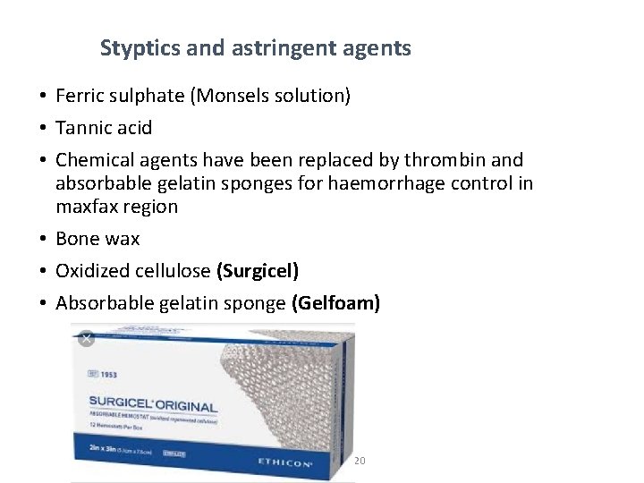
Styptics and astringent agents • Ferric sulphate (Monsels solution) • Tannic acid • Chemical agents have been replaced by thrombin and absorbable gelatin sponges for haemorrhage control in maxfax region • Bone wax • Oxidized cellulose (Surgicel) • Absorbable gelatin sponge (Gelfoam) III year BDS 29/2/20
• • • Topical application of Thrombin Microfibrillar collagen (Avitene) LA with Adr Tissue adhesive - Cyanoacrylate Tranexamic acid III year BDS 29/2/20
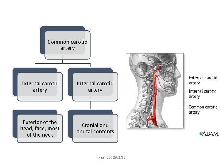
Common carotid artery External carotid artery Internal carotid artery Exterior of the head, face, most of the neck Cranial and orbital contents III year BDS 29/2/20
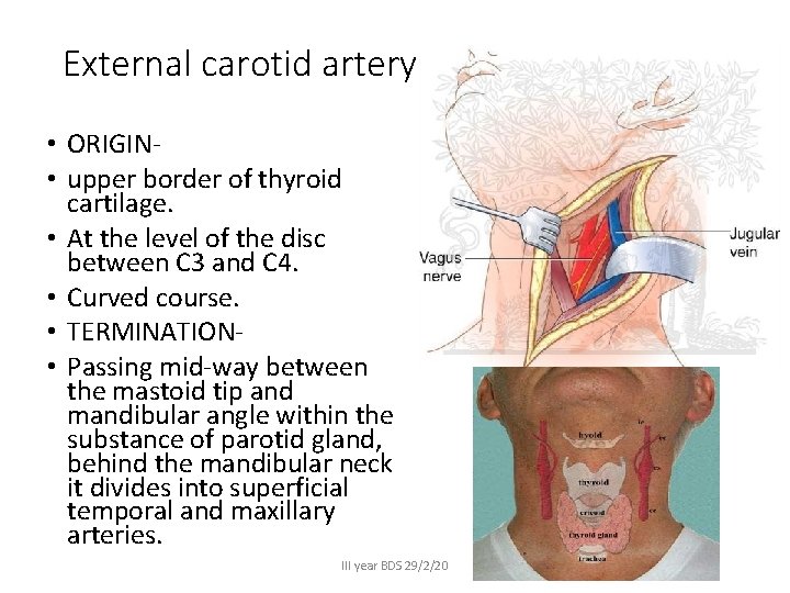
External carotid artery • ORIGIN • upper border of thyroid cartilage. • At the level of the disc between C 3 and C 4. • Curved course. • TERMINATION • Passing mid-way between the mastoid tip and mandibular angle within the substance of parotid gland, behind the mandibular neck it divides into superficial temporal and maxillary arteries. III year BDS 29/2/20
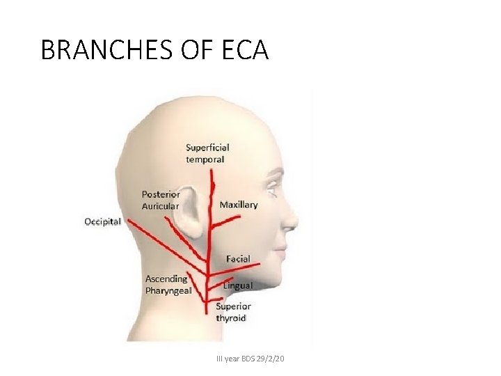
BRANCHES OF ECA III year BDS 29/2/20
III year BDS 29/2/20
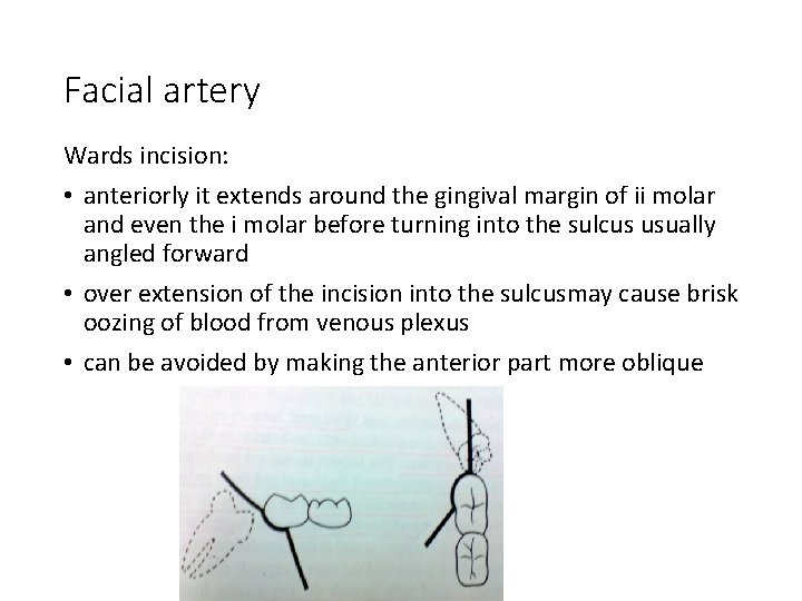
Facial artery Wards incision: • anteriorly it extends around the gingival margin of ii molar and even the i molar before turning into the sulcus usually angled forward • over extension of the incision into the sulcusmay cause brisk oozing of blood from venous plexus • can be avoided by making the anterior part more oblique III year BDS 29/2/20
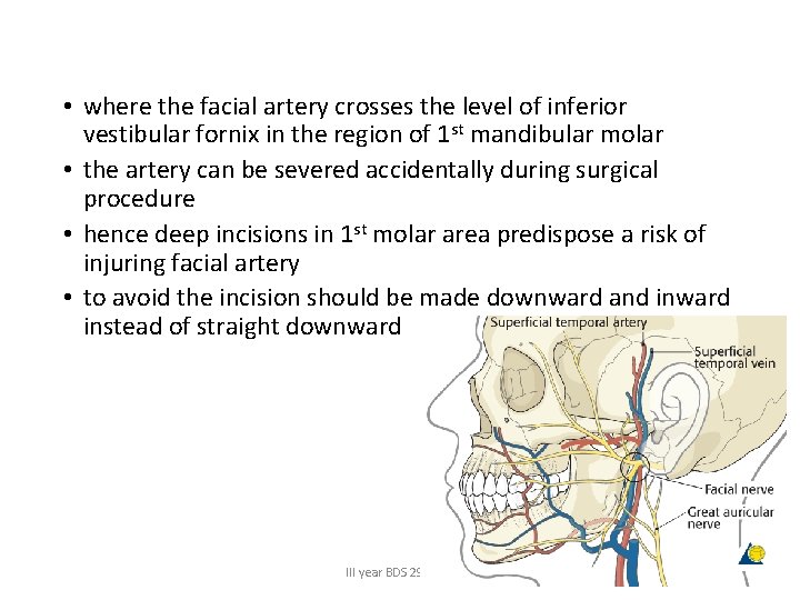
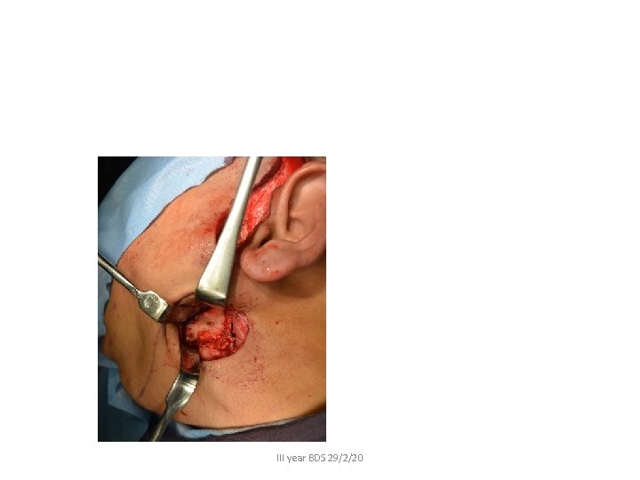
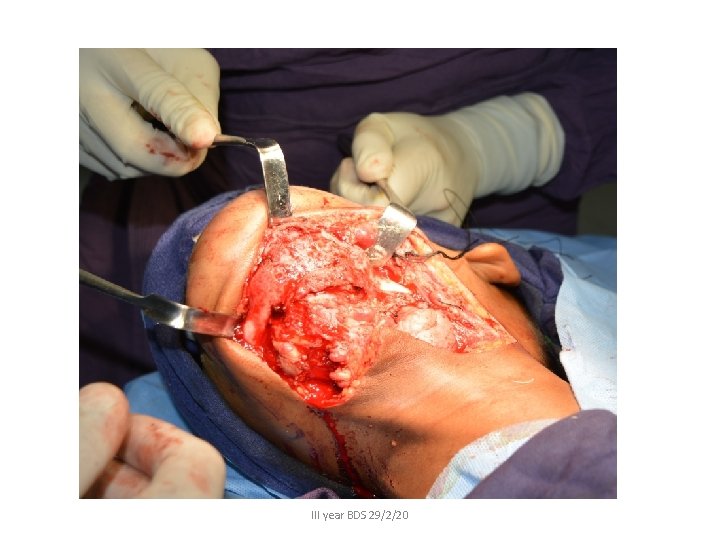
• where the facial artery crosses the level of inferior vestibular fornix in the region of 1 st mandibular molar • the artery can be severed accidentally during surgical procedure • hence deep incisions in 1 st molar area predispose a risk of injuring facial artery • to avoid the incision should be made downward and inward instead of straight downward III year BDS 29/2/20
III year BDS 29/2/20
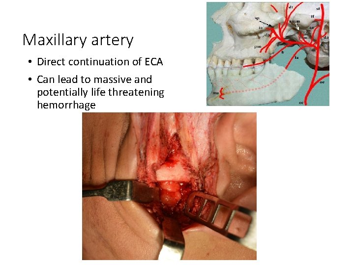
Maxillary artery • Direct continuation of ECA • Can lead to massive and potentially life threatening hemorrhage III year BDS 29/2/20
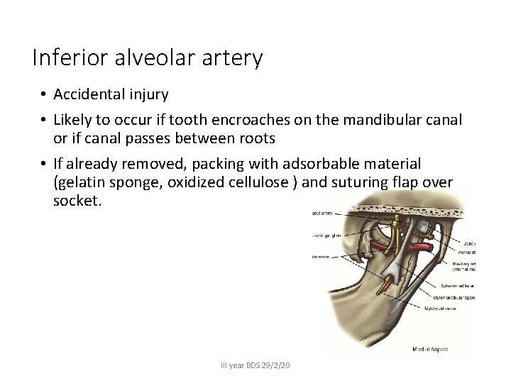
Inferior alveolar artery • Accidental injury • Likely to occur if tooth encroaches on the mandibular canal or if canal passes between roots • If already removed, packing with adsorbable material (gelatin sponge, oxidized cellulose ) and suturing flap over socket. III year BDS 29/2/20
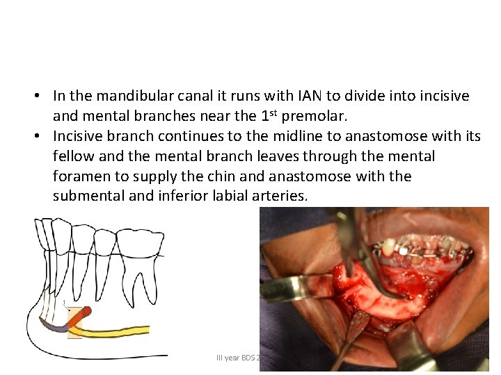
• In the mandibular canal it runs with IAN to divide into incisive and mental branches near the 1 st premolar. • Incisive branch continues to the midline to anastomose with its fellow and the mental branch leaves through the mental foramen to supply the chin and anastomose with the submental and inferior labial arteries. III year BDS 29/2/20
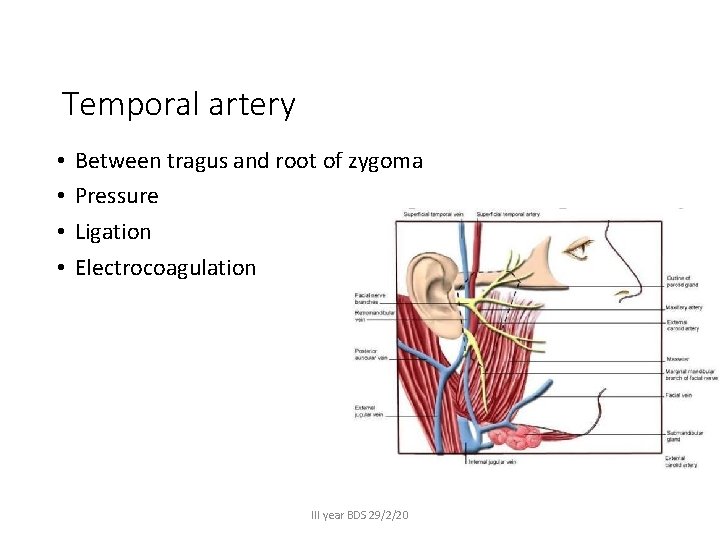
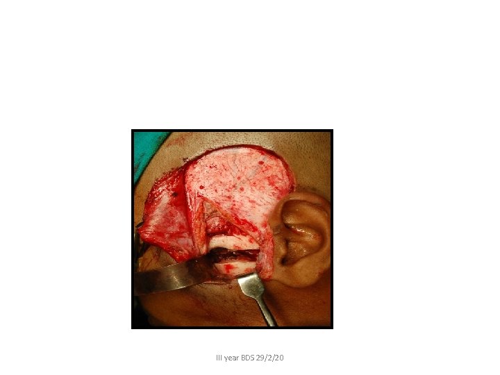
Temporal artery • • Between tragus and root of zygoma Pressure Ligation Electrocoagulation III year BDS 29/2/20
III year BDS 29/2/20
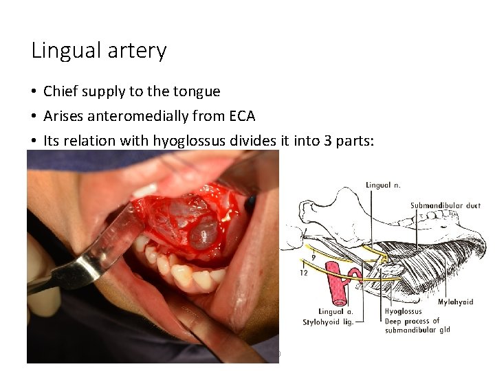
Lingual artery • Chief supply to the tongue • Arises anteromedially from ECA • Its relation with hyoglossus divides it into 3 parts: III year BDS 29/2/20
Bleeding: • Presents a severe risk to the airway • The main vessels involved are an ethmoid artery, ophthalmic, vidian branch of internal carotid, and maxillary artery. • Increased risk of aspiration exists in patients with altered level of consciousness. • A gastric tube may be used to decompress the stomach and reduce the risk of vomiting and aspiration. • If massive bleeding continues, it may be necessary to place an endotracheal airway.
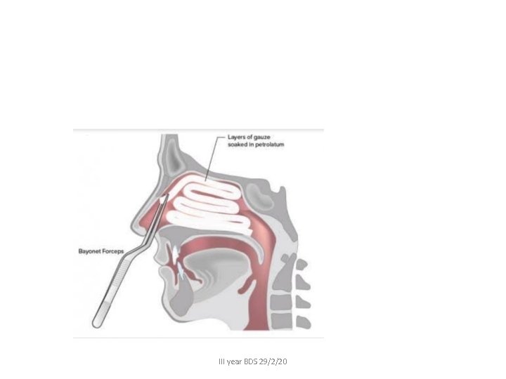
Hemostasis may be obtained with: • Control of blood pressure • Vasoconstrictor based nasal decongestants • Injection of vasoconstrictors • Anterior and/or posterior nasal packing • Cauterization • Selective embolization
III year BDS 29/2/20
• Massive bleeding (not controlled) can be treated by head elevation, ice packs, tamponade using nasal packing, or a balloon device. • Massive bleeding, uncontrolled- treatment begins with tamponade. • If bleeding persists after removal of tamponadeelectrocoagulation. • Massive nasal bleeding is also controlled using anterior and posterior packing (eg, Belloque tamponade, the balloon of a Foley catheter)
- Slides: 32