Hemophilia Presented by Mrs Rama Shukla Hemophilia Xlinked

Hemophilia Presented by Mrs. Rama Shukla

Hemophilia � X-linked recessive disorder characterized by deficiency of coagulation factor. Hemophilia A Hemophilia B (Chrismas disease) Deficiency of coagulation factor

X linked recessive Males ♂ Female ♀ Patient Carrier

Queen Victoria

Hemophilia = Royal disease The Romanovs Russia’s last royal family

Bleeding: � Intraarticular, � Intramuscular, � Intraosseous, � Subperiosteal. Life threatening hemorrhage: � Intracranial. � Throacic. � Abdominal.

Hemophilia Hemophilic arthropathy Hemophilic pseudotumor

Hemophilic arthropathy Etiology: � Repeated hemarthrosis. Pathology: Repeated hemarthrosis � chronic synoviitis, synovial hemo-siderosis � synovial proliferation & pannus formation � cartilage destruction � bone erosions and subcortical cysts.

Hemophilic arthropathy Demographics: � Age: 1 st or 2 nd decade. � Sex: it is a disease of males.

Hemophilic arthropathy Clinical presentation: 3 types of arthropathy

Acute arthropathy � Red � hot � tender � joint swelling. � Fever � Leukocystosis. DDx: septic arthritis

Subacute arthropathy � Limitation of joint motion &muscle wasting

Chronic arthropathy � Sever joint contracture. � DDx: juvenile rheumatoid arthritis.

Hemophilic arthropathy � Location: Knee �elbow �ankle �hip � shoulder. � Distribution: Unilateral or bilateral asymmetrical (DDx with JRA).

Radiological manifestiation of hemophilic arthropathy Dense joint effusion Juxta-articular osteoporosis. Hemoarthrosis Hyperemia of the epiphysis Enlargement of the epiphysis Hyperemia of the epiphysis Diffuse narrowing of the joint space Cartilage destruction Irregular articular surfaces Bone erosion Subchondral sclerosis, cystic Premature 2 ry osteoarthritis. changes and marginal osteophytic lipping.

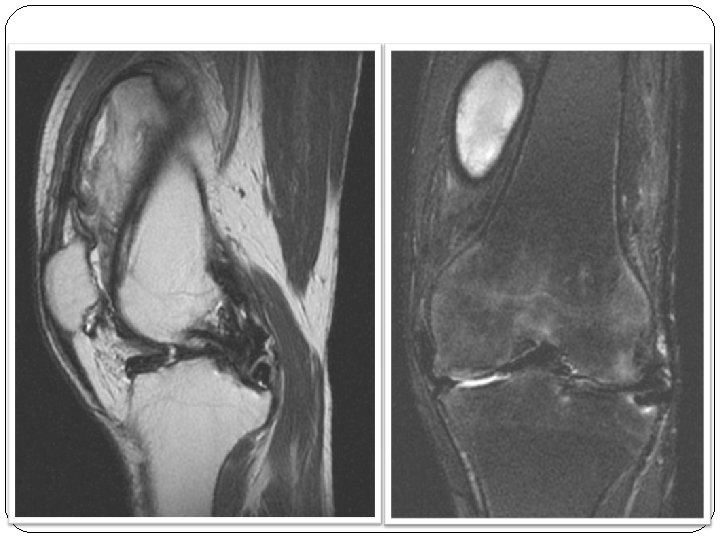
MRI � Low signal intensity of the hypertrophied synovium on all pulse sequences due to siderotic synoviitis.

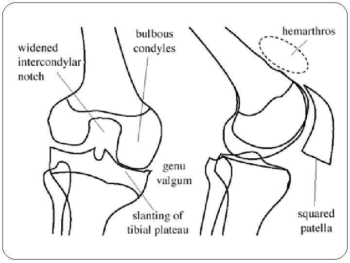
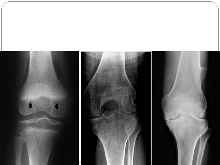
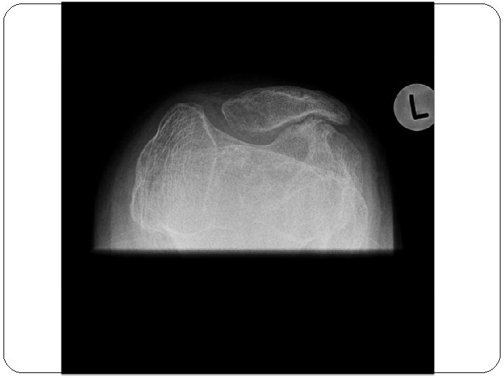
Knee � 1� 6+ � Bulbous femoral condyles with flattened inferior surface. � Slanting of the tibial plateau. � Widening of the intercondylar notch (due to repeated hemorrhage in the attachments of the cruciate ligaments). � Squaring of the lower pole of the patella.

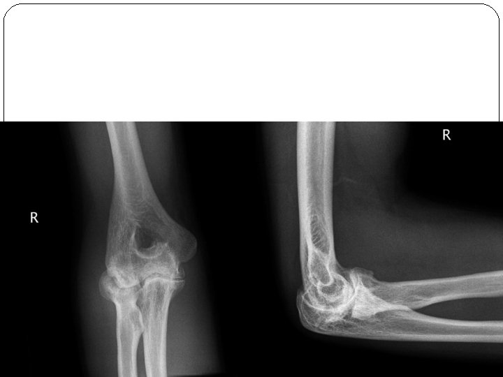
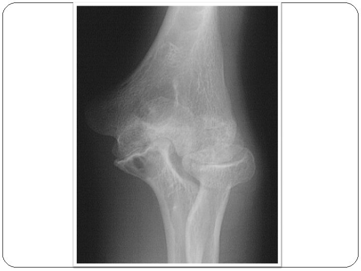
Elbow � 1� 6+ � Widening of the trochlear &ulnar notches. � Enlargement of the radial head.
")
Dense joint effusion (sail sign)

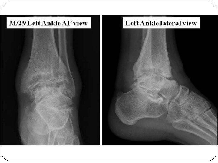
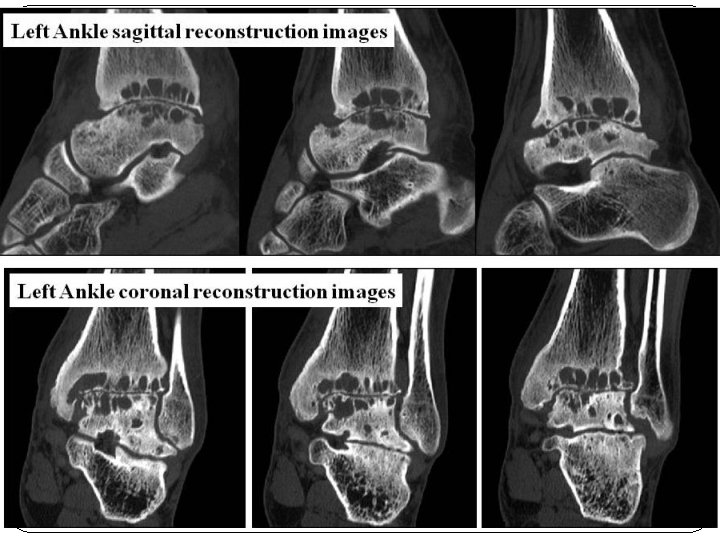
Ankle � 1� 6+ � Talar tilt: medial slanting of the tibiotalar joint with alteration of ankle mortise.

MRI 1. 2. 3. 4. Hemorrhagic joint effusion. Synovial hemosiderin deposition (low signal intensity on all pulse sequences with blooming artifact on gradient echo images). Destruction of the articular surface. Bone marrow edema of the epiphysis.

Complications of hemophilic arthropathy: 1. 2. 3. 4. Premature 2 ry osteoarthritis. Premature fusion of the epiphysis with limb shortening. Avascular bone necrosis. Septic arthritis.

DD of hemophilia � JRA � Neuromuscular disorders. Neuromuscular disordres immobilization Synovial atrophy Decreased cartilage nutrition Vascular stasis Epiphyseal enlargement

Paraplegia

Poliomyelitis

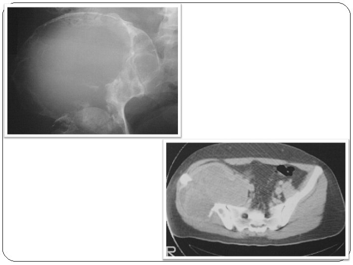
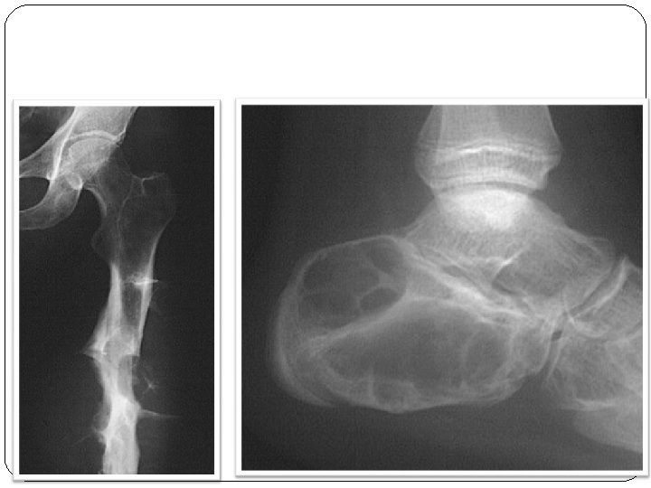
Hemophilic pseudotumor � Repeated intramusular, subperiosteal or intraosseous hemorrhage.

Pathology: � Subperiosteal hemorrhage �destruction of the underlying cortex &medulla &elevation of the overlying periosteum. � Intraosseous hemorrhage �intraosseous lytic lesion.

Location: � Iliac wings. � Femoral diaphysis. � Calcaneus.
 � Inherited hemophilia Bfrom her")
Special Case � Female patient with Turner syndrome (XO) � Inherited hemophilia Bfrom her father. � X ray ankle shows talar tilt.

Bleeding into kidney

Intracranial hemorrhage 1/3 of deaths.
- Slides: 41