HEMOPHILIA HEMOPHILIA bleeding disorders due to inherited deficiencies

HEMOPHILIA
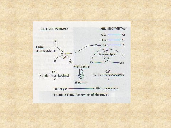

HEMOPHILIA bleeding disorders due to inherited deficiencies in coagulation factors Ø Types: 1. Haemophilia A (Classic) Factor VIII deficiency 2. Haemophilia B (Christmas Disease) Factor IX deficiency Ø

HEMOPHILIA A & B Ø clinically similar: Ø occur in approximately 1 in 5, 000 male births Ø account for 90% of congenital bleeding disorders Ø Hemophilia A is approximately 5 times more common than B

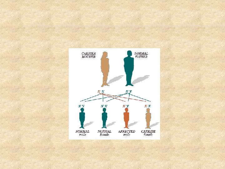
ETIOLOGY Ø Ø Inherited as a sex linked recessive trait with bleeding manifestations only in males genes which control factor VIII and IX production are located on the x chromosome; if the gene is defective synthesis of these proteins is defective female carriers transmit the abnormal gene A disease of males

CLASSIFICATION Severe % normal factor level < 1% Moderate 1 - 5% Mild 6 - 20 % Causes of bleeding after trivial injury or spontaneous bleeding after minor injury; occasional spontaneous bleeds following major trauma, surgical or dental procedures

DIAGNOSIS � Atypical bleeding at circumcision or bruising at neonatal vaccines � Toddlers with lip bleeding or unusual bruising when learning to walk � Hx of affected males on mother’s side � Elevated PTT � Factor assays
 § Knees, ankles and")
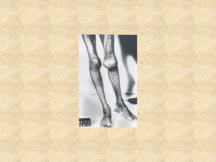
CLINICAL FEATURES – JOINT BLEEDS Ø Ø Ø Joints (Hemarthrosis) § Knees, ankles and elbows most common sites § begin as the child begins to crawl and walk Single joint bleed: stiffness, swelling, pain With repeated bleeding into same jt---arthropathy-> stiffness and contractures

SUB ACUTE HEMARTHROSIS Develops after repeated bleeds into the joint Ø Synovium becomes inflamed Ø Hypertrophy, hyperplasia and increased vascularity of synovial membrane Ø Hemosiderosis: hemoglobin of intra articular blood is degraded and iron deposited into the joint space Ø
, & enzymes begin")
CHRONIC ARTHROPATHY Progressive destruction of a joint Ø Pannus (inflammed synovium), & enzymes begin to destroy articular cartilage Ø Microfracture and cyst formation in subchondral bone Ø End stage: firbrous joint contracture, and disorganization of articular surfaces Ø

CLINICAL FEATURES – MUSCLE BLEEDS Bleeding into muscle or soft tissue Ø Sites: iliopsoas, calf Ø Symptoms: pain, swelling, muscle spasm Ø Complications: nerve compression, contracture Ø

OTHER SITES OF HEMORRHAGE Abdomen Ø GI tract Ø Intracranial bleeds Ø Around vital structures in the neck Ø Can cause death…

� They have high risk of HIV, Hep B and Hep C due to repeated transfusion of blood products

MANAGEMENT � Specific Hemophillia A Fac viii preparations Cryo DDAVP Hemophillia B Fac ix CPP

GENERAL Ø Ø Ø Avoid NSAIDs Avoid contact sports Avoid IM injections Good dental care Education – life long management Acute and long term management of musculoskeletal problems

MUSCULOSKELETAL MANAGEMENT Ø Acute Bleeds: § § § Immediate replacement of factor Immobilize joint No weight bearing Ice Analgesics Once acute condition resolves gradually resume exercises -PT

MUSCULOSKELETAL MANAGEMENT Ø After 24 hours: § § § Continue minimal or no weight bearing for lower extremity bleed Active range of motion; gentle stretching Isometric strengthening; progress to isotonic Continue use of ice Hydrotherapy if available

THANK YOU…
- Slides: 20