Hemoglobin An overview and more Prof Mamoun Ahram

Hemoglobin An overview and more Prof. Mamoun Ahram Blood module 2019
. The")
Heme structure It is a complex of protoporphyrin IX + Iron (Fe 2+). The porphyrin is planar and consists of four rings (designated A-D) called pyrrole rings. Each pyrrole can bind two substituents. Two rings have a propionate group each. Note: the molecule is hydrophobic. Fe has six coordinates of binding. A B C D

Hemoproteins Many proteins have heme as a prosthetic group called hemoproteins.

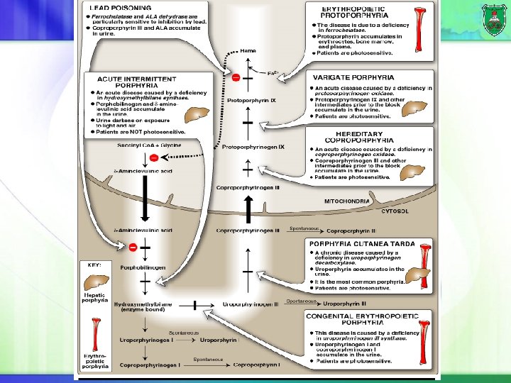
Synthesis of heme Liver and erythroid tissues are main sites of synthesis. The first reaction is the rate limiting and committed step. It requires vitamin B 6 (pyrodoxal phosphate). It is regulated by hemin. The last reaction is spontaneous, but can be catalyzed by ferrochelatase.

Hydroxymethylbilane

Treatment Intravenous injection of hemin and glucose Protection from sunlight Ingestion of beta-carotene.

Hemoglobin

Structure of hemoglobin Hb is a globular protein. Amino acid distribution Positions of two histidine residues (proximal and distal) It is an allosteric protein. Multiple subunit (2 + 2β) Altered structure depending on bound molecules Positive cooperativity towards oxygen Regulated by allosteric effectors

How are the subunits bound? A dimer of a dimer (I made up this term) ( -β)2 Note how they interact with each other.

Heme binding to hemoglobin Exterior of the protein Propionate Interior of the protein

Oxygen distribution in blood versus tissues

Oxygen saturation curve The saturation curve of hemoglobin binding to O 2 has a sigmoidal shape. It is allosteric. At 100 mm Hg, hemoglobin is 95 -98% saturated (oxyhemoglobin). As the oxygen pressure falls, oxygen is released to the cells. Note: at high altitude (~5000 m), alveolar p. O 2 = 75 mm. Hg.

p. O 2 at different altitudes

Structural amplification change 0. 4 A
 This movement triggers changes in tertiary structure of")
How does the structure change? (2) This movement triggers changes in tertiary structure of individual hemoglobin subunits breakage of the electrostatic bonds at the other oxygen-free hemoglobin chains. In myoglobin, movement of the helix does not affect the function of the protein.

Electrostatic interactions are broken

Positive cooperativity
 Most accurate The protein exists in two states in")
The concerted model (MWC model) Most accurate The protein exists in two states in equilibrium: T (taut, tense) state with low affinity and R (relaxed) state with high affinity. Increasing ligand concentration drives the equilibrium between R and T toward the R state (positive cooperativity) sigmoidal curve The effect of ligand concentration on the conformational equilibrium is a homotropic effect (oxygen). Other effector molecules that bind at sites distinct from the ligand binding site and thereby affect the R and T equilibrium in either direction are called heterotropic effectors (to be discussed). Note direction of arrows

The sequential, induced fit, or KNF model Less accurate The subunits go through conformational changes independently of each other, but they make the other subunits more likely to change, by reducing the energy needed for subsequent subunits to undergo the same conformational change. Ligand binding may also result in negative cooperativity. The MWC model only suggests only positive cooperativity.

Itisnotonlyonehemoglobin

Developmental transition of hemoglobins

The embryonic stage Hemoglobin synthesis begins in the first few weeks of embryonic development within the yolk sac. The major hemoglobin (Hb. E Gower 1) is a tetramer composed of 2 zeta ( ) chains and 2 epsilon ( ) chains Other forms exist: Hb. E Gower 2 (α 2ε 2), Hb. E Portland 1 (ζ 2γ), Hb. E Portland 2 (ζ 2β).

Beginning of fetal stage By 6 -8 weeks of gestation, the expression of embryonic hemoglobin declines dramatically and fetal hemoglobin synthesis starts from the liver. Fetal hemoglobin consists of two polypeptides and two gamma ( ) polypeptides ( 2 2) The polypeptides remain on throughout life.

Beginning of adult stage Shortly before birth, there is a gradual switch from to adult globin. Still, Hb. F makes up 60% of the hemoglobin at birth, but 1% of adults. At birth, both synthesis of and chains occurs in the bone marrow.

Adult hemoglobins The major hemoglobin is Hb. A 1 (a tetramer of 2 and 2 chains). A minor adult hemoglobin, Hb. A 2, is a tetramer of 2 chains and 2 delta ( ) chains. Hb. A can be glycosylated with a hexose and is designated as Hb. Ac. The major form (Hb. A 1 c) is attached to glucose attached to valines of chains. Hb. A 1 c is present at higher levels in patients with diabetes mellitus.

Advantages of Hb. A 1 c testing Hb. A 1 c provides a longer-term trend, similar to an average, of how high your blood sugar levels have been over a period of time (2 -3 months). Blood fasting glucose level is the concentration of glucose in your blood at a single point in time, i. e. the very moment of the test. Hb. A 1 c can be expressed as a percentage (DCCT unit) or as a value in mmol/mol (IFCC unit). IFCC is new. Limitations of Hb. A 1 c test: It does not capture short-term variations in blood glucose, exposure to hypoglycemia and hyperglycemia, or the impact of blood glucose variations on individuals’ quality of life.

Table

Geneticsofglobinsynthesis
 and gene. The")
The genes The gene cluster contains two genes ( 1 2) and gene. The gene cluster contains gene in addition to gene, two genes, and gene. The gene order parallels order of expression. Genetic switching is controlled by a transcription factor-dependent developmental clock, independent of the environment. Premature newborns follow their gestational age.
. The β-globin")
Locus structure Each gene has its promoter and regulatory sequences (activators, silencers). The β-globin cluster is controlled by a master enhancer called locus control region (LCR).

The mechanism of regulation The mechanism requires timed expression of regulatory transcription factors for each gene, epigenetic regulation (e. g. acetylation, methylation), and chromatin looping. Note: treatment!!
- Slides: 32