HEMODYNAMIC MONITORING Outline 1 Basic of cardiovascular system

HEMODYNAMIC MONITORING

Outline 1 Basic of cardiovascular system 2 Principle of physics 3 Non invasive hemodynamic monitoring

The Cardiovascular System • A closed system of the heart and blood vessels (capillaries , arteries , veins) o The heart pumps blood o Blood vessels allow blood to circulate to all parts of the body • The function of the cardiovascular system is to deliver oxygen and nutrients and to remove carbon dioxide and other waste products

Principle of physics • Poiseuille low : the rate of flow of a fluid in vessel is determined by pressure different between the two end of vessels and resistance within the lumen • Flow (Q)= Pressure difference / resistance

Definition o Hemodynamic monitoring refers to measurement of pressure, flow and oxygenation of blood within the cardiovascular system. OR o Using invasive technology to provide quantitative information about vascular capacity, blood volume, pump effectiveness and tissue perfusion.

Indications: • Continuous, real-time blood pressure monitoring • Planned pharmacologic or mechanical cardiovascular manipulation • Repeated blood sampling • Determination of volume responsiveness from systolic pressure or pulse -pressure variation • Supplementary diagnostic information from the arterial waveform • Failure of indirect arterial blood pressure measurement

cardiac out put • Cardiac output is the amount of blood that is pumped out of the ventricles in one minute. A normal cardiac output is between 4 -8 L/minute. • Cardiac output = heart rate X stroke volume

The critical care nurse should recognize and understand the building blocks of hemodynamic. • • Heart rate Stroke volume Cardiac output Blood pressure Systemic vascular resistance/Pulmonary vascular resistance • Preload o After load Contractility Mixed venous oxygen saturation (SVO 2)

• Heart rate: Not only is the rate important, the rhythm can cause hemodynamic imbalances • Stroke volume: Stroke volume (SV) is the amount of blood pumped out of the ventricle with each beat, • normal stroke volume is between 60 – 130 m. L/beat. • Preload: Preload is the volume required to STRETCH the cardiac muscle fibers in the atria and ventricles. • Afterload: is the RESISTANCE or pressure the ventricular heart muscle must overcome to open aortic valve and eject volume. • Contractility: is the strength of the cardiac muscle to push blood from the ventricles against the systemic vascular resistance/blood pressure of the patient.

HEMODYNAMIC MONITER

Company Logo www. themegallery. com

Company Logo www. themegallery. com

Type of Invasive monitoring • Invasive o Arterial pressure monitoring o Central venous pressure monitoring o Pulmonary artery pressure monitoring Company Logo www. themegallery. com

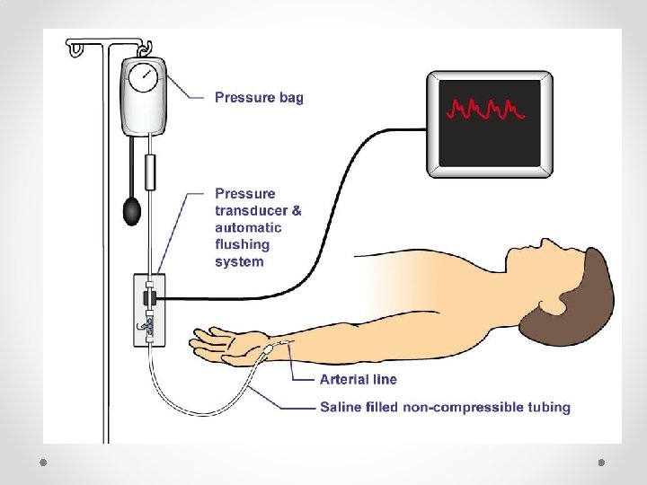
Indications for Arterial Catheterization • • • Need for continuous blood pressure measurement Hemodynamic instability Vasopressor requirement Respiratory failure Frequent arterial blood gas assessments Most common locations: radial, femoral, axillary, and dorsalis pedis Company Logo www. themegallery. com
 The CVP, the pressure in the vena cava or")
CENTRAL VENOUS PRESSURE MONITORING (CVP) The CVP, the pressure in the vena cava or right atrium, is used to assess right ventricular function and venous blood return to the right side of the heart. The CVP can be continuously measured by connecting either a catheter positioned in the vena cava or the proximal port of a pulmonary artery catheter to a pressure monitoring system Normal CVP is 2 -6 mm/Hg

Company Logo www. themegallery. com

Central Venous Pressure Waveform Company Logo www. themegallery. com

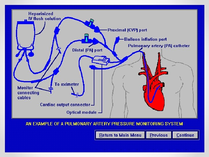
Indications for Pulmonary Artery Catheterization • Identification of the type of shock o Cardiogenic (acute MI) o Hypovolemic (hemorrhagic) o Obstructive (PE, cardiac tamponade) o Distributive (septic) o Many critically ill patients exhibit elements of more than 1 shock classification • Monitoring the effectiveness of therapy
")
Pulmonary Artery Catheter (Swan-Ganz catheter)

PULMONARY ARTERY Catheter

Measuring hemodynamic parameters • Before obtaining hemodynamic parameters, the transducer is leveled and zeroed. 1. Explain the procedure to the patient to gain informed consent. 2. If IV fluid is not running, ensure that the CVC is patent by flushing the catheter. 3. Place the patient flat in a supine position if possible. • Zero reference point is: mid-axillary line at the fourth intercostal space (Phlebostatic axis) Company Logo www. themegallery. com

Company Logo www. themegallery. com

Company Logo www. themegallery. com

Company Logo www. themegallery. com

Catheter displacement If the CVC moves into the chambers of the heart then cardiac arrhythmias may be noted, and should be reported. If the CVC is no longer in the correct position, CVP readings and medication administration will be affected. Company Logo www. themegallery. com
 70 -90 mm Hg Cardiac")
Normal value Parameter Normal value Mean Arterial Pressure (MAP) 70 -90 mm Hg Cardiac Index (CI) 2. 8 -4. 2 L/min/m 2 Cardiac Output (CO) 4 -8 L/min Central Venous Pressure (CVP) 2 -6 mm. Hg Atrial Pressure (RA)) 2 -8 mm H 2 O Pulmonary Artery Pressure (PA) Systolic 20 -30 mm. Hg (PAS) Diastolic 8 -12 mm. Hg (PAD) Mean 15 -25 mm. Hg Pulmonary Capillary Wedge Pressure (PWCP) 8 -12 mm. Hg

NURSING INTERVENTIONS • Once catheter is inserted, it is secured and a dry, sterile dressing is applied. • Catheter placement is confirmed by a chest x-ray, and the site is inspected daily for signs of infection. The dressing and pressure monitoring system or water manometer are changed according to hospital policy. • In general, the dressing is to be kept dry and air occlusive. • • Dressing changes are performed with the use of sterile technique. CVP catheters can be used for infusing intravenous fluids, administering intravenous medications, and drawing blood specimens in addition to monitoring pressure.

Continue • the transducer must be positioned at the phlebostatic axis to ensure accurate readings. • The nurse who obtains the wedge reading ensures that the catheter has returned to its normal position in the pulmonary artery by evaluating the pulmonary artery pressure waveform. • The pulmonary artery diastolic reading and the wedge pressure reflect the pressure in the ventricle at end-diastole and are particularly important to monitor in critically ill patients, because they are used to evaluate left ventricular filling pressures (preload) At end-diastole, when the mitral valve is open, the wedge pressure is the same as the pressure in the left atrium and the left ventricle, unless the patient has mitral valve disease or pulmonary hypertension. • • Critically ill patients usually require higher left ventricular filling pressures to optimize cardiac output. These patients may need to have their wedge pressure maintained as high as 18 mm Hg.

NURSING RESPONSIBILITIES Site Care and Catheter Safety: • A sterile dressing is placed over the insertion site and the catheter is taped in place. The insertion site should be assessed for infection and the dressing changed every 72 hours and prn. • The placement of the catheter, stated in centimeters, should be documented and assessed every shift. • The integrity of the sterile sleeve must be maintained so the catheter can be advanced or pulled back without contamination. • The catheter tubing should be labeled and all the connections secure. • The balloon should always be deflated and the syringe closed and locked unless you are taking a PCWP measurement

Patient Activity and Positioning: • Many physicians allow stable patients who have PA catheters, such as post CABG patients, to get out of bed and sit. The nurse must position the patient in a manner that avoids dislodging the catheter. • Proper positioning during hemodynamic readings will ensure accuracy.

www. themegallery. com
- Slides: 33