Hemodynamic monitoring of neurocritically ill patients Outline Hemodynamics
 of neurocritically ill patients 雙和醫院 神經內科 紀乃方醫師")


 •")

 × Heart Rate • SV =")
 – Heart")


 – Cardiac output/ Body surface area – Normal =3")
 – CT perfusion – MR perfusion")

 (PWI/")

 correlates best with CBF • MFV = (FVs")

: 547 -55.")
- Slides: 25
Hemodynamic monitoring (血流動力監測) of neurocritically ill patients 雙和醫院 神經內科 紀乃方醫師
Outline • Hemodynamics的簡介 • 監測hemodynamics的目的 • 監測hemodynamics的方法
腦的代謝 • 大腦: 2 -3% 體重,佔用 20%心輸出量 • 高代謝率 – 50% 能量維持神經細胞電性活動 – 25%能量維持離子通道功能 – 25%能量進行生化合成作用 http: //www. brighamandwomens. org/departments_and_services/radiology/services/nuclearmedicine/patient/petscan. aspx
Hemodynamic monitoring • Cardiac monitoring – Cardiac rhythm – Cardiac output • Cerebral monitoring – Cerebral blood flow (CBF)
Hemodynamic disturbance • 腦中風 – Ischemic stroke • Penumbra – Subarachnoid hemorrhage (SAH) • Cerebral vasospasm – Traumatic brain injury (TBI) – Status epilepticus Neurogenic stress cardiomyopathy Neurogenic pulmonary edema Delayed ischemia
Cardiac monitoring • Cardiac rhythm: EKG monitor • Circulation function assessment: – Input/ output record – Body weight – Instrumental assessment • Echo • Pulmonary artery catheter
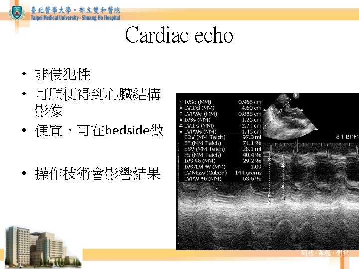
Cardiac echo • Cardiac Output = Stroke volume(SV) × Heart Rate • SV = VTI × CSA – CSA = valve orifice cross sectional area – VTI = the velocity time integral of the trace of the Doppler flow profile
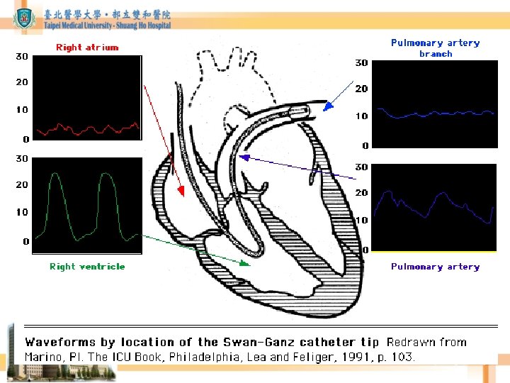
Pulmonary artery catheter • Measuring – Cardiac output (by thermal -dilution method) – Heart preload (fluid status) • Controvesies of its benefit http: //en. wikipedia. org/wiki/File: Pulmonary_artery_catheter_english. JPG
Table 30 -1 in: The Washington manual of surgery.
Crit Care Nurse October 2004 vol. 24 no. 5 74 -78
• Cardiac Index (CI) – Cardiac output/ Body surface area – Normal =3 -5 L/ min/ m 2 – If < 1. 8 L/ min/ m 2 cardiogenic shock
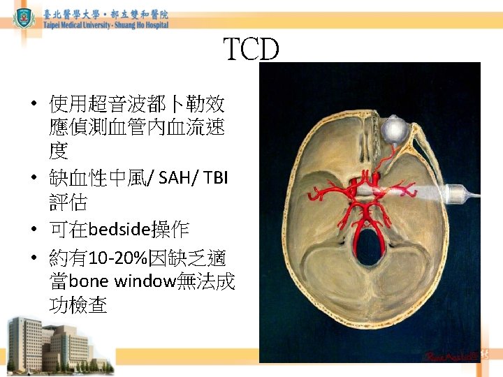
Brain hemodynamic monitoring • Cerebral blood perfusion (CBF) – CT perfusion – MR perfusion • Cerebral blood flow velocity – Transcranial Doppler (TCD)
CT/ MR perfusion DWI TMax ADC CBV CBF MTT
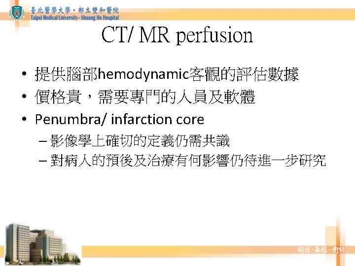
CT/ MR perfusion • Infarction core size evaluation – 代表不可逆腦組織損傷的大小 • Penumbra (缺血半影區) (PWI/ DWI mismatch area) – 大的penumbra代表如果不積極治療,症狀可能 惡化
Images from RAPID system
TCD • Mean Flow velocity (平均血流速度) correlates best with CBF • MFV = (FVs + 2 FVd)/3
TCD in SAH • MCA diameter <1. 5 mm (normal: 2. 3 -3. 4 mm) correlated with FV>140 cm/s. • Lindegaard ratio: MCA / ICA mean velocities (N: 1. 1 -2. 3) – LR > 3: vasospasm (mean FV >120 cm/sec) – LR > 6: severe vasospasm (mean FV >200 cm/sec) (Lindeggard KF et al. Acta Radiol Suppl. 1986; 369: 96 -98. )
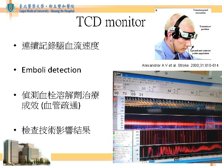
TCD monitor after IV t. PA Rinsho Shinkeigaku. 2010 Aug; 50(8): 547 -55.