Hemodynamic Disorders Hyperemia and congestion both refer to

Hemodynamic Disorders

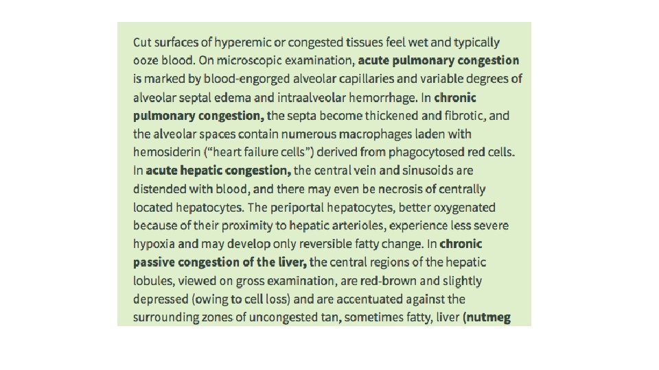
Hyperemia and congestion both refer to an increase in blood volume within a tissue, but have different underlying mechanisms. Hyperemia is an active process resulting from arteriolar dilation and increased blood inflow, as occurs at sites of inflammation or in exercising skeletal muscle. Hyperemic tissues are redder than normal because of engorgement with oxygenated blood.

Congestion is a passive process resulting from impaired outflow of venous blood from a tissue. It can occur systemically, as in cardiac failure, or locally as a consequence of an isolated venous obstruction. Congested tissues have an abnormal blue red color (cyanosis) that stems from the accumulation of deoxygenated hemoglobin in the affected area. In long standing chronic congestion, inadequate tissue perfusion and persistent hypoxia may lead to parenchymal cell death and secondary tissue fibrosis, and the elevated intravascular pressures may cause edema or sometimes rupture capillaries, producing focal hemorrhages.
 In this autopsy specimen, central")
Liver with chronic passive congestion and hemorrhagic necrosis. (A) In this autopsy specimen, central areas are red and slightly depressed compared with the surrounding tan viable parenchyma, creating “nutmeg liver” (so called because it resembles the cut surface of a nutmeg). (B) Microscopic preparation shows centrilobular hepatic necrosis with hemorrhage and scattered inflammatory cells.

Edema Approximately 60% of lean body weight is water, two-thirds of which is intracellular. Most of the remaining water is found in extracellular compartments in the form of interstitial fluid; only 5% of the body’s water is in blood plasma. Edema is an accumulation of interstitial fluid within tissues. Extravascular fluid can also collect in body cavities and such accumulations are often referred to collectively as effusions. Examples include effusions in the pleural cavity (hydrothorax), the pericardial cavity (hydropericardium), or the peritoneal cavity (hydroperitoneum, or ascites). Anasarca is severe, generalized edema marked by profound swelling of subcutaneous tissues and accumulation of fluid in body cavities.

Factors influencing fluid movement across capillary walls Capillary hydrostatic and osmotic forces are normally balanced so there is little net movement of fluid into the interstitium. However, increased hydrostatic pressure or diminished plasma osmotic pressure leads to extravascular fluid accumulation (edema). Tissue lymphatics drain much of the excess fluid back to the circulation by way of the thoracic duct; however, if the capacity for lymphatic drainage is exceeded, tissue edema results.

Fluid movement between the vascular and interstitial spaces is governed mainly by two opposing forces—the vascular hydrostatic pressure and the colloid osmotic pressure produced by plasma proteins. Normally, the outflow of fluid produced by hydrostatic pressure at the arteriolar end of the microcirculation is nearly balanced by inflow at the venular end owing to slightly elevated osmotic pressure; hence there is only a small net outflow of fluid into the interstitial space, which is drained by lymphatic vessels. Either increased hydrostatic pressure or diminished colloid osmotic pressure causes increased movement of water into the interstitium Increased Hydrostatic Pressure Increases in hydrostatic pressure are mainly caused by disorders that impair venous return. Local increases in intravascular pressure caused, for example, by deep venous thrombosis in the lower extremity can cause edema restricted to the distal portion of the affected leg. Generalized increases in venous pressure, with resultant systemic edema, occur most commonly in congestive heart failure

Pathways leading to systemic edema resulting from heart failure, renal failure, or reduced plasma osmotic pressure.

Reduced Plasma Osmotic Pressure Reduction of plasma albumin concentrations leads to decreased colloid osmotic pressure of the blood and loss of fluid from the circulation. Under normal circumstances, albumin accounts for almost half of the total plasma protein. Therefore, conditions in which albumin is either lost from the circulation or synthesized in inadequate amounts are common causes of reduced plasma osmotic pressure. Nephrotic syndrome is the most important cause of albumin loss from the blood

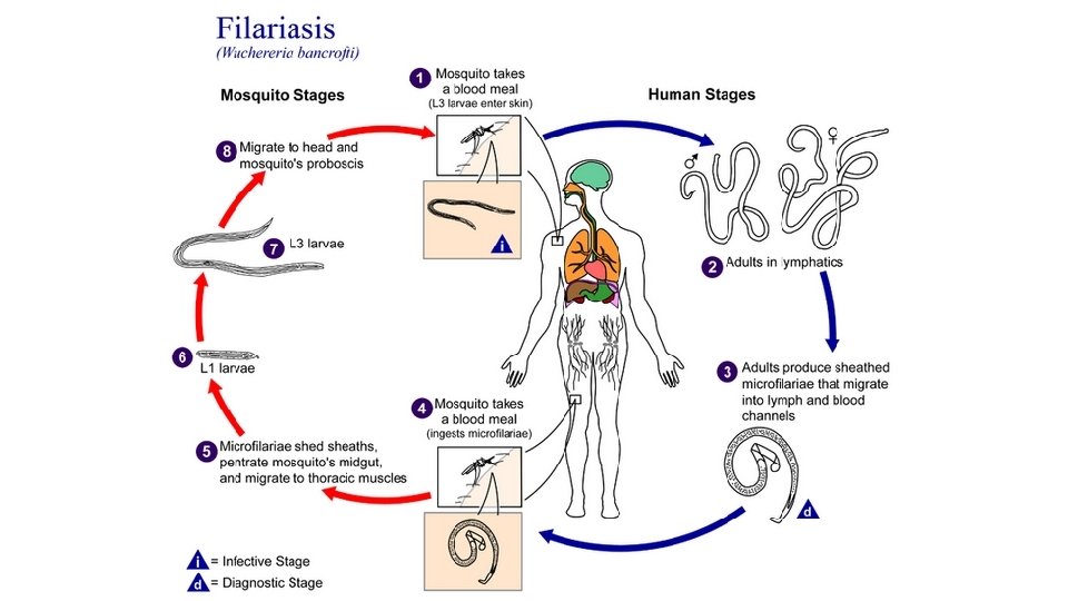
Lymphatic Obstruction Edema may result from lymphatic obstruction that compromises resorption of fluid from interstitial spaces. Impaired lymphatic drainage and consequent lymphedema usually results from a localized obstruction caused by an inflammatory or neoplastic condition. For example, the parasitic infection filariasis can cause massive edema of the lower extremity and external genitalia (so called “elephantiasis”) by producing inguinal lymphatic and lymph node fibrosis. Infiltration and obstruction of superficial lymphatics by breast cancer may cause edema of the overlying skin; the characteristic finely pitted appearance of the skin of the affected breast is called peau d’orange (orange peel)
 can")
Sodium and Water Retention Excessive retention of salt (and its obligate associated water) can lead to edema by increasing hydrostatic pressure (because of expansion of the intravascular volume) and reducing plasma osmotic pressure. Excessive salt and water retention are seen in a wide variety of diseases that compromise renal function, including poststreptococcal glomerulonephritis and acute renal failure.

Clinical Features The effects of edema vary, ranging from merely annoying to rapidly fatal. Subcutaneous edema is important to recognize primarily because it signals potential underlying cardiac or renal disease; however, when significant, it also can impair wound healing and the clearance of infections Pulmonary edema is a common clinical problem. It is seen most frequently in the setting of left ventricular failure, but also may occur in renal failure, acute respiratory distress syndrome, and inflammatory and infectious disorders of the lung. It can cause death by interfering with normal ventilatory function; besides impeding oxygen diffusion, alveolar edema fluid also creates a favorable environment for infections. Brain edema is life threatening; if the swelling is severe, the brain can herniate (extrude) through the foramen magnum. With increased intracranial pressure, the brain stem vascular supply can be compressed, leading to death due to injury to the medullary centers controlling respiration and other vital functions

Hemorrhage, defined as the extravasation of blood from vessels, is most often the result of damage to blood vessels or defective clot formation. Capillary bleeding can occur in chronically congested tissues. Trauma, atherosclerosis, or inflammatory or neoplastic erosion of a vessel wall also may lead to hemorrhage, which may be extensive if the affected vessel is a large vein or artery. The risk of hemorrhage (often after a seemingly insignificant injury) is increased in a wide variety of clinical disorders collectively called hemorrhagic diatheses. These have diverse causes, including inherited or acquired defects in vessel walls, platelets, or coagulation factors, all of which must function properly to ensure homeostasis.

Hemorrhage may be manifested by different appearances and clinical consequences. Hemorrhage may be external or accumulate within a tissue as a hematoma, which ranges in significance from trivial (e. g. , a bruise) to fatal (e. g. , a massive retroperitoneal hematoma resulting from rupture of a dissecting aortic aneurysm). Large bleeds into body cavities are described variously according to location—hemothorax, hemopericardium, hemoperitoneum, or hemarthrosis (in joints). Extensive hemorrhages can occasionally result in jaundice from the massive breakdown of red cells and hemoglobin. Petechiae are minute (1 to 2 m in diameter) hemorrhages into skin, mucous membranes, or serosal surfaces; causes include low platelet counts (thrombocytopenia), defective platelet function, and loss of vascular wall support, as in vitamin C deficiency. Purpura are slightly larger (3 to 5 m) hemorrhages. Purpura can result from the same disorders that cause petechiae, as well as trauma, vascular inflammation (vasculitis), and increased vascular fragility. Ecchymoses are larger (1 to 2 m) subcutaneous hematomas (colloquially called bruises). Extravasated red cells are phagocytosed and degraded by macrophages; the characteristic color changes of a bruise result from the enzymatic conversion of hemoglobin (red-blue color) to bilirubin (blue green color) and eventually hemosiderin (golden brown).
 Fatal intracerebral")
Punctate petechial hemorrhages of the colonic mucosa, a consequence of thrombocytopenia. (B) Fatal intracerebral hemorrhage.

The clinical significance of any particular hemorrhage depends on the volume of blood that is lost and the rate of bleeding. Rapid loss of up to 20% of the blood volume, or slow losses of even larger amounts, may have little impact in healthy adults; greater losses, however, can cause hemorrhagic (hypovolemic) shock. The site of hemorrhage also is important; bleeding that would be trivial in the subcutaneous tissues can cause death if located in the brain. Finally, chronic or recurrent external blood loss (e. g. , due to peptic ulcer or menstrual bleeding) frequently culminates in iron deficiency anemia as a consequence of a loss of iron in hemoglobin. By contrast, iron is efficiently recycled from phagocytosed red cells, so internal bleeding (e. g. , a hematoma) does not lead to iron deficiency.

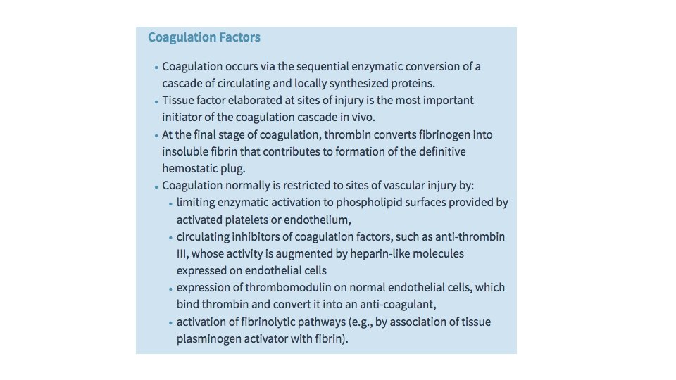
Normal Hemostasis is a precisely orchestrated process involving platelets, clotting factors, and endothelium that occurs at the site of vascular injury and culminates in the formation of a blood clot, which serves to prevent or limit the extent of bleeding.
 After vascular injury, local neurohumoral factors induce a transient vasoconstriction.")
Normal hemostasis. (A) After vascular injury, local neurohumoral factors induce a transient vasoconstriction.
 Platelets bind via glycoprotein Ib (Gp. Ib) receptors to von Willebrand factor (VWF)")
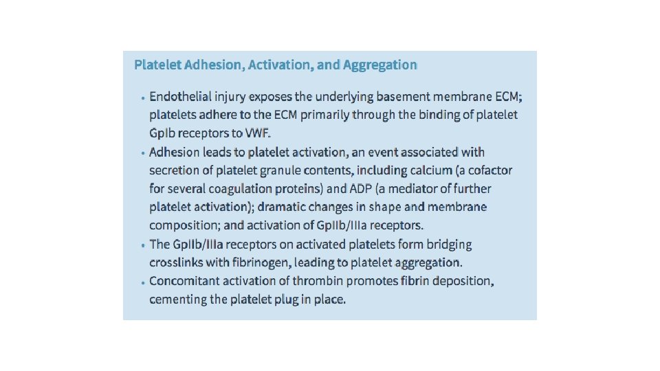
(B) Platelets bind via glycoprotein Ib (Gp. Ib) receptors to von Willebrand factor (VWF) on exposed ECM and are activated, undergoing a shape change and granule release. Released ADP and thromboxane A 2 (TXA 2) induce additional platelet aggregation through platelet Gp. IIb IIIa receptor binding to fibrinogen, and form the primary hemostatic plug.
 Local activation of the coagulation cascade (involving tissue factor and platelet phospholipids) results")
(C) Local activation of the coagulation cascade (involving tissue factor and platelet phospholipids) results in fibrin polymerization, “cementing” the platelets into a definitive secondary hemostatic plug.
 Counterregulatory mechanisms, mediated by tissue plasminogen activator (t PA, a fibrinolytic product) and")
(D) Counterregulatory mechanisms, mediated by tissue plasminogen activator (t PA, a fibrinolytic product) and thrombomodulin, confine the hemostatic process to the site of injury.

It should be emphasized that endothelial cells are central regulators of hemostasis; the balance between the anti thrombic and prothrombotic activities of endo thelium determines whether thrombus formation, propagation, or dissolution occurs. Normal endothelial cells express a variety of anticoagulant factors that inhibit platelet aggregation and coagulation and promote fibrinolysis; after injury or activation, however, this balance shifts, and endothelial cells acquire numerous procoagulant activities (activation of platelets and clotting factor). Besides trauma, endothelium can be activated by microbial pathogens, hemodynamic forces, and a number of pro inflammatory mediators.

Platelets play a critical role in hemostasis by forming the primary plug that initially seals vascular defects and by providing a surface that binds and concentrates activated coagulation factors. Platelets are disc shaped anucleate cell fragments that are shed from megakaryocytes in the bone marrow into the bloodstream.

Their function depends on several glycoprotein receptors, a contractile cytoskeleton, and two types of cytoplasmic granules. α-Granules have the adhesion molecule P selectin on their membranes and contain proteins involved in coagulation, such as fibrinogen, coagulation factor V, and v. WF, as well as protein factors that may be involved in wound healing, such as fibronectin, platelet factor 4 (a heparin binding chemokine), platelet derived growth factor (PDGF), and transforming growth factor β. Dense (or δ) granules contain adenosine diphosphate (ADP) and adenosine triphosphate, ionized calcium, serotonin, and epinephrine.

Platelet adhesion and aggregation. VWF functions as an adhesion bridge between subendothelial collagen and the glycoprotein Ib (Gp. Ib) platelet receptor Platelet aggregation is accomplished by fibrinogen binding to platelet Gp. IIb IIIa receptors on different platelets. Congenital deficiencies in the various receptors or bridging molecules lead to the diseases indicated in the colored boxes. ADP, Adenosine diphosphate.

The coagulation cascade is a series of amplifying enzymatic reactions that lead to the deposition of an insoluble fibrin clot. (A) Clotting is initiated in the laboratory by adding phospholipids, calcium, and either a negative charged substance such as glass beads (intrinsic pathway) or a source of tissue factor (extrinsic pathway).
 In vivo, tissue factor is the major initiator of coagulation, which is amplified")
(B) In vivo, tissue factor is the major initiator of coagulation, which is amplified by feedback loops involving thrombin (dotted lines). The red polypeptides are inactive factors, the dark green polypeptides are active factors, whereas the light green polypeptides correspond to cofactors.

The cascade of reactions in the pathway can be likened to a “dance, ” in which coagulation factors are passed from one partner to the next. Each reaction step involves an enzyme (an activated coagulation factor), a substrate (an inactive proenzyme form of a coagulation factor), and a cofactor (a reaction accelerator). These components are assembled on a negatively charged phospholipid surface, which is provided by activated platelets. Assembly of reaction complexes also depends on calcium, which binds to γ carboxylated glutamic acid residues that are present in factors II, VII, IX, and X. The enzymatic reactions that produce γ carboxylated glutamic acid use vitamin K as a cofactor and are antagonized by drugs such as Coumadin, a widely used anti coagulant.
 assay assesses the function of the proteins in the extrinsic")
The prothrombin time (PT) assay assesses the function of the proteins in the extrinsic pathway (factors VII, X, V, II (prothrombin), and fibrinogen). In brief, tissue factor, phospholipids, and calcium are added to plasma and the time for a fibrin clot to form is recorded. The partial thromboplastin time (PTT) assay screens the function of the proteins in the intrinsic pathway (factors XII, XI, IX, VIII, X, V, II, and fibrinogen). In this assay, clotting of plasma is initiated by the addition of negative charged particles (e. g. , ground glass) that activate factor XII (Hageman factor) together with phospholipids and calcium, and the time to fibrin clot formation is recorded.

Sequential conversion of factor X to factor Xa by way of the extrinsic pathway, followed by conversion of factor II (prothrombin) to factor IIa (thrombin). The initial reaction complex consists of a protease (factor VIIa), a substrate (factor X), and a reaction accelerator (tissue factor) assembled on a platelet phospholipid surface. Calcium ions hold the assembled components together and are essential for the reaction. Activated factor Xa then becomes the protease component of the next complex in the cascade, converting prothrombin to thrombin (factor IIa) in the presence of a different reaction accelerator, factor Va.

Among the coagulation factors, thrombin is the most important, because its various enzymatic activities control diverse aspects of hemostasis and link clotting to inflammation and repair. Among thrombin’s most important activities are the following: Conversion of fibrinogen into crosslinked fibrin. Thrombin directly converts soluble fibrinogen into fibrin monomers that polymerize into an insoluble fibril, and also amplifies the coagulation process, not only by activating factor XI, but also by activating two critical cofactors: factors V and VIII. It also stabilizes the secondary hemostatic plug by activating factor XIII, which covalently crosslinks fibrin. Platelet activation. Thrombin is a potent inducer of platelet activation and aggregation through its ability to activate Protease Activated Receptors (PARs), thereby linking platelet function to coagulation. Proinflammatory effects. PARs also are expressed on inflammatory cells, endothelium, and other cell types and activation of these receptors by thrombin is believed to mediate proinflammatory effects that contribute to tissue repair and angiogenesis. Anti-coagulant effects. Remarkably, through mechanisms described later, on encountering normal endothelium, thrombin changes from a procoagulant to an anti coagulant; this reversal in function prevents clots from extending beyond the site of the vascular injury.

Role of thrombin in hemostasis and cellular activation. Thrombin generates fibrin by cleaving fibrinogen, activates factor XIII (which is responsible for crosslinking fibrin into an insoluble clot), and also activates several other coagulation factors, thereby amplifying the coagulation cascade Through protease activated receptors (PARs), thrombin activates (1) platelet aggregation and Tx. A 2 secretion; (2) endothelium, which responds by generating leukocyte adhesion molecules and a variety of fibrinolytic (t PA), vasoactive (NO, PGI 2), or cytokine (PDGF) mediators; and (3) leukocytes, increasing their adhesion to activated endothelium. ECM, Extracellular matrix; NO, nitric oxide; PDGF, platelet derived growth factor; PGI 2, prostaglandin I 2 (prostacyclin); TXA 2, thromboxane A 2; t PA, tissue type plasminogen activator

Activation of the coagulation cascade also sets into motion a fibrinolytic cascade that limits the size of the clot and contributes to its later dissolution. Fibrino lysis is largely accomplished through the enzymatic activity ofplasmin, which breaks down fibrin and interferes with its polymerization. An elevated level of breakdown products of fibrinogen (often called fibrin split products), most notably fibrin derived D dimers, are a useful clinical markers of several thrombotic states. Plasmin is generated by enzymatic catabolism of the inactive circulating precursor plasminogen, either by a factor XII–dependent pathway (possibly explaining the association of factor XII deficiency and thrombosis) or by plasminogen activators. The most important plasminogen activator is t PA; it is synthesized principally by endothelium and is most active when bound to fibrin. This characteristic makes t PA a useful therapeutic agent, since its fibrinolytic activity is largely confined to sites of recent thrombosis. Once activated, plasmin is in turn tightly controlled by counterregulatory factors such as α 2 plasmin inhibitor, a plasma protein that binds and rapidly inhibits free plasmin.

The fibrinolytic system, illustrating various plasminogen activators and inhibitors

The antithrombotic properties of endothelium can be divided into activities directed at platelets, coagulation factors, and fibrinolysis. Platelet inhibitory effects. An obvious effect of intact endothelium is to serve as a barrier that shields platelets from subendothelial v. WF and collagen. However, normal endothelium also releases a number of factors that inhibit platelet activation and aggregation. Among the most important are prostacyclin (PGI 2), nitric oxide (NO), and adenosine diphosphatase; the latter degrades ADP, a potent activator of platelet aggregation. Finally, endothelial cells bind alter the activity of thrombin, which is one of the most potent activators of platelets.

Anticoagulant effects Normal endothelium shields coagulation factors from tissue factor in vessel walls and expresses multiple factors that actively oppose coag ulation, most notablythrombomodulin, endothelial protein C receptor, heparin-like molecules, and tissue factor pathway inhibitor. Thrombomodulin and endothelial protein C receptor bind thrombin and protein C, respectively, in a complex on the endothelial cell surface. When bound in this complex, thrombin loses its ability to activate coagulation factors and platelets, and instead cleaves and activates protein C, a vitamin K–dependent protease that requires a cofactor, protein S.

Activated protein C/protein S complex is a potent inhibitor of coagulation factors Va and VIIIa. Heparin-like molecules on the surface of endothelium bind activate antithrombin III, which then inhibits thrombin and factors IXa, XIa, and XIIa. The clinical utility of heparin and related drugs is based on their ability to stimulate antithrombin III activity. Tissue factor pathway inhibitor (TFPI), like protein C, requires protein S as a cofactor and, as the name implies, binds and inhibits tissue factor/factor VIIa complexes. Fibrinolytic effects. Normal endothelial cells synthesize t PA, a key component of the fibrinolytic pathway.
 and procoagulant properties of injured or activated endothelium")
Anti-coagulant properties of normal endothelium (top) and procoagulant properties of injured or activated endothelium (bottom). NO, Nitric oxide; PGI 2, prostaglandin I 2 (prostacyclin); t PA, tissue plasminogen activator; VWF, von Willebrand factor. Thrombin receptors are also called protease activated receptors (PARs).
 endothelial injury, (2)")
Thrombosis The primary abnormalities that lead to intravascular thrombosis are (1) endothelial injury, (2) stasis or turbulent blood flow, and (3) hypercoagulability of the blood (the so called “Virchow triad”). Thrombosis is one of the scourges of modern man, because it underlies the most serious and common forms of cardiovascular disease. Here, the focus is on its causes and consequences Virchow's triad in thrombosis. Endothelial integrity is the most important factor. Abnormalities of procoagulants or anti coagulants can tip the balance in favor of thrombosis. Abnormal blood flow (stasis or turbulence) can lead to hypercoagulability directly and also indirectly through endothelial dysfunction.

Major prothrombotic alterations: Procoagulant changes. Endothelial cells activated by cytokines downregulate the expression of thrombomodulin, already described as a key modulator of thrombin activity. This may result in sustained activation of thrombin, which can in turn stimulate platelets and augment inflammation through PARs expressed on platelets and inflammatory cells. In addition, inflamed endothelium also downregulates the expression of other anti coagulants, such as protein C and tissue factor protein inhibitor, changes that further promote a procoagulant state. Anti-fibrinolytic effects. Activated endothelial cells secrete Plasminogen activator inhibitors (PAI), which limit fibrinolysis and downregulate the expression of t PA, alterations that also favor the development of thrombi.
 contributes to arterial and cardiac thrombosis by")
Abnormal Blood Flow Turbulence (chaotic blood flow) contributes to arterial and cardiac thrombosis by causing endothelial injury or dysfunction, as well as by forming countercurrents and local pockets of stasis. Stasis is a major factor in the development of venous thrombi. Under conditions of normal laminar blood flow, platelets (and other blood cells) are found mainly in the center of the vessel lumen, separated from the endothelium by a slower moving layer of plasma. By contrast, stasis and turbulence have the following deleterious effects: Both promote endothelial cell activation and enhanced procoagulant activity, in part through flow induced changes in endothelial gene expression. Stasis allows platelets and leukocytes to come into contact with the endothelium when the flow is sluggish. Stasis also slows the washout of activated clotting factors and impedes the inflow of clotting factor inhibitors.

Hypercoagulability refers to an abnormally high tendency of the blood to clot, and is typically caused by alterations in coagulation factors
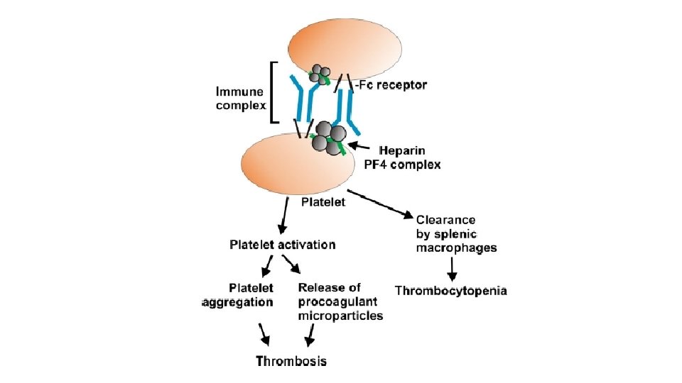
 syndrome. This syndrome occurs in up to 5% of patients treated")
Heparin-induced thrombocytopenia (HIT) syndrome. This syndrome occurs in up to 5% of patients treated with unfractionated heparin (for therapeutic anti coagulation). It is marked by the development of autoantibodies that bind complexes of heparin and platelet membrane protein (platelet factor-4). Although the mechanism is unclear, it appears that these antibodies may also bind similar complexes present on platelet and endothelial surfaces, resulting in platelet activation, aggregation, and consumption (hence thrombocytopenia), as well as causing endothelial cell injury. The overall result is a prothrombotic state, even in the face of heparin administration and low platelet counts. Newer low molecular weight fractionated heparin preparations induce autoantibodies less frequently but can still cause thrombosis if antibodies have already formed.
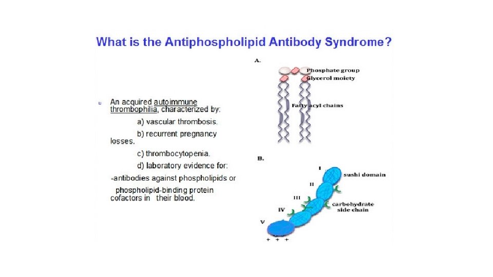
 has protean")
Anti-phospholipid antibody syndrome. This syndrome (previously called the lupus anti coagulant syndrome) has protean clinical manifestations, including recurrent thromboses, repeated miscarriages, cardiac valve vegetations, and thrombocytopenia. Depending on the vascular bed involved, the clinical presentations can include pulmonary embolism (following lower extremity venous thrombosis), pulmonary hypertension (from recurrent subclinical pulmonary emboli), stroke, bowel infarction, or renovascular hypertension. Fetal loss does not appear to be explained by thrombosis, but rather seems to stem from antibody mediated interference with the growth and differentiation of trophoblasts, leading to a failure of placentation. Anti phospholipid antibody syndrome is also a cause of renal microangiopathy, resulting in renal failure associated with multiple capillary and arterial thromboses
- Slides: 51