Hematopoiesis Lecture Objectives Name organs responsible for hematopoiesis

Hematopoiesis

Lecture Objectives • Name organs responsible for hematopoiesis in the fetus. • List the developmental stages of hematopoiesis both prenatally and postnatally. • Outline the major steps of post‐natal development of blood formed elements (erythropoiesis, granulopoiesis, monocytopoiesis and megakaryopoiesis. • Identify characteristic features of these cells.

Formation of Blood Cells • Negative feedback systems regulate the total number of RBCs and platelets in circulation • Abundance of WBC types based of response to invading pathogens or foreign antigens • Hemopoiesis or hematopoiesis • Red bone marrow primary site • Pluripotent stem cells have the ability to develop into many different types of cells
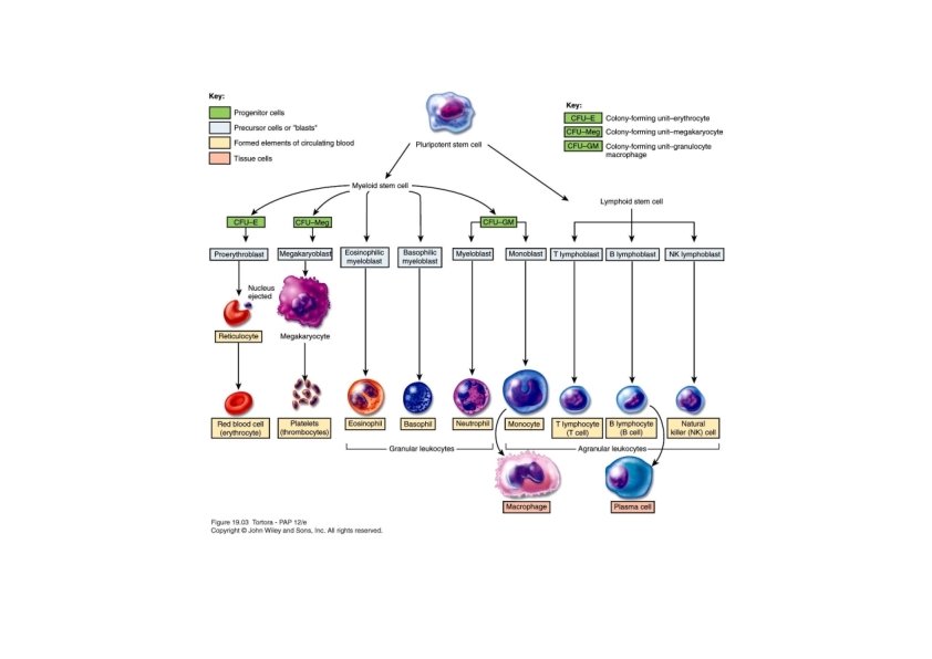

Formation of Blood Cells • Stem cells in bone marrow • Reproduce themselves • Proliferate and differentiate • Cells enter blood stream through sinusoids • Formed elements do not divide once they leave red bone marrow • Exception is lymphocytes

Formation of Blood Cells • Pluripotent stem cells produce • Myeloid stem cells • Give rise to red blood cells, platelets, monocytes, neutrophils, eosinophils and basophils • Lymphoid stem cells give rise to • Lymphocytes • Hemopoietic growth factors regulate differentiation and proliferation • Erythropoietin – RBCs • Thrombopoietin – platelets • Colony‐stimulating factors (CSFs) and interleukins – WBCs

Bone Marrow • Bone marrow found in the medullary canals of long bones and cavities of cancellous bone • Tow types: • Red bone marrow, color due to blood and blood forming cells • Yellow bone marrow, color due to great number of adipose cells • In newborns all bone marrow is red

Red Bone Marrow • Functions • Production of blood cells • Destruction of worn‐out RBCs • Storage (in macrophages) of iron derived from breakdown of hemoglobin

Red Bone Marrow • Structure • Stroma • Meshwork of reticular cells in delicate web of reticular fibers • Collagen type I and III • Fibronectin, laminin, and hemonectin • Interact with cell receptors to bind cells to stroma • Hematopoietic cords • Hematopoietic cells • Sinusoidal capillaries • Reinforced by external discontinuous layer of reticular cells and a loose net of reticular fibers

Maturation of Erythrocytes • The basic process of maturation is synthesis of hemoglobin and formation of enucleated, biconcave erythrocyte • Changes during maturation • Decrease in cell volume • Nucleoli diminish in size until invisible in scope • Nuclear diameter decrease and chromatin become denser until nucleus have apyknotic appearance and finally extruded • Decrease number of polyribosomes (basophilia decrease) with increase in hemoglobin (acidophilic) • Cell organelles gradually disappear • Development of erythrocyte from the first cell of the series until released to blood take 7 days

Differentiation of Erythrocytes • Proerythroblast • First recognizable cell in the series • Large cell with loose chromatin and visible nucleoli • Its cytoplasm is basophilic • Basophilic erythroblast • Strongly basophilic cytoplasm • Condensed nucleus that has no visible nucleolus • Polychromatophilic erythroblast • Decrease polyribosoms and increase hemoglobin • Staining causes several colors to appear • Orthochromatophilic erythroblast • Nucleus continue to condense • No basophilia is evident and uniformly acidophilic cytoplasm • Reticulocyte • Nucleus is expelled and engulfed by macrophages • Small number of polyribosoms
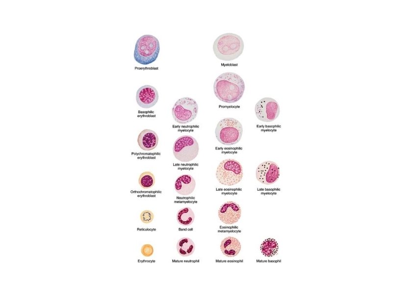

Granulopoiesis • Maturation process involves synthesis of protein that are backed in granules • First stage results in the development of the azurophilic granules that contain enzymes of lysosomal system • Stain with basic dyes • In the second stage results in the development of the specific granules • several types of proteins are backed in these granules depending on the type of the granulocytes

Maturation of Granulocytes • Myeloblast is the most immature recognizable cell in the myeloid series • Dispersed chromatin with visible nucleoli • Promyelocyte • Basophilic cytoplasm and azurophilic granules • Give rise to the three known types of granulocytes • Myelocyte • Specific granules appear and increase • Nutrophilic, basophilic and eosinophilic myelocyte • Further condensation of nucleus • Considerable increase in their specific granules • Neutrophilic granulocyte before maturation passes through an intermediate stage where nucleus has curved rod shape (band cell)


Maturation of Lymphocytes & Monocytes • Precursor cells do not contain specific cytoplasmic granules or nuclear lobulation • Lymphocytes and monocytes are distinguished on the basis of size, chromatin structure and the presence of nucleoli • As lymphocyte mature • Chromatin becomes more compact • Nucleoli become less visible • Cell decrease in size

Lymphocytes • Circulating lymphocytes originate in lymphoid organs • Lymphocyte progenitor cells originate in the bone marrow • Lymphoblast • First identifiable progenitor of lymphoid cells • Large cells • Polymorphocytes • Smaller and more condensed chromatin

Monocytes • Monoblast • Committed progenitor cell that looks like myeloplast • Promonocyte • • • Large cell (up to 18 µm in diameter) Basophilic cytoplasm Slightly indented nucleus Chromatin is lacy and visible nucleoli Divide twice leading to monocytes • Fine azurophilic granules can be seen in the blood monocyte • Mature monocyte circulate blood for 8 h, then enter the CT and mature into macrophages, which function for several months

Origin of Platelets • Megakaryoblasts • 15‐ 50 µm in diameter • Large ovoid or kidney‐shaped nucleus with numerous nucleoli • Cytoplasm is homogenous and intensely basophilic • Megakaryocytes • • Giant cell, 35‐ 150 µm in diameter Irregularly lobulated nucleus Coarse chromatin, no visible nucleoli Cytoplasm contain well developed organelles
- Slides: 20