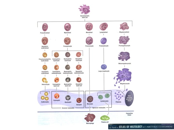
Hematopoeisis VIBS 443602 113 Peripheral blood smear MayGrunwaldGiemsa

Hematopoeisis VIBS 443/602
")
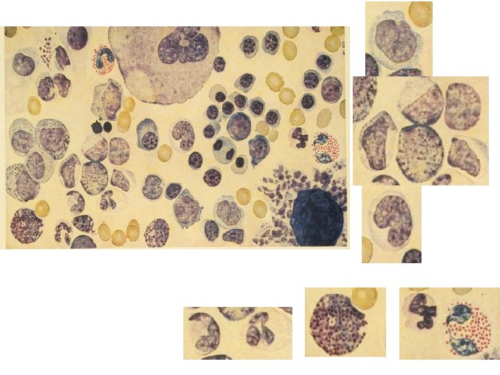
113 Peripheral blood smear (May-Grunwald-Giemsa)
 eosinophil, basophil, and neutrophils")
110 Peripheral blood smear (Leishman-Giemsa) eosinophil, basophil, and neutrophils

19761 Bone marrow 19761
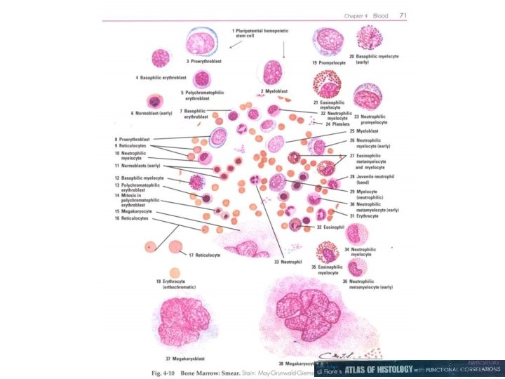

Bone marrow smear; low power view, showing negative images of fat globules

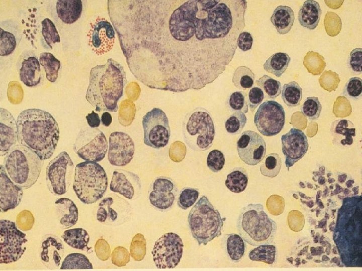
Megakaryocyte: contain many joined nuclei. Each probably contain 8 -32 times a normal diploid amount of DNA 1. Nuclei of megakaryocyte 2. Red blood cell

Macrophage: contains greenish, iron-containing granules due to poor quality. Differentiating cells have been torn away during preparation. 1. Macrophage 2. Lymphocyte 3. Neutrophil

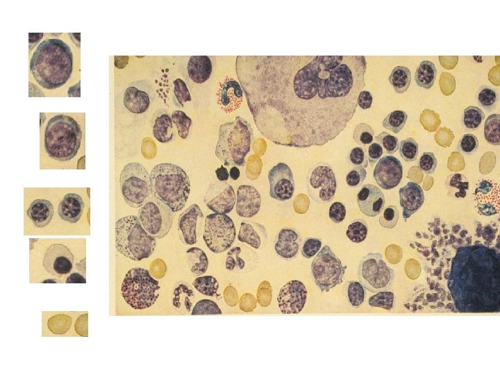
1. Pronormoblast 2. Basophilic normoblast 3. Polychromatophilic normoblast 4. Orthochromatic normoblast 5. Myelocyte 6. Late metamyelocytes

1. Pronormoblast 2. Basophilic normoblast 3. Polychromatophilic normoblast 4. Orthochromatic normoblast 5. Myelocyte 6. Late metamyelocytes

1. Late promyelocyte 2. Late myelocyte/early metamyelocyte 3. Nearly mature PMN

1. Smudge cell 2. Promyelocyte 3. Myelocyte 4. Metamyelocytes

1. Pronormoblast 2. Metamyelocytes 3. Band cell

Bone marrow: EM 12 a • Reticular cell • Developing red blood cell

Bone marrow; EM 8 a 1. Lymphocyte 2. PNM 3. Red blood cell 4. eosinophil

Bone marrow; EM 12 b 1. PMN 2. Immature neutrophil 3. Red blood cell

Routine H&E of bone • Marrow spicules • Lipid in fat cells

H&E of bone marrow 1. megakaryocytes

19761

H&E of bone marrow: • Red blood cell

420 Rib

420 Rib

421 Tibia, fetal

583

83
")
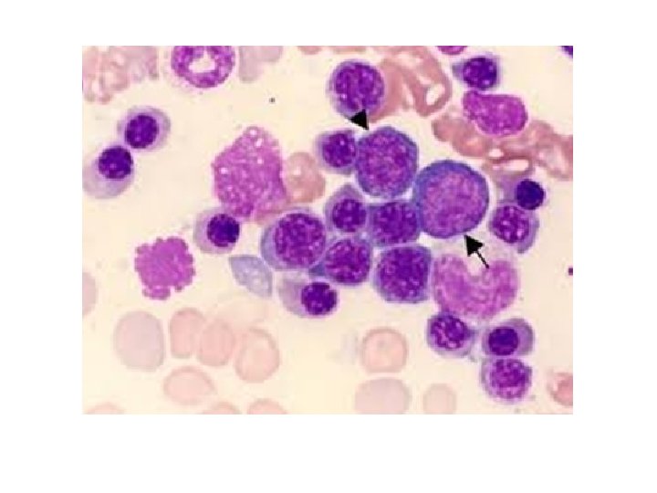
112 Red bone marrow smear (May-Grunwald-Giemsa)

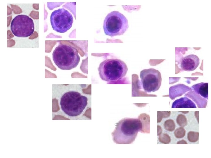
Cells of Erythropoisesis Pronormoblast round nucleus and thin rim of deep blue cytoplasm Basophilic normoblast nuclear condensation ("fractured nucleus") with a deep blue cytoplasm

Polychromatophilic normoblast cytoplasm has accumulated enough hemoglobin so that it is no longer blue but a bluish-gray to bluish-red Orthochromatic normoblast must have an almost mature cytoplasm with only the faintest hint of blue: the nucleus is fully

Reticulocyte no nucleus, mature cytoplasm with blue tint Red blood cell typical of mature cell seen in circulation


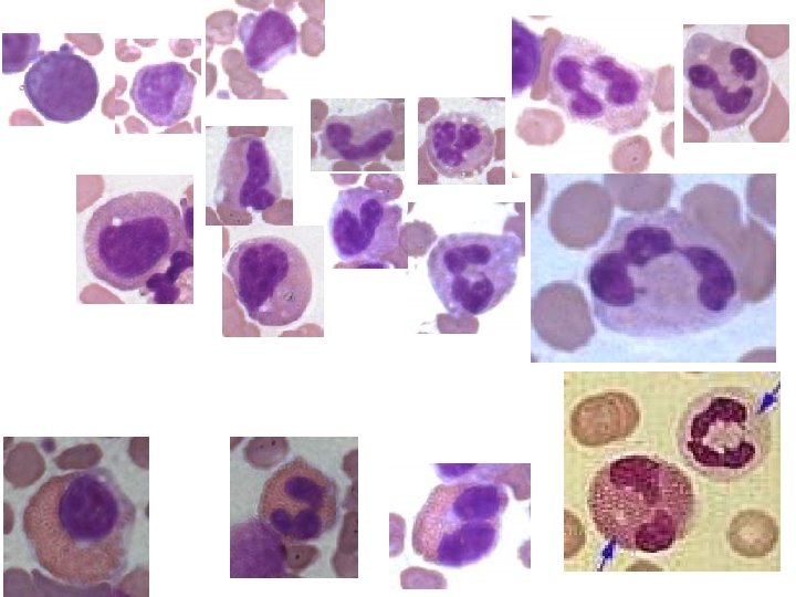
Cells of Granulopoiesis Promyelocyte oval nucleus with deep blue cytoplasm and azurophilic granules Myelocyte cytoplasm becomes increasingly mature (reddish/clear) but still contains blue, nucleus of variable shape
 Band cell nucleus")
Metamyelocyte deeply indented nucleus with nearly mature cytoplasm (no longer blue) Band cell nucleus just prior to lobulation with fully mature cytoplasm Neutrophil (mature) lobulated nucleus with fully mature cytoplasm

- Slides: 40