HEMATOPOEISIS Dr Bastiana Bermawi Sp PK 1 Hematopoiesis

HEMATOPOEISIS Dr. Bastiana Bermawi, Sp. PK 1
 Pembentukan sel darah (Production of Blood cell) Where? ? 2")
Hematopoiesis (hemopoiesis) Pembentukan sel darah (Production of Blood cell) Where? ? 2
 : tulang pipih. tulang tengkorak, clavicula ,")
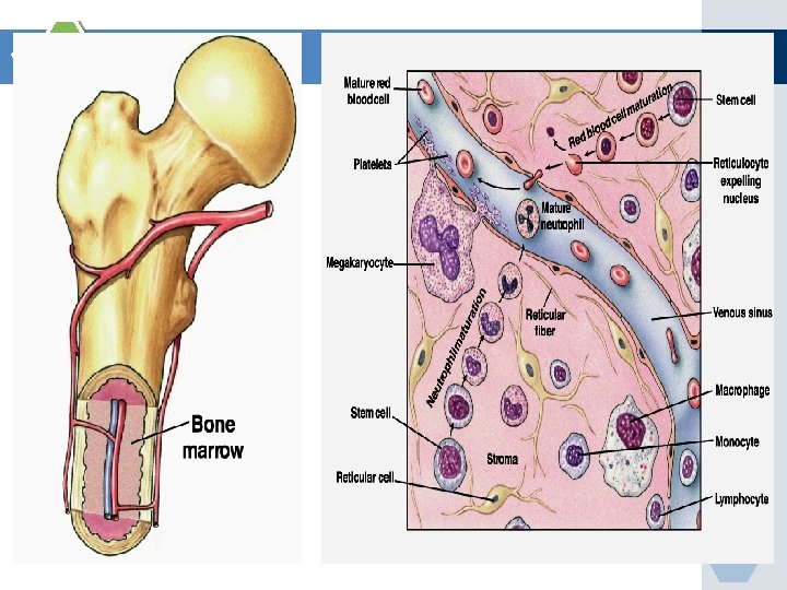
Tempat Hematopoeisis Neonatus (semua sumsum merah tulang) : tulang pipih. tulang tengkorak, clavicula , sternum, tulang rusuk, vertebra dan tulang pelvis Dewasa vertebra, tulang rusuk, sternum, tulang kepala, tulang pelvis, proksimal dan distal femur 50% sutul terisi lemak dan dapat diubah jadi tempat hematopoisis pada keadaan tertentu dapat terjadi hematopoisis ekstramedular ( hati , limpa ). 3

Artery White blood cells Platelets Red blood cells 4

ERITROSIT 5

eosinophil neutrophil monocyte RBC neutrophil monocyte lymphocyte basophil lymphocyte 6

PLATELET/ TROMBOSIT 7

DEFINISI v Hematopoeisis: Proses pembentukan darah yang teregulasi dan berkesinambungan, meliputi cell renewal, proliferasi, diferensiasi dan maturasi v Cell renewal: kemampuan sel memperbarui diri sendiri shg tidak akan habis meski terus membelah v Proliferasi: kemampuan membelah /memperbanyak diri v Difrensiasi: kemampuan berkembang menjadi sel dgn fungsi tertentu v Maturasi: kemampuan mematangkan diri 8
 Hematopoeisis patologis")
v Pada orang dewasa, hematopoisis fisiologis hanya terjadi di sumsum tulang (sutul) Hematopoeisis patologis terjadi di luar sutul, disebut hematopoeisis ekstramedularis v Selama perkembangan janin, proses ini terjadi di beberapa tempat, terdiri dari 3 fase, yaitu: - fase mesoblastik (yolk sac); - fase hepatik; - fase medularis (mieloid) 9

10

v Hematopoisis: 1. Yolk sac: 0 -3 bulan intra uterine 2. Hati dan Lien: ~3 - 6 bulan intra uterine 3. sumsum tulang: ~ 4 bln intrauterine - dws 11
 2. Lingkungan")
PROSES HEMATOPOEISIS v Proses hematopoeisis memerlukan: 1. Sel induk hematopoisis (hematopoeiticstem cell) 2. Lingkungan mikro (microenvironment) 3. Bahan pembentuk darah 4. Mekanisme regulasi 12
 v Sel induk hematopoetik (SIH): Sel yg akan")
1. Sel induk hematopoesis (Hematopoetic stemsel) v Sel induk hematopoetik (SIH): Sel yg akan berkembang mjd sel darah, yaitu: eritrosit, lekosit, trombosit dan sel lain dlm sutul, spt fibroblas v SIH paling primitif: Pluripoten/totipoten stem-cell, mempunyai sifat: * self renewal * proliferatif * differensiatif * maturatif 13

Pluripotential stem cells rangsangan Kemampuan: v memperbaharui diri v proliferasi v diferensiasi v maturasi 14
 v berdasarkan sifat diffrensiatif, SIH terbagi atas: - pluripotent/totipoten")
Lanjutan…. Sel induk hematopoesis (SIH) v berdasarkan sifat diffrensiatif, SIH terbagi atas: - pluripotent/totipoten stemcell: mampu berdiffrensiasi jd seluruh jenis sel - Commited stemcell: komitmen berdifrensiasi menjadi satu cell line (cth : SIH mieloid, SIH Limfoid) - Oligopotent stemcell: hanya mampu berdifrensiasi jd beberapa jenis sel, cth: CFU-GM (Colonyforming unit- Granulocyte/monocyte hy menjadi sel granulosit dan sel monosit. - Unipotent stemcell: hanya mampu berdifrensiasi jd satu jenis sel saja, cth: CFU-E eritrosit 15

v Pluripotent stemcells tingkat proliferasinya rendah v Unipotent stemcells tingkat proliferasinya tinggi v Jumlah Pluripotent hematopoietic stem cells di sumsum tulang dan di peredaran darah sedikit. 16

17

2. Lingkungan mikro Sutul: - Fungsi: - - menyediakan nutrisi & bhn hemopoisis - komunikasi antar sel: Adhesi molekul - Menghasilkan zat yg mengatur hematopoeisis: sitokin, hemopoetik growth factor. - Meliputi: a. mikrosirkulasi dlm sutul b. sel stroma: sel endothel, makrofag, sel lemak, fibroblast, sel retikulum. c. matriks ekstraseluler: haemonektin, kolagen, fibronektin, glikosaminoglikan. 18

3. Bahan Pembentuk darah: * as folat, vit B 12 * Fe * Co, Mg, Cu, Zn * As amino * vitamin: vitamin B komplek, Vitamin C 20

4. Mekanisme regulasi Mekanisme Regulasi: - Untuk mengatur arah & kuantitas pertumbuhan & pelepasan sel darah matur dari sutul darah tepi. - sistim ‘feed back mechanism’ 21

Zat yg berpengaruh dlm mekanisme Regulasi 1. Faktor pertumbuhan Hemopoisis: - GM-CSF - G-CSF - M-CSF, dll. 2. Sitokin: - IL-3, IL-4, IL-6, IL-11 dll. - Ada yg merangsang & menekan pertumbuhan sel induk. 3. Hormon hemopoitik spesifik: Erytropoetin 4. Hormon non spesifik (dlm jumlah kecil) - androgen - estrogen - Hormon tiroid. - glukokortikoid - growth hormon 22

Hematopoitic growth factors and some of their characteristics Factor Cell stimulated Production sources M-CSF Monocytes Endothelial cells, monocytes, fibroblasts GM-CSF All granulocytes, Tcells, endothelial cells, megakaryocytes, erythrocyt fibroblast es, stem cell, leukemic blast 23

Factor Cell stimulated Production sources G-CSF Granulocytes, megakaryocytes, erythrocytes, stem cell, leukemic blast Endotelial cells, placenta, monocytes IL-3 Granulocytes, erythroid cells, multipotential progenitors, leukemic blasts T cells 1 L-4 B, T cells 24

Factor Cell stimulated Production sources IL-5 B cells, CFU-EO IL-6 B, T cells, CFU-GM Fibroblasts, GFU-GEMM, BFU-E, leucocytes, endo. Macrophages, neural thelial cells, hepatocytes IL-7 B cells leucocytes IL-8 T cells, neutrophils leucocytes T cells 25

Factor Cell stimulated Production sources IL-9 BFU-E, CFU-GEMM Lymphocytes IL-11 B, T cells, CFU-GEMM, macrophages Erythropoitin CFU-E, BFU-E Kidney , liver 26

Sel progenitor mempunyai reseptor untuk faktor humoral tertentu yang sesuai Invitro v BFU-E-, CFU-E + Eritropoitin Eritropoisis v CFU-MEG. + MEG-CSF Megakariosit v CFU-GM + GM-CSF Netrofil, monosit v CFU-EO + EO. CSF Eosinofil. 27

28

29

30

31

32
: keganasan, disfungsi/ defisiensi SIH (an.")
Gangguan hematopoeisis 1. Gangguan pada sel induk hematopoeisis (SIH): keganasan, disfungsi/ defisiensi SIH (an. aplastik) 2. Gangguan Organ tempat hematopoisis: semua kerusakan sutul (fibrosis, infeksi metastase) 3. Gangguan pada bahan yang diperlukan: (faktor nutrisi, gangguan Hormon, bahan beracun). 33

Eritropoesis 34
 § Hematopoetic stem-cell berdifrensiasi menjadi")
Eritropoeisis v Eritropoeisis: Pembentukan/produksi eritrosit (red blood cell production) § Hematopoetic stem-cell berdifrensiasi menjadi proerythroblast § Proerythroblasts early erythroblasts § Fase Perkembangan selanjutnya: 1. Ribosome synthesis 2. Hemoglobin accumulation 3. Ejection of the nucleus and formation of reticulocytes § Retikulosit eritrosit matur 35

Stem cell Hemocytoblast Committed cell Developmental pathway Proerythroblast Early Late erythroblast Phase 1 Ribosome synthesis Phase 2 Hemoglobin accumulation Phase 3 Ejection of nucleus Normoblast Reticulo- Erythrocyte 36 Figure 17. 5
 Vitamin ( B")
Eritropoisis Membutuhkan: v Nutrisi : Mineral ( Fe, mangan, cobalt ) Vitamin ( B 12, C, B 6, B 1, asam folat, dll) Asam amino v Faktor perangsang : Eritropoitin, tiroksin, androgen v Keutuhan jaringan sumsum tulang (microenvironment ) 37

Tiroksin: meningkatkan kebutuhan jaringan terhadap oksigen Androgen : merangsang eritropoitin meningkatkan sensitivitas stem cell terhadap eritropoitin. mempengaruhi langsung eritropoisis 38

39

Proerythroblast v No hemoglobin v Nucleus 12 um v Contain nucleoli 40

Basophil erythroblast v Early normoblast v Nucleoli disappear v Show mitosis v Cytoplasm deep blue § Increase in RNA v Hemoglobin starts appearing – Little Hb 41

Polychromatophil erythroblast v Late normoblast v Nucleus smaller v Coarse Chromatin v Hemoglobin increase § Eosinophil Stain v RNA – Basophil stain 42

Orthochromatic Erythroblast v Normoblast v Nucleus smaller § Pyknosis v Nuclear lysis and v Nuclear extrusion 43

Reticulocyte v Reticulum v Remnant of ER § Synthesize Hb v Few Mitochondria v Young RBCs (34% Hb) v 1 % of Red Cells 44

GRANULOPOESIS 45
 Mieloblas Promielosit segmen netrofil mielosit N metamielosit N stab")
Contoh GRANULOPOESIS GRANULOPOEISIS (Line: neutrofil) Mieloblas Promielosit segmen netrofil mielosit N metamielosit N stab N 46

Fungsi utama netrofil: v Pertahanan tubuh migrasi ke tempat infeksi. v Pengenalan & pengolahan antigen asing v Fagositosis 47

Distribusi netrofil setelah masuk sirkulasi darah Sutul Sirkulasi darah Jaringan MGP CGP T 1/2 = 6, 3 -7, 6 jam MGP CGP = Circulating granulocyte pool MGP = Marginated granulocyte pool TGP = Total blood granulocyte pool = CGP + MGP 48

Perubahan pola distribusi Netrofil : v Olah-raga v Epinefrin v Hipoksia v Stres → me↑ CGP sampai 50% , me↓ MGP, tapi TGP tetap → pseudonetrofilia v Endotoksin Kortikosteroid mobilisasi MGP ke CGP, mobilisasi dari sutul ke CGP → TGP ↑ 49

Faktor yg mempengaruhi mobilisasi netrofil dari sutul ke sirkulasi: v Bakteri / organisme v Endotoksin → merusak dinding sinusoid. v Besarnya pori-pori dinding sinusoid. v Tingkat maturasi sel. Netrofil batang / segmen dapat lewat pori 2 dgn Ø 1 μm ; promielosit dapat lewat pori 2 dgn Ø 8 μm. 50

51

Nama monosit di jaringan v Di hati ---- sel Kupfer v Di limpa ---- sel retikulum v Di tulang ---- osteoclast v Di jaringan ikat ---- histiosit v Di sutul ---- makrofag v Di otak ---- neurog. Iia 52

LIMFOPOESIS 53

54
 tak perlu rangsangan v Di organ sekunder")
Limfopoisis v Di organ primer (sutul, timus) tak perlu rangsangan v Di organ sekunder (kelenjar getah bening, limpa, perlu rangsangan antigen). Limfosit T v Di sirkulasi darah v Umur lebih v Resirkulasi + v Rangsangan antigen atipis v Fungsi imunitas seluler Limfosit B 15 -20% + blast transform (sel plasma) humoral 55

Rangkuman v Hematopoisis: Pembentukan, perkembangan, dan proses pegkhususan/spesialisasi sel darah cara fungsional. v Fase hematopoesis: mesoblastik, hepatik dan medularis v Organ tempat hematopoesis: Hati, Limpa, lymphnodus, Timus dan Sumsum tulang v Sumsum tulang: tempat utama hematopoeisis mulai dari lahir sampai seumur hidup 56
,")
Rangkuman v Pada keadaan tertentu, produksi sel darah bisa terjadi luar sumsum tulang (sutul), disebut hematopoeisis ekstramedular v Lingkungan mikro pada sutul penting untuk proliferasi dan difrensiasi stemsel v Sel matur, karakteristik umum: penurunan ukuran sel dan inti sel, hilangnya nukleoli, kondensasi kromatin inti, pengurangan basofilia di sitoplasma. 57

58

DARAH v merupakan komponen esensial makhluk hidup, yang berfungsi, a. l: 1. sebagai pembawa oksigen, 2. mekanisme pertahanan tubuh thd infeksi 3. mekanisme hemostasis v terdiri dari 2 komponen utama, yakni: 1. Plasma: bgn cair darah: air, elektrolit, protein 2. Sel darah (blood corpuscles): a. eritrosit (sel darah merah) b. leukosit (sel darah putih) c. trombosit sel /butir pembeku darah) 59
 • Membuang produk sisa metabolisma tubuh")
• Membawa O 2 (Deliver O 2) • Membuang produk sisa metabolisma tubuh • Mengatur suhu, p. H, dan volume cairan • Melindungi dari kehilangan darah/sumbat perdarahan-trombosit (Protection from blood loss- platelets) • Mencegah infeksi- antibodi dan leukosit • Transpor hormon 60

Erythrocyte 7. 5 m in diameter · Anucleate- so can't reproduce; however, repro · · · · in red bone marrow Hematopoiesis- production of RBC Function- transport respiratory gases Hemoglobin- quaternary structure, 2 chains and 2 chains 1 RBC contains 280 million hemoglobin molecules Men- 5 million cells/mm 3 Women- 4. 5 million cells/mm 3 Life span 100 -120 days and then destroyed in spleen (RBC graveyard) 61

RBC Formation before birth v Mesoblastic stage § Nucleated RBCs - Yolk sac and Mesothelial layers of the placenta – 3 rd week v Hepatic stage • At 6 weeks - Liver form blood cells § Spleen + lymphoid tissues form blood cells. 62

RBC Formation before birth v Myeloid stage • From the third month onwards - the bone marrow gradually becomes the principal source of the RBCs • Last month – Bone marrow exclusively 63
- Slides: 63