HEMATOLOGY Seminar Notes HEMATOLOGY OVERVIEW of Blood properties

HEMATOLOGY Seminar Notes

HEMATOLOGY • OVERVIEW of Blood properties – Blood provides a transport medium for • nutrients - O 2, amino acids, sugars, lipids absorbed via G. I. system • hormones - secreted from endocrine glands and elsewhere • antibodies - secreted by plasma cells derived from B lymphocytes • Wastes – to liver for secretion in the bile – to kidneys for excretion in the urine – CO 2 to the lungs for exhalation

Overview continued – Oxygen transport • In 100 mls of whole blood, 20 mls is O 2 • Hemoglobin (Hb) = 4 polypeptides = 2 alpha chains + 2 beta chains each chain has a heme group with a Fe atom capable of bonding to an O 2

Hemoglobin
 – respire anaerobically and do not use the")
HEMATOLOGY • Red Blood Cells (RBCs) – respire anaerobically and do not use the oxygen they carry – have no nucleus and so protein replacement cannot take place = short life span of 120 days – produced in the bone marrow that is regulated by the hormone erythropoietin which is secreted by liver and kidney cells. Increased production is in response to decreased O 2 levels.
 continued – destruction is by phagocytic cells of")
HEMATOLOGY • Red Blood Cells (RBCs) continued – destruction is by phagocytic cells of the reticuloendothelial system in the spleen, liver and marrow. • RBC → protein → amino acid pool • → iron → bone marrow • → heme → bilirubin → liver → bile pigment – JAUNDICE - An increase in bilirubin in tissues as a result of increased RBC destruction, liver dysfunction (as in hepatitis) and/or bile duct obstruction produces a yellow skin color.
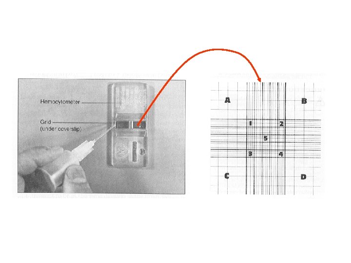
 Use a hemocytometer • 2)")
RBC Counts • To calculate RBCs/mm 3 • 1) Use a hemocytometer • 2) More easily done if the blood is first diluted. The final dilution in the Ery-TIC kit was 1: 200, so the dilution factor is 200 • 3) You counted cells in 5 representative squares of the 25 so you have to multiply by 5 to get an estimate of the cells in the whole area • 4) the grid on the hemocytometer is 1 mm 2 x 0. 1 mm deep so: 1 mm 2 X 0. 1 mm = 0. 1 mm 3 and X 10 = 1 mm 3. total multiplication factor = 5 X 10 X 200 = 10, 000 • Normal for adult male 4. 5 - 6. 0 X 106/ mm 3 • Normal for adult female 4. 0 - 5. 5 X 10⁶/ mm 3

Hematocrit & Hemoglobin • HEMATOCRIT = ratio of volume of packed RBCs to the total blood volume – Normal adult male 47 + 7 % – Normal adult female 42 + 5 % • HEMOGLOBIN – Fe-heme complex gives a red color to the blood and this is used to determine the Hb. – Hb concentration expressed as grams/ 100 mls (d. L) • Normal 12 -15 g % or 12 -15 g/dl for females • Normal 13 -16 g % or 13 -16 g/dl for males • Anemias - decreased oxygen carrying capacity of blood - decreased Hb content of blood - decreased number of RBCs/ volume of blood

Hematocrit Measurement 100 % 90 80 70 60 50 40 30 20 Plasma 10 Packed RBCs

CALCULATIONS • MCV = mean corpuscular volume – measure of the volume of an erythrocyte Normal value 82 -92 mm 3 • MCHC = mean corpuscular hemoglobin concentration – measure of average RBC [Hb] Normal value = 32 -36%
 Macrocytic: MCV>")
ANEMIAS • The reduced oxygen carrying capacity of the blood. • 1) Macrocytic: MCV> 94 MCHC normal – folic acid deficiency – B 12 deficiency develops from a lack of intrinsic factor secretion in the stomach. This factor is needed for the absorption of B 12 and folic acid are needed for RBC maturation • 2) Normocytic normochromic: MCV & MCHC normal – blood loss via hemorrhage – decreased marrow function • 3) Microcytic hypochromic: low MCV; low MCHC – decreased iron

WHITE BLOOD CELL DIFFERENTIAL COUNT • WBCs leave the vasculature during inflammatory responses. This process is called DIAPEDESIS. This process is aided by the secretion of histamine from basophils that causes the capillary wall to become more leaky. • Certain WBCs increase in number during certain disease conditions. • Two general kinds of WBCs based on their cytoplasmic granules – Granulocytes – Agranulocytes

Neutrophil • Granulocyte • Phagocytosis of pathogens • 50 -75% of WBC’s • 10 -12 um in diameter • Three-lobed nucleus • Purple nucleus, light, small granules

Eosinophil • Granulocyte • Secretions destroy parasites • 1 -6% of WBC’s • 13 um diameter • Bilobed nucleus • Large, red-orange granules, blue nucleus

Basophil • Granulocyte • Release of histamine during inflammatory response • 0. 5% of WBC’s • Bilobed nucleus • Dark blue or purple granules, granules usually cover the nucleus • Will become Mast cells

Small Lymphocytes • • • Agranulocyte T and B cells 30% of WBC’s 7 µm in diameter Large nucleus, with small band of cytoplasm • Light blue cytoplasm, purple nucleus • B cells will become plasma cells

Monocytes • • • Agranulocyte Tissue macrophage 2 -8% of WBC’s 15 µm in diameter Blue-gray cytoplasm, with kidney-shaped dark nucleus • Will become macrophage

Blood Typing • Agglutination Rxn = clumping caused by reaction between RBC surface Antigen (Ag) and plasma antibodies (Ab) – each RBC has specific Ag molecules on its surface A, B, Rh – each plasma Ab can bind to two Ag at a time and so to two RBCs at a time. This results in clumping of cells. – Rh factors are found on 85% of the population (Rh+). – Rh is a dominant trait so RR or Rr expresses, rr does not.

Erythroblastosis Fetalis • When a female who is Rh- carries an Rh+ fetus, birthing may allow fetal blood to enter her circulation. She will then make Ab to the Rh factor. These Ab could attack the RBCs of any subsequent fetus resulting in a BLUE BABY. A preventative measure is to administer Rh antibodies to the mother to destroy fetal RBCs.

Blood Typing • • • Each individual inherits two genes each of which may be in multiple forms (alleles) Genotype Phenotype (Blood Type) RBC Ag Plasma Ab AA A A Anti B AO A A Anti B BB B B Anti A BO B B Anti A AB AB AB NONE* OO O NONE Anti A & B** * Universal Acceptor: When an AB type receives a transfusion the donor's Abs are diluted out ** Universal Donor: When type OO donates blood their plasma Abs are diluted out

Clotting Pathways • Extrinsic pathway – Primary pathway - this pathway is activated when tissue damage causes the release of thromboplastin

Clotting Pathways • Intrinsic pathway – secondary pathway - this pathway is activated when blood comes into contact with collagen from a damaged vessel or by contact with glass and this activates the Hageman factor (XII). This pathway keeps one from bleeding internally.

Figure 12. 76 Hageman factor Thromboplastin fibrinogen fibrin

Important • Calcium is needed for these pathways to be activated • Vitamin K is a vitamin necessary for the formation of prothrombin, • Factor VII, factor IX, and factor Xdeficiency will cause an increase in the time for fibrin threads to be formed and a clot produced
- Slides: 25