Hematology Oncology Division of Hematology Oncology TriService General


, or CD 8+ is a")


: 1 -2%of all RBCs “retic count” helps determine")


")


, 1 -4): size or direct extent")
 (n=427)")


- Slides: 48
Hematology & Oncology 何景良 Division of Hematology /Oncology Tri-Service General Hospital National Defense Medical Center 1
Hematology 2
Hematopoiesis Formation of blood cells Occurs mostly in red bone marrow All cells arise from same blood stem cell (pluripotent hematopoietic stem cells) Recently some have been found in adults which are mesenchymal stem cells, which can also form fat cells, osteoblasts, chondrocytes, fibroblasts and muscle cells 3
Blood stem cells divide into: 1. myeloid stem cells or 2. lymphoid stem cells All except for lymphocytes arise from myeloid stem cells All originate in the bone marrow Not shown are mast cells, osteoclasts, dendritic cells 4
CBC is probably commonest test done (“complete blood count”-how much of each type of cell) Hemoglobin (gm/dl) usually 15 Hematocrit (%) RBC count WBC in thousands/cumm Differential if ordered: broken down to amount of each type WBC Platelet count in thousands/cumm 5
Erythrocytes Also called RBCs or red blood cells Biconcave discs and flexible Plasma membrane but no nuclei or organelles Packed with hemoglobin molecules Oxygen carrying protein 4 chains of amino acids, each with iron which is binding site for heme oxygen; CO 2 carried also Young ones still containing ribosomes are called reticulocytes Live 100 -120 days iron atom 6
Leukocytes WBCs: white blood cells 7
__RBC Leukocytes neutrophil eosinophil WBCs: white blood cells basophil Note the size difference compared to erythrocytes small lymphocyte monocyte 8
Leukocyte types Artificial division into granulocytes and agranulocytes Granulocytes: neutrophils, eosinophils, basophils (according to how stain) Granules Lobed nuclei All are phagocytic Agranulocytes: lymphocytes, monocytes 9
Neutrophils 60% of all WBCs Nuclei of 2 -6 lobes Other names: Polymorphonuclear cells (PMNs, polys, segs) Granules have enzymes Can damage tissue if severe or prolonged Pus 10
Eosinophils 1 -4 % of leukocytes Bilobed Granules have digestive enzymes Role in ending allergic reactions and in fighting parasitic infections 11
Basophils Rarest WBC Bilobed nucleus Dark purple granules Later stages of reaction to allergies and parasitic infections 12
Lymphocytes* * Most important WBC 20 -45% Most are enmeshed in lymphoid connective tissue, e. g. lymph nodes, tonsils, spleen 13
Lymphocytes: nucleus occupies most of the cell volume Response to antigens (foreign proteins or parts of cells) is specific Two main types attack antigens in different ways T cells B cells plus “natural killer cells” 14
T cells attack foreign cells directly Killer cells (“cytotoxic”), or CD 8+ is a main type 15
B cells Differentiate into plasma cells Plasma cells secrete antibodies Antibodies flag cells for destruction by macrophages (see stem cell chart) 16
Monocytes* 4 -8% of WBCs In connective tissue they transform into macrophages (phagocytic cells with pseudopods) * 17
Platelets* Not cells Small fragments broken off from megakaryocytes Important in forming clots in damaged vessels * 18
Significant young cells Reticulocytes* (young erythrocytes): 1 -2%of all RBCs “retic count” helps determine if producing RBCs at accelerated rate (anemia, move to a high climate, etc. ) * * Bands* (young neutrophils): 1 -2% of all WBCs Increases with acute bacterial infections 19
Disorders of Erythrocytes Polycythemia: too many cells Anemia: not enough cells Hemoglobinopathy Sickle cell disease: genetic disease AR 1/400 African Americans Defect in hemoglobin 20
Disorders of Leukocytes Leukemia: too many, abnormal, crowd out normal marrow Classified into Lymphoblastic or myeloblastic Acute or chronic Disorders of Platelets Thrombocytopenia Causes internal bleeding Many causes 21
Oncology 22
WHO Statistics n 2020 15 million people will die from cancer n Causes ¨ Ageing population ¨ Obesity ¨ Smoking
What is Cancer? n Division – uncontrolled cell division n Growth – formation of a lump (tumour) or large numbers of abnormal white cells in the blood n Mutation – changes to how the cell is viewed by the immune system n Spread – ability to move within the body and survive in another part
Division – uncontrolled cell division n Oncogenes n Tumour suppressor genes – p 53 n Suicide genes – apoptosis n DNA repair genes
Growth n Tumour ¨ Pressure on nerves ¨ Blocking organs ¨ Stopping normal function ¨ Altering nerve signals ¨ Fungating
Mutation and Spread n Invasion n Angiogenesis
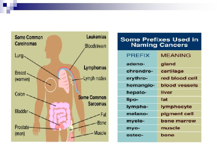
Types of Cancer Carcinomas n Sarcomas n Lymphomas n Leukaemias n Adenomas n n Often prefixed by the specific cell
What are the differences in the features of normal and cancer cells?
Malignant versus benign tumours
Normal and abnormal cell growth
Normal cell growth
Cancerous growth
Metastatic cancer
What causes cancer?
Carcinogenesis. Some factors to consider… n n n n Heredity Immunity Chemical Physical Viral Bacterial Lifestyle
Heredity n Genes isolated for several classic familial cancer syndromes: ¨ RB 1 (retinoblastoma) ¨ APC (familial polyposis) ¨ Human Non Polyposis Colon Cancer (HNPCC) ¨ BRCA ¨ p 53 1&2 (breast cancer) (many cancers)
Immunity n HIV / AIDS n Immunosuppression
Virus’s n Hepatitis B n Human T-cell Leukaemia virus n Epstein Barr Virus n Human Papilloma Virus (HPV)
Bacterial n H. pylori n Other Parasites: ¨ Schistosoma spp ¨ Clonorchis sinensis
TNM Staging n n T (a, is, (0), 1 -4): size or direct extent of the primary tumor N (0 -3): degree of spread to regional lymph nodes N 0: tumor cells absent from regional lymph nodes ¨ N 1: tumor cells spread to closest or small number of regional lymph nodes ¨ N 2: tumor cells spread to an extent between N 1 and N 3. ¨ N 3: tumor cells spread to most distant or numerous regional lymph nodes ¨ n M (0/1): presence of metastasis M 0: no distant metastasis ¨ M 1: metastasis to distant organs (beyond regional lymph nodes) ¨
轉譯醫學-肺癌的生物標記 BR. 21: summary of significant clinical predictors of response Tarceva patients (%) (n=427) Gender Female (146) Male (281) Histology Ethnicity Ever smoked Adenocarcinoma (209) 14. 4 6. 1 13. 9 Other (218) 4. 1 Asian (53) 18. 9 Other (374) 7. 5 Yes (311) 3. 8 No (93) 24. 7 Unknown (23) 13. 0 *Significance between subgroups p value* 0. 006 <0. 001 0. 02 <0. 001
Biomarker- EGFR-TKI Exons Activating mutations exon 19 deletion G 719 X Overall incidence in population ~10% in Western countries 18 3. 2% P-loop L 858 R EGFR Deletion 19 48. 2% Insertion 3. 7% C helix ~30% in Asian countries Kinase domain 20 K A-loop L 858 R 21 42. 7% Mitsudomi T, et al. Int J Clin Oncol 2006
轉譯醫學-大腸癌的危險因子 Distal intramural spread CEA Surgical skill Socioeconomic status Dendritic cell counts Alleles: CCND 1 870 A BRCA 1 LOH Invasion: Depth Lymphatic Venous Perineural Serosal • Multifactorial • Role of many markers unclear Perforation Occlusion No. of nodes examined Invasive front budding Differentiation Mutations: K-ras DNA ploidy Watanabe T et al. NEJM 2001; 344: 1196 -206
n = 399 DLBCL, untreated 60– 80 years, stage II–IV, PS: 0– 2 aa-IPI 0– 1: 40% aa-IPI 2– 3: 60% Mabthera (Rituximab) Probability 1. 0 EFS 0. 8 0. 6 R-CHOP 0. 4 OS R-CHOP 0. 4 CHOP 0. 2 0. 0 0. 2 Median: 3. 8 years vs 1. 1 years P = 0. 00002 0 1 2 3 4 Years 5 6 0. 0 Median: not reached vs 3. 1 years P = 0. 007 0 1 2 3 4 Years Feugier P, et al. J Clin Oncol. 2005; 23: 4117– 4126. Coiffier B, et al. New Engl J Med 2002; 346: 235– 242. 5 6
• 轉譯醫學-CML的生物標記 P 190 b 2 a 2, b 2 a 3 P 210 P 230