
helium Connective T Cells closely aggregated tight by

helium Connective T. • Cells: closely aggregated, tight by junctional complexes • Intercellular substance: very little • No fibers in the ground substance • Blood vessels: Avascular • Origin: ectodermal, endodermal, mesodermal • Function: covering or secretion (glands) • Cells: few, widely separated • Intercellular substance: abundant • fibers are present in the ground substance • Blood vessels: rich Origin: mesodermal • Function: support, defence and nutrition

General Properties of Connective Tissue One of the four basic types of tissues (epithelium, connective tissue, muscle, and nervous tissue) Composition: – cells – Extracellular matrix • fibers • ground substance 3. Functions: –Architectural framework of the body –Bind together and provide mechanical support for other tissue (metabolic, defense, transport, storage) –Nutrition –Wound repair –Protection: inflammatory response

Extracellular Matrix (ECM) Ground Substance of the 1. Glycosaminoglycans (GAG) • linear (unbranched) polysaccharides, e. g. heparan sulfate, condroitin sulfate, keratan sulfate, hyaluronic acid • attract sodium & hold water • very hydrophilic due to abundant negative charges. • except for hyaluronic acid, are usually bound covalently to protein core as part of a proteoglycan 2. Proteoglycans: Forms thick gel that slows the spread of pathogens • core protein + GAG side chains (like a bottle brush) • bind cells, other proteins, and/or ECM components 3. Multiadhesive glycoproteins • small glycosylated proteins containing NUMEROUS binding sites to cells, signaling molecules, and other ECM components • e. g. fibronectin and laminin: important for adhesion of epithelial cells to the basal lamina via transmembrane integrin receptors. Tissue fluid: Similar to blood plasma FUNCTION: - Nutrition - Barrier to bacterial penetration


Connective Tissue Fibers • Collagen – most abundant protein in human body – Several types: fibril-forming or fibril-associated (in skin, tendon, cartilage, bone, dentin, blood vessels); crosslinked networks (in all basement membranes) • Reticular Fibers – specialized type of collagen (Type III; reticulin) associated with smooth muscle in organs subjected to changes in volume, forms the stroma in lymphatic and hematopoietic organs • Elastic Fibers –thin fibers or fenestrated sheets composed of various glycoproteins, including the protein elastin, providing elastic properties to tissues that experience repeated deformation (in skin, blood vessels, lung, bladder)

C. T. fibers The proportion of different fiber types varies depending on the structural needs or function of the particular CT. v Collagen fibers are the most abundant fiber type. All connective tissues have collagen fibers, but the number and organization varies in different types. They are flexible and have a high tensile strength. v Reticular fibers means ‘network’. are especially abundant in arteries, spleen, liver, uterus and intestinal muscle layers, and they form the primary framework for hematopoietic organs, the parencyma of cellular organs such as liver and endocrine glands. Reticular fibers are also associated with the reticular lamina of basement membrane. v Elastic fibers are especially abundant in elastic ligaments (ligamenta flava of the vertebral column, ligamentum nuchae of the neck, and vocal folds of the larynx, and in the walls of arteries (where elastic fibers are produced by smooth muscle cells, not fibroblasts)

Collagen fibers (white fibers) n • • • n n The most numerous are extremely tough. Formed of collagen protein They are stronger than steel fibers of the same size. They provide high tensile strength, which is the ability to resist longitudinal stress. Since fresh collagen fibers have a glistening white appearance they are sometime called "white fibers". Fibers do not branch but bundles can do Stain pink with eosin

Collagen Fibers H&E fibers fibrils

Collagen Synthesis DISULFIDE BONDS ALDOL CONDENSATION


Assembly of collagen fiber bundles

Major Collagen Fiber Types (out of at least 20) According to the chemical composition of collagen molecules Collagen Type Tissues Function Fibril-forming collagens (these are visible) I (most abundant) Skin, tendon, bone, dentin Resistance to tension II Cartilage, vitreous of eye Resistance to pressure III (reticulin) Skin, muscle, blood vessels, liver, etc. Structural framework and stability Network-forming collagens IV All basement membranes Support and filtration Fibril-associated collagens with interrupted triple helices (FACIT) VI, IX Assoc. w/ type I and II fibrils Fibril-fibril / fibril-ECM binding Anchoring filament collagens VII Epithelia Epidermis to basal lamina

Reticular (Reticulin) Fibers • Form a delicate supporting framework for highly cellular tissues (endocrine glands, lymph nodes, liver, bone marrow, spleen, smooth muscle). • Composed mainly of Type III collagen, with a carbohydrate moiety that reduces Ag+ to metallic sliver = argyrophilic. • Special stain: silver impregnation to visualize. • Thinner than type I collagen (Type III fibrils are 30 -40 nm diameter; type I fibrils are ~200 nm diameter) • made by reticular cells (specialized fibroblasts) and vascular smooth muscle cells

Elastic Fibers Elastic fibers (yellow fibers): can be stretched to one and one-half times their length, but recoil to their initial length when released. Fresh elastic fibers appear yellow and are also called yellow fibers. Stain : H&E , Orcein , VVG Elastin: is rich in glycine and proline, but it contains little or no hydroxyproline and hydroxylysine. uniquely contains desmosine and isodesmosine, which are thought to cross-link the molecules into a network of randomly coiled chains. This cross-linking is responsible for its rubber-like properties. They are found where greater elasticity is needed present in large amounts in ligaments, lung, skin, bladder, and walls of blood vessels.

Network of elastin molecules can stretch and recoil like a rubber band

Fibers n n n n The most numerous White if in great number (white fibers) Strong and flexible Fibers do not branch but bundles can do Formed of collagen protein Stain pink with eosin Types of Collagen Fibres Yellow if in great number (Yellow fibers) Elastic and stretchable Fibers can branch and unit Formed of elastin protein Stained weakly by H&E Stain brick red by orcein Stain dark violet with V. V. G stain. Thin branching n Not stained by H&E n Stained dark brown with silver stain n Consist of type III collagen n Supportive function n

Fibers Fiber Components Location Collagenous Fibers Alpha polypeptide chains tendon, ligament, skin, cornea, cartilage, bone, blood vessels, gut, and intervertebral disc. Elastic fibers elastic microfibril & elastin extracellular matrix (blood vessels) Reticular fibers Type-III collagen liver, bone marrow, lymphatic organs

Connective Tissue Cells Resident cells Fibroblasts Adipocytes Pigment cells Immigrant cells Monocytes/ histocytes/ v macrophag es Plasmocytes v Mast cells v Leucocytes v derived from hematopoietic stem cells and involved in immune function and inflammation Specialised cells Reticular cells (reticular tissue) Pericytes (blood vessels) Schwan cells (nerve fibers) Glial cells (CNS) CHondriocytes (cartillage) Osteocytes (bone) Blood cells

Connective Cell Lineages

C. T. CELLS • Origin • Structure LM EM Function
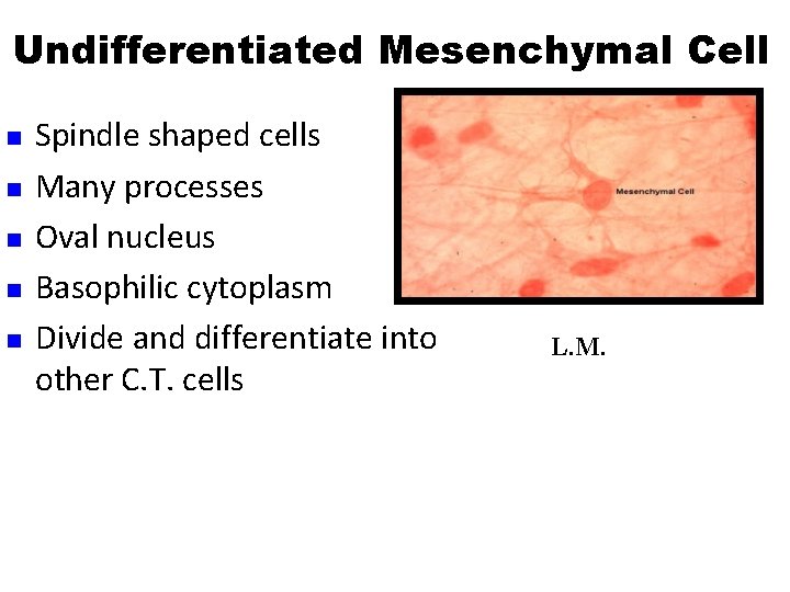
Undifferentiated Mesenchymal Cell n n n Spindle shaped cells Many processes Oval nucleus Basophilic cytoplasm Divide and differentiate into other C. T. cells L. M.

Fibroblasts the most common type §Origin : from UDMC mature (Fibrocytes): § 2 types Young fibroblast: §Small in size §Large in size §Fusiform with processes § oval central paler nucleus §Basophilic cytoplasm Function § synthesis of C. T. fibers and matrix §Fusiform smaller § oval central darker nucleus §acidophilic cytoplasm


Fibroblasts are the most common cells in connective tissue • Active and quiescent stages (when quiescent sometimes called fibrocytes or mature fibroblasts). • Synthesize and secrete components of the ECM: fibers and ground substance. • Synthesize growth factors. • Rarely undergo cell division unless tissue is injured, which activates the quiescent cells. • Play a major role in the process of wound healing and respond to an injury by proliferating and enhanced fiber formation.

Fat Cell §Origin : UDMC §large, sperical or polyhedral §Single or several lipid droplets § flattened peripheral nucleus E Function : § storage of fat M Lipocytes stained black with osmic acid

Adipocytes • Fat cells or adipocytes are fixed cells in loose connective tissue. Their main function is the storage of lipids. The cytoplasm only forms a very narrow rim around a large central lipid droplet. The flattened nucleus may be found in a slightly thickened part of this cytoplasmic rim - if it is present in the section, which may not be the case since the diameter of an adipocyte (up to 100 µm). • A "starving" adipocyte may contain multiple small lipid droplets and gradually comes to resemble a fibrocyte. • Lipid storage/mobilisation is under nervous (sympathetic) and hormonal (insulin) control. • Adipocytes also have an endocrine function - they secrete the protein leptin which provides brain centers which regulate appetite with feedback about the bodies fat reserves. • Adipocytes are very long-lived cells. Their number is determined by the number of preadipocytes (or lipoblast) generated during foetal and early postnatal development.

Pigment Cells § branched cells §Contain pigment granules: Melanin (melanocytes) Function : § gives the color of skin and iris of the eye

Macrophages Origin : From blood monocytes Three types: §Resident : resting §Elicited: moving to a stimulus §Activated: active in phagcytosis: -pseudopodea -Kidny shaped eccentric nucleus-large number of lysosomes Function : Phagocytosis

Mast Cells §Origin : from haemopoetic stem cell in B. M § the largest of free cells §Contain basophilic granules § granules stain red with toluidine blue (metachromatic staining) Function : secretion of histamine and heparin

Mast Cells • Principal function is storage in secretory granules and REGULATED release (degranulation) of histamine and other vasoactive mediators of inflammation. • Responsible for the immediate hypersensitivity response characteristic of allergies, asthma and anaphylactic shock. • Connective tissue mast cells are found in skin (dermis) and peritoneal cavity; mucosal mast cells are in the mucosa of the digestive and respiratory Metachromasia – when stained with tracts. toluidine blue, the granules bind the dye and change its color to red.

Plasma Cells q Origin : from activated B – lymphocytes q large ovoid cells q Basophilic cytoplasm (r. ER) q spherical eccentric nucleus q Cart wheel appearance of the nucleus q Large negative golgi image Function : § formation of antibodies

- Slides: 32