Heart Rhythms Normal or Abnormal Arrhythmias Anatomy Physiology
 Anatomy & Physiology L 3")
Heart Rhythms: Normal or Abnormal (Arrhythmias) Anatomy & Physiology L 3
 Group of specialized myocytes near Sup. Vena")
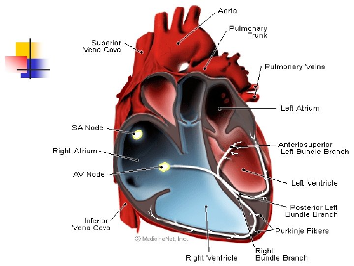
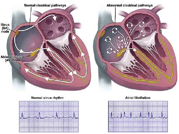
Electrical Conduction n n SA Node (sinoatrial) Group of specialized myocytes near Sup. Vena cava Create action potential Creates an action potential approx 70 -80 bpm Innervated by Brain stem n n Sympathetic: Increase HR Vagus: decrease HR n n n AV node (atrioventricl. ) “Decremental conduction” (AV delay = 0. 01 sec) Prevents premature contraction of ventricles Alone, can create a beat approx 40 bpm NOT strong enough to sustain life

Interior anatomy of heart n n Bundle of His Collection of cardiac cells creating the AV node Conduct impulses along septum to the Purkinje fibers in walls of the ventricles Takes impulse about 0. 03 -0. 04 sec to travel to the ventricles n n Purkinje fibers: thin filaments Located on inner wall of endocardium Carry electrical impulse from SA and AV nodes Move from left to right

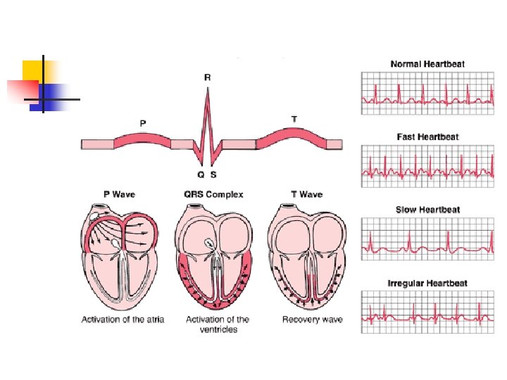
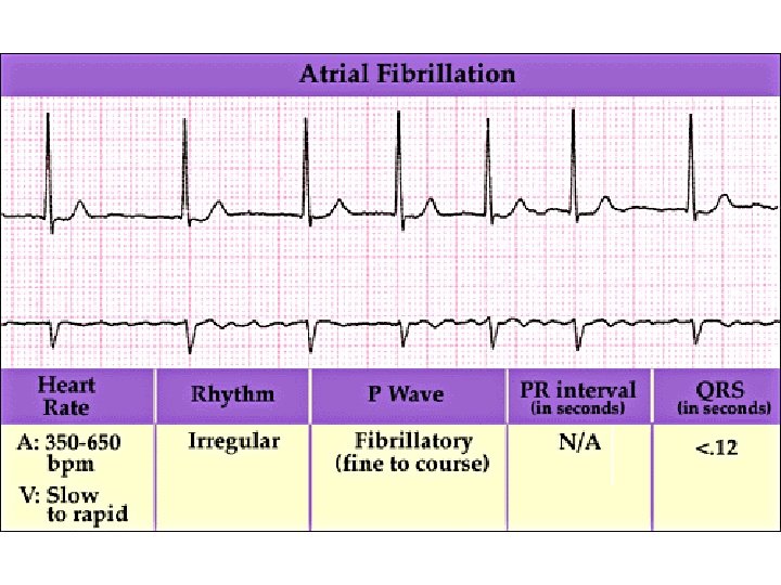
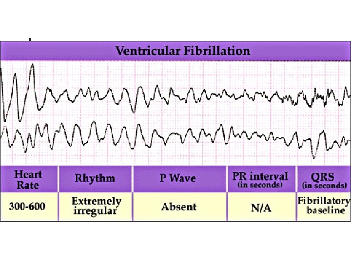
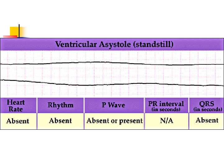
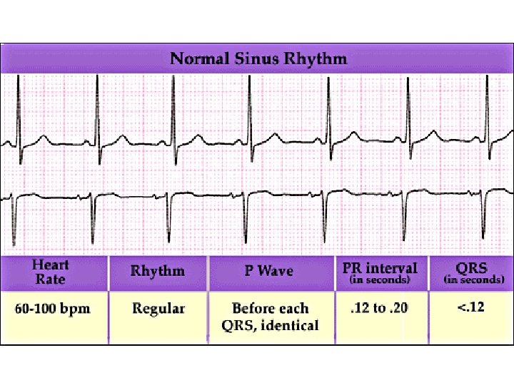
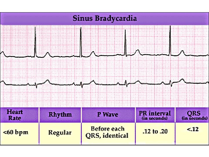
Normal sinus rhythm
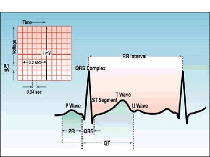
 n 12 -lead (multi-angle)")
Electrocardiogram (ECG or EKG) n 12 -lead (multi-angle)

Acute MI
 n n Mortality: 500 K – 700 K per year")
Myocardial Infarction (heart attack) n n Mortality: 500 K – 700 K per year in US 1. 3 million reported cases of MI 600 per 100 K Complete obstruction of coronary artery(s) leading to portion of heart muscle to die n n n Signs & Symptoms Persistent chest pain Tightness in chest Difficulty breathing Jaw, neck, shoulder pain (referred pain) Sweating

What do I Do? n n n Recognize the signs Get immediate medical help BLS, Meds, Hospital Try to stabilize in the ER Time for “Dr Burke” n n Surgery: Angioplasty Use of a stent Coronary Bypass

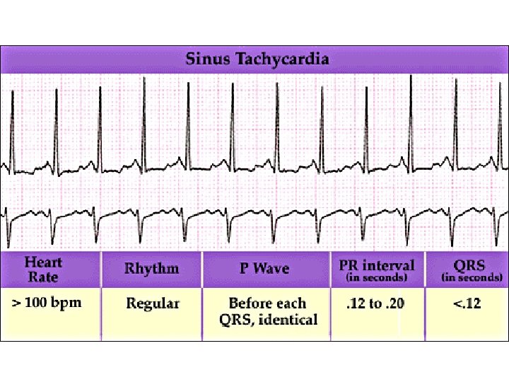
TEST TIME! Good Luck! n n n Anatomy of the Heart Pathway of blood flow Electrical conduction Common problems (ex; atherosclerosis, pulmonary embolism, coronary thrombosis) Myocardial Infarction and the treatment of Common ECG readings
- Slides: 19