
Heart Pulses Jennifer Coleman MSN Health Assessment Circulation
- Slides: 47
Heart & Pulses Jennifer Coleman MSN Health Assessment
Circulation
Quick Facts • One day – Heart beats 100, 000 times – Heart pumps about 2, 000 gallons • 70 years – Heart beats more than 2. 5 billion times
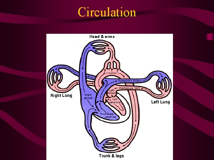
The double pump • Heart is truly two pumps working together – Right side pumps blood into the lungs – Left side pumps blood into the body – Pump is systole and to fill is diastole • Pumps separated by impermeable wall (septum) • Each side has two chambers – Atrium – thin-walled, holds blood – Ventricle – thick-walled, muscular, pumps blood • Chambers separated by valves – Unidirectional (prevents backflow/regurgitation) – Passively open and close with pressure gradients
Heart anatomy review • What is the order of the flow of blood from the Superior/Inferior Vena Cava to Aorta? – Superior/Inferior Vena Cava – Right Atrium – Tricuspid Valve (right atrioventricular valve) – Right Ventricle – Pulmonary Valve (right semilunar valve) – Pulmonary Artery – Lungs
Blood flow direction continued • • • Pulmonary Vein Left Atrium Mitral Valve (left atrioventricular valve) Left Ventricle Aortic Valve (left semilunar valve) Aorta
The cardiac cycle: 2 phases • 2/3 Diastole – Ventricles relax, pressure is high in atria, AV valves open & blood pours into the ventricles • 1/3 Systole – The heart’s contraction
Heart Landmarks • Base = “top” • Apex = “bottom” – Apical impulse/PMI (point of maximal impulse) – 4 th-5 th L ICS, midclavicular line (MCL) – Palpated in ~ ½ of all adults – Felt as a local “tap”, 1 cm or less in diameter – High diaphragm of pregnancy changes the location to the lateral part of the MCL – Impulse strengthens with anxiety, fever, anemia, hyperthyroidism & HTN
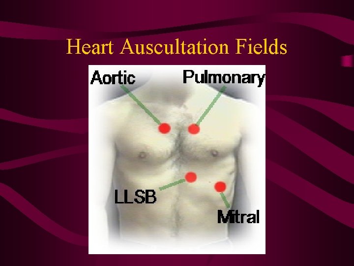
Heart Auscultation Fields
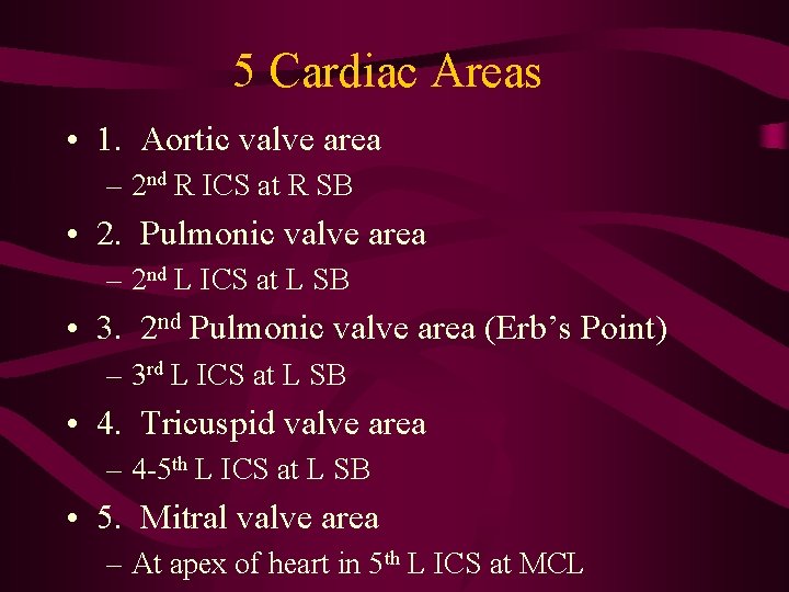
5 Cardiac Areas • 1. Aortic valve area – 2 nd R ICS at R SB • 2. Pulmonic valve area – 2 nd L ICS at L SB • 3. 2 nd Pulmonic valve area (Erb’s Point) – 3 rd L ICS at L SB • 4. Tricuspid valve area – 4 -5 th L ICS at L SB • 5. Mitral valve area – At apex of heart in 5 th L ICS at MCL
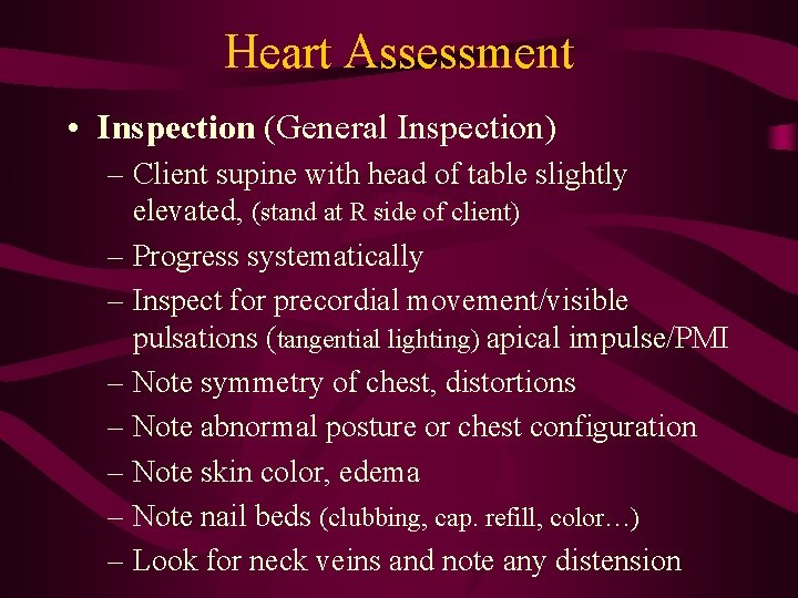
Heart Assessment • Inspection (General Inspection) – Client supine with head of table slightly elevated, (stand at R side of client) – Progress systematically – Inspect for precordial movement/visible pulsations (tangential lighting) apical impulse/PMI – Note symmetry of chest, distortions – Note abnormal posture or chest configuration – Note skin color, edema – Note nail beds (clubbing, cap. refill, color…) – Look for neck veins and note any distension
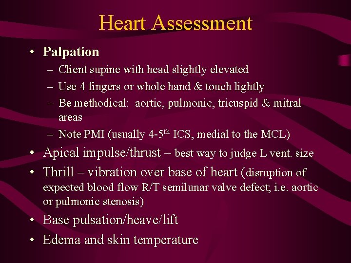
Heart Assessment • Palpation – Client supine with head slightly elevated – Use 4 fingers or whole hand & touch lightly – Be methodical: aortic, pulmonic, tricuspid & mitral areas – Note PMI (usually 4 -5 th ICS, medial to the MCL) • Apical impulse/thrust – best way to judge L vent. size • Thrill – vibration over base of heart (disruption of expected blood flow R/T semilunar valve defect; i. e. aortic or pulmonic stenosis) • Base pulsation/heave/lift • Edema and skin temperature
Heart Assessment • Percussion – May est. size of heart but of limited value – Begin percussion at anterior axillary line, 5 th ICS, moving medially along ICS toward SB – Should change from resonant to dull – note left border of cardiac dullness (LBCD) • Measure (7 -9 cm) from mid-sternal line – Loss of resonance usually close to PMI at apex of heart – MCL, 5 th ICS – Distortion may occur d/t obesity, unusual muscular development, pathological (air, fluid)
Auscultation • Sound is transmitted in direction of blood flow, so specific sounds are best heard over areas where blood flows after it passes through a valve • Client supine with head slightly elevated, stand at right side in a quiet room (concentrated listening) • Begin by identifying S 1 & S 2 – Palpate carotid while auscultating the heart – Carotid pulsation & S 1 are synonymous • Be methodical – use diaphragm 1 st over each of the cardiac areas then repeat with the bell (S 3 & S 4 are heard best with bell)
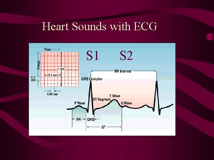
Systole and S 1 So much blood has been pumped into the ventricles that ventricular pressure is higher than atrial pressure (maximal ventricular volume) The mitral and tricuspid (AV valves) swing shut (S 1), this signals the beginning of systole Loudest at the apex (mitral area) 5 th ICS The ventricles contract and blood fills pulmonary and systemic arteries QRS complex
Systole and S 2 • After the ventricle’s have ejected the blood, it’s pressure falls • When the pressure is below that of the aorta, the aortic and pulmonic (semilunar valves) valves close creating the second heart sound S 2 • Loudest at base (aortic area) • When the pulmonic valve is delayed = physiological splitting of S 2 (with inspiration) • This signals the end of systole/beginning of diastole • End of T wave
Heart Sounds with ECG S 1 S 2
Auscultation • Always auscultate in at least 2 positions • Listen to client supine first • Have client roll on L side (L lateral recumbent position) – listen with bell at apex – S 3 and mitral murmurs • Have client sit up, lean forward and hold their breath in exhalation – Listen with diaphragm at the L 3 rd & 4 th ICS at L SB – Aortic murmurs
Heart Sounds • S 1 – Closing of mitral & tricuspid (AV) valves – Heard best at apex, 5 th intercostal space – Beginning of systole • S 2 - Closing of aortic and pulmonic (semilunar) valves – Heard best at base (top of heart) – End of systole • S 3 – Sound of rapid filling the ventricles – Normally silent (at apex & at beginning of expiration, best L side lying with bell) – May be normal/physiological third heart sound – Ventricular gallop rhythm over 40 yo – CHF • S 4 - Vibration of left ventricle & contraction of atria – Heard best at apex with bell – right before S 1 – May be a normal finding – Atrial (S 4) gallop – CAD, Hx of MI, HTN
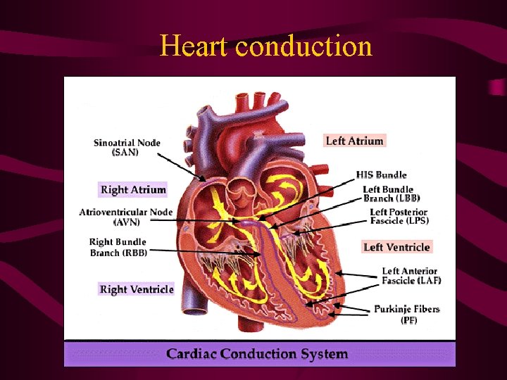
Heart conduction
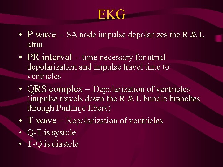
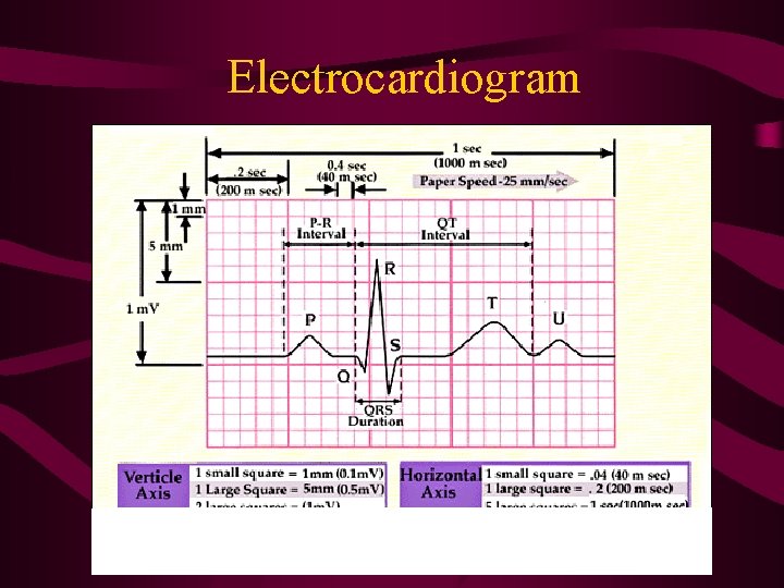
EKG • P wave – SA node impulse depolarizes the R & L atria • PR interval – time necessary for atrial • • depolarization and impulse travel time to ventricles QRS complex – Depolarization of ventricles (impulse travels down the R & L bundle branches through Purkinje fibers) T wave – Repolarization of ventricles Q-T is systole T-Q is diastole
Electrocardiogram
Murmurs • An extra sound d/t turbulent blood flow or valvular vibration. – Velocity of blood increases (flow murmur), high flow through normal or abnormal valve – Viscosity of blood decreases (anemia) – Structural defects (constriction or irregular valve) – Unusual opening in the chambers (septal defect, patent ductis arteriosis)
Characteristics of heart sounds • • • Frequency (pitch) – high or low pitched Intensity – loudness (Grades I-VI) Duration – very short for heart sounds Timing – during systole, diastole or continuous Position – Effect of client maneuvers (respirations, sitting, squatting, exercise, Valsalva maneuver) • Quality (murmur) – Crescendo, decrescendo, crescendo/decrescendo • Location and radiation – Where it is heard best and where does it radiate to “mitral area that radiates to the left axilla”
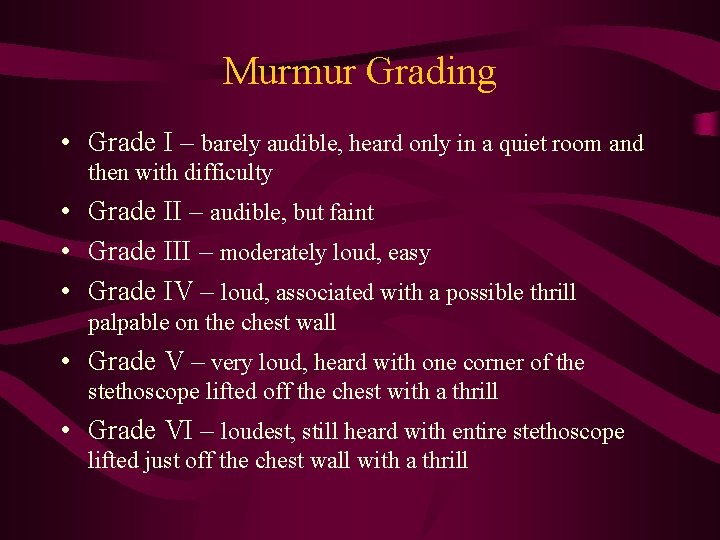
Murmur Grading • Grade I – barely audible, heard only in a quiet room and then with difficulty • Grade II – audible, but faint • Grade III – moderately loud, easy • Grade IV – loud, associated with a possible thrill palpable on the chest wall • Grade V – very loud, heard with one corner of the stethoscope lifted off the chest with a thrill • Grade VI – loudest, still heard with entire stethoscope lifted just off the chest wall with a thrill
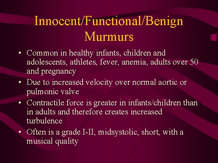
Innocent/Functional/Benign Murmurs • Common in healthy infants, children and adolescents, athletes, fever, anemia, adults over 50 and pregnancy • Due to increased velocity over normal aortic or pulmonic valve • Contractile force is greater in infants/children than in adults and therefore creates increased turbulence • Often is a grade I-II, midsystolic, short, with a musical quality
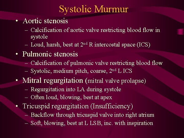
Systolic Murmur • Aortic stenosis – Calcification of aortic valve restricting blood flow in systole – Loud, harsh, best at 2 nd R intercostal space (ICS) • Pulmonic stenosis – Calcification of pulmonic valve restricting blood flow – Systolic, medium pitch, coarse, 2 nd L ICS • Mitral regurgitation (mitral valve prolapse) – Regurgitation into LA during systole – Often loud, blowing, best at apex • Tricuspid regurgitation (Insufficiency) – Backflow through tricuspid valve into right atrium – Soft, blowing, best at L LSB, inc. with inspiration
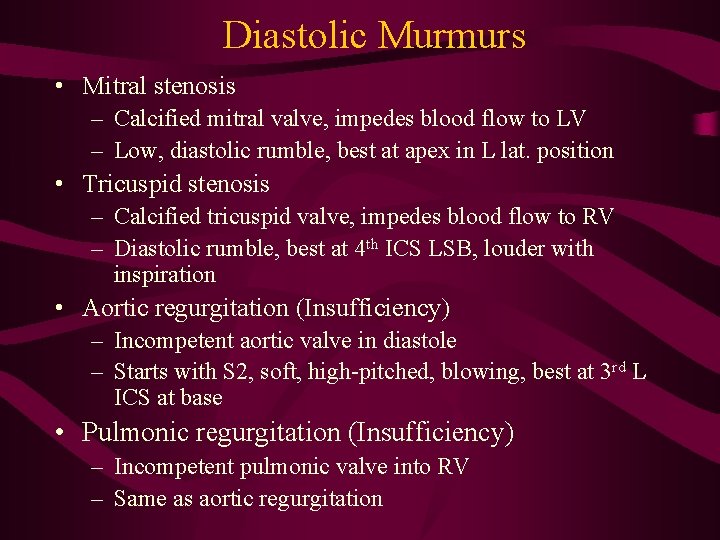
Diastolic Murmurs • Mitral stenosis – Calcified mitral valve, impedes blood flow to LV – Low, diastolic rumble, best at apex in L lat. position • Tricuspid stenosis – Calcified tricuspid valve, impedes blood flow to RV – Diastolic rumble, best at 4 th ICS LSB, louder with inspiration • Aortic regurgitation (Insufficiency) – Incompetent aortic valve in diastole – Starts with S 2, soft, high-pitched, blowing, best at 3 rd L ICS at base • Pulmonic regurgitation (Insufficiency) – Incompetent pulmonic valve into RV – Same as aortic regurgitation
Extra heart sounds • • • Ejection click Opening snap Friction rub Thrills Bruits
Ejection Click • Early in systole at start of ejection • Results from the opening of the semilunar valves (aortic and pulmonic) • With SLV stenosis their opening makes a sound • Best heard at: – 2 nd RICS, apex – aortic – 2 nd LICS - pulmonic
Opening Snap • Sound produced by the opening of stenotic AV valves (tricuspid & mitral) • Increased atrial pressure is required to open the valve (mitral stenosis c murmur) • Sharp, high-pitched with a snapping quality • Best heard with diaphragm at 3 rd or 4 th ICS at LSB/apex – after S 2
Friction rub • D/t inflammation of the pericardial sac • High-pitched, scratchy (sandpaper rubbing together) • Best heard: – Client sitting and leaning forward – Diaphragm – Hold breath in expiration – Apex and LLSB – Systolic &/or diastolic • Common in 1 st week after MI
Thrills • • A palpable vibration (like purring cat) Turbulent blood flow Accompanies a loud murmur Also follows an MI with pericordial inflammation
Bruits • D/t turbulent, vascular, blood flow (atherosclerotic narrowing, aortic valve disease) • Auscultate at carotid and femoral (should be done with abdominal assessment) • Best heard with bell – at three places • Murmur-like sound • May be able to palpate a slight thrill
Charting Record finding from inspection, palpation and percussion • Include S 1, S 2, (S 3), (S 4) as well as the grade and configuration of any murmurs – “two over six” or “ 2/6”, “pansystolic” or “crescendo” – Record bilateral arterial pulses • Consistency, rate, rhythm, quality, equality, bruit
Locations of Palpable Pulses • • Carotid: Brachial: Radial: Femoral: bottom 1/3 (synonymous with S 1) medial to biceps tendon medial/ventral side of wrist inferior and medial to inguinal ligament • Popliteal: popliteal fossae with knee flexed • Dorsalis pedis: medial side of dorsum of foot • Posterior tibial: behind & slightly inferior to medial malleolus of ankle
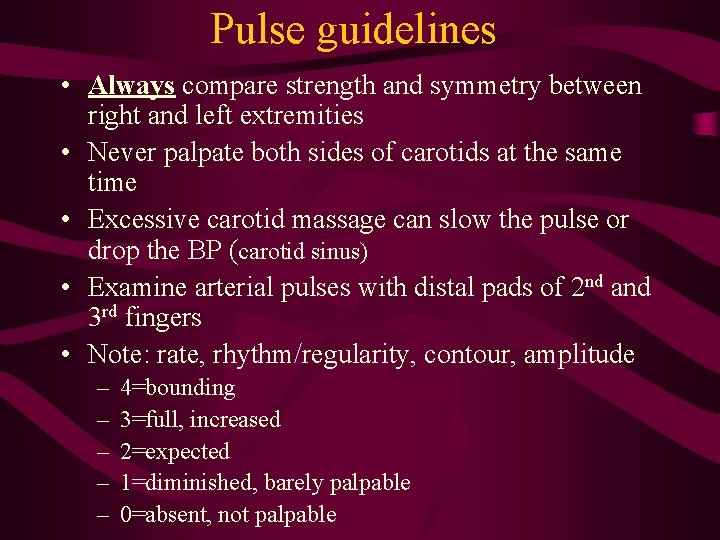
Pulse guidelines • Always compare strength and symmetry between right and left extremities • Never palpate both sides of carotids at the same time • Excessive carotid massage can slow the pulse or drop the BP (carotid sinus) • Examine arterial pulses with distal pads of 2 nd and 3 rd fingers • Note: rate, rhythm/regularity, contour, amplitude – – – 4=bounding 3=full, increased 2=expected 1=diminished, barely palpable 0=absent, not palpable
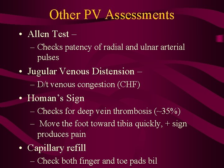
Other PV Assessments • Allen Test – – Checks patency of radial and ulnar arterial pulses • Jugular Venous Distension – – D/t venous congestion (CHF) • Homan’s Sign – Checks for deep vein thrombosis (~35%) – Move the foot toward tibia quickly, + sign produces pain • Capillary refill – Check both finger and toe pads bil
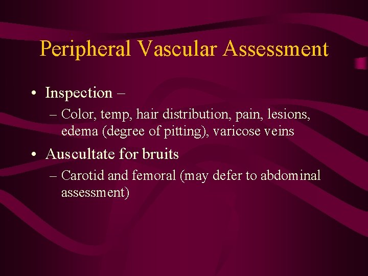
Peripheral Vascular Assessment • Inspection – – Color, temp, hair distribution, pain, lesions, edema (degree of pitting), varicose veins • Auscultate for bruits – Carotid and femoral (may defer to abdominal assessment)
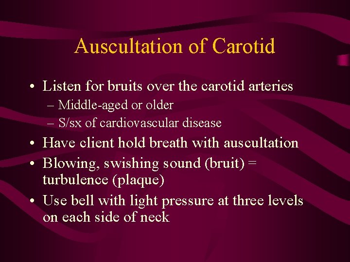
Auscultation of Carotid • Listen for bruits over the carotid arteries – Middle-aged or older – S/sx of cardiovascular disease • Have client hold breath with auscultation • Blowing, swishing sound (bruit) = turbulence (plaque) • Use bell with light pressure at three levels on each side of neck
Variables contributing to pulse characteristics • 1. Stroke volume (volume of blood ejected) • 2. Distensibility of aorta and large arteries – Elastic recoil • 3. Viscosity of blood • 4. Rate of ejection/Cardiac Output – CO normally is 4 -6 L/min • 5. Peripheral arterial resistance • 6. Venous insufficiency – Decreased venous blood return to heart – decreases peripheral pulses
Arterial and Venous Insufficiency • Arterial Insufficiency – Arteriosclerosis – ischemic ulcers – Pulse diminished/absent – Cool and pale – Claudication (pain with walking/exercise) • Venous Insufficiency – Venous (stasis) Ulcer – Pulse present – Brawny, firm edema – Aching pain, worse at end of day (standing or sitting)
Blood Pressure • BP generally increases with age • Pulse pressure – difference between systolic and diastolic pressures (nl: 30 -40 mm. Hg) – Normal systolic – 100 -120 mm. Hg – Normal diastolic – 60 -80 mm. Hg • Readings between L & R arms vary as much as 10 mm. Hg (tend to be higher in R arm) • Better to use a larger cuff than smaller cuff
Prehypertensive • New 2003 Guidelines – see handout • Above 115/75 increases your risks • Prehypertensive (warning zone) – 120 -139/80 -89
Hypertension • Stage 1: – 140 -159/90 -99 • Stage 2: – >159/99
Postural hypotension • AKA – “orthostatic hypotension” • Significant decrease in systolic pressure (>15 mm. Hg) and a decrease in diastolic pressure – Minor blood loss – Drugs – Autonomic nervous system disorders – Prolonged stay in recumbent position
Arterial Pulse & Pressure • Arterial pulse (ventricular systole) produces pressure wave throughout arterial system • Arterial pressure – force exerted by blood against wall of an artery as ventricles contract and relax