HEART PHYSIOLOGY RHYTHMICAL EXCITATION OF HEART By Dr

HEART PHYSIOLOGY RHYTHMICAL EXCITATION OF HEART By Dr. Mudassar Ali Roomi (M. B; B. S, M. PHIL. )

CONDUCTION SYSTEM OF HEART SA node Internodal pathways AV node AV bundle of His Right and left bundle branches • Purkinje fibers • • •

• SA node: sinoatrial node. The pacemaker. – Specialized cardiac muscle cells. – Generate spontaneous action potentials (autorhythmic tissue). – Action potentials pass to atrial muscle cells and to the AV node Conduction System of the Heart
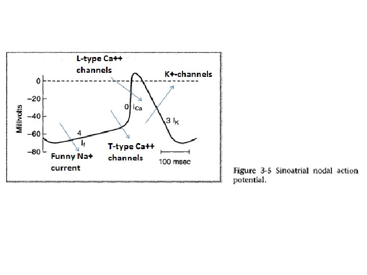

Conduction System of the Heart • AV node: atrioventricular node. – Action potentials conducted more slowly here than in any other part of system. • Function: Ensures ventricles receive signal to contract after atria have contracted

Conduction System of the Heart • AV bundle: passes through hole in cardiac connective tissue skeleton to reach interventricular septum

Conduction System of the Heart • Right and left bundle branches: extend beneath endocardium to apices of right and left ventricles

Conduction System of the Heart • Purkinje fibers: – Large diameter cardiac muscle cells with few myofibrils. – Many gap junctions. – Conduct action potential to ventricular muscle cells (myocardium) very rapidly

Rate of impulse generation in heart: • SA NODE: 70 -80/min • AV NODE: 40 -60/min • AV BUNDLE, BRANCHES & VENTRICLES: 15 -40/min SA NODE normal PACEMAKER OTHERS ECTOPIC FOCI

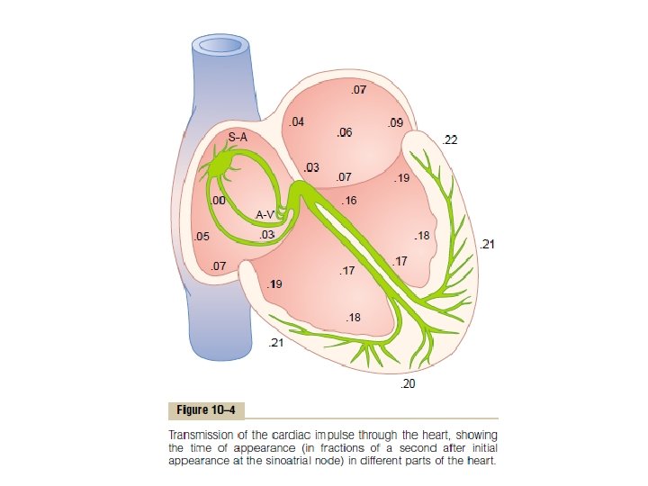
AV Node, and Delay of Impulse Conduction from the Atria to the Ventricles • Location of the A-V node: in the posterior wall of the right atrium immediately behind the tricuspid valve • there is a delay of 0. 09 second in the A-V node • A delay of another 0. 04 second occurs mainly in A-V bundle • Thus, the total delay in the A-V nodal and A-V bundle system is about 0. 13 second.

AV Node, and Delay of Impulse Conduction from the Atria to the Ventricles Cause of the Slow Conduction: • caused mainly by decreased numbers of gap junctions b/w successive cells in the conducting pathways Importance of AV nodal delay: • this delay allows time for the atria to empty their blood into the ventricles before ventricular contraction begins.

• They are very large fibers, even larger than the normal ventricular muscle fibers • and they transmit action potentials at a velocity of 1. 5 to 4. 0 m/sec, a velocity about 6 times that in the usual ventricular muscle and 150 times that in some of the A-V nodal fibers. Rapid Transmission in the Ventricular Purkinje System

Cause of rapid transmission: • The rapid transmission of action potentials by Purkinje fibers is believed to be caused by a very high level of permeability of the gap junctions at the intercalated discs between the successive cells that make up the Purkinje fibers. Rapid Transmission in the Ventricular Purkinje System

FUNCTION: rapid transmission in Purkinje fibers is responsible for synchronous contraction of ventricular muscle. Rapid Transmission in the Ventricular Purkinje System

SUMMARY OF SPREAD OF THE CARDIAC IMPULSE THROUGH THE HERAT Conduction velocities of heart tissues: • ATRIAL MUSCLE= 0. 3 m/sec • INTERNODAL PATHWAYS= 1 m/sec • AV NODE: slowest 0. 050. 1 m/sec • AV BUNDLE & BRANCHES/PURKINJE SYSTEM: Maximum velocity= 1. 54 m/sec • VENTRICULAR MUSCLE= 0. 5 m/sec

ECTOPIC PACEMAKER OF HEART • Normal pacemaker of heart? • Ectopic pacemaker? Causes of ectopic pacemaker of heart? 1. Excessive excitability of some part of the heart other than SA node 2. Heart blocks
- Slides: 17