HEART FAILURE Institute of Pathological Physiology Martin Vokurka


 -")

 = heart rate (HR) × stroke volume (SV) 70 /min 70")
 heart rhythm - has impact of")
 = heart rate (HR) × stroke volume (SV) 70 /min 70")


 preload changes in the ventricle geometry dilatation increased wall tension increased")


")


 - cardiac output (ultrasound")


 CO 1. 2.")


 Periphery vasoconstriction increase of afterload CO/blood flow")

 and to neurohumoral stimuli")


 - apoptosis")


 - reduction of")


- Slides: 81
HEART FAILURE
Institute of Pathological Physiology Martin Vokurka mvoku@lf 1. cuni. cz WS 2008/09
normal situation heart failure congestion backward
normal situation heart failure congestion backward decreased cardiac output forward Insufficient organ perfusion – muscles, kidneys, skin, GIT…
heart insufficiently filled decreased venous return e. g. shock decreased cardiac output bad filling of the ventricles (e. g. constrictive pericarditis)
heart changes heart failure congestion decreased cardiac output compensatory events
HEART FAILURE pathophysiologic state in which an abnormality and/or can do so only from an of cardiac function is responsible for the abnormally elevated failure of the heart to pump blood at a rate diastolic volume commensurate with the requirements of the metabolizing tissues decrease of cardiac output increase of the ventricular filling pressure (enddiastolic pressure, EDP)
Heart failure is not only failing of the heart as a pump, but it is systemic disorder with activation of hormonal processes, with changed metabolism, changed regulation of water-mineral balance, with cytokines involved, heart changes, changes of gene expression etc. HEMODYNAMIC ASPECTS NEUROHUMORAL ASPECTS CELLULAR AND GENE EXPECT
Frequency of heart failure In the Czech Rep. the prevalence is about 1 -2 % (i. e. 100 000 of patients) The number of patients is increasing – among others due to successful treatment of other heart diseases
TYPES OF HEART FAILURE - LEFT-SIDED - RIGHT-SIDED - BOTH-SIDED according to the failing ventricle
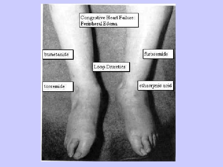
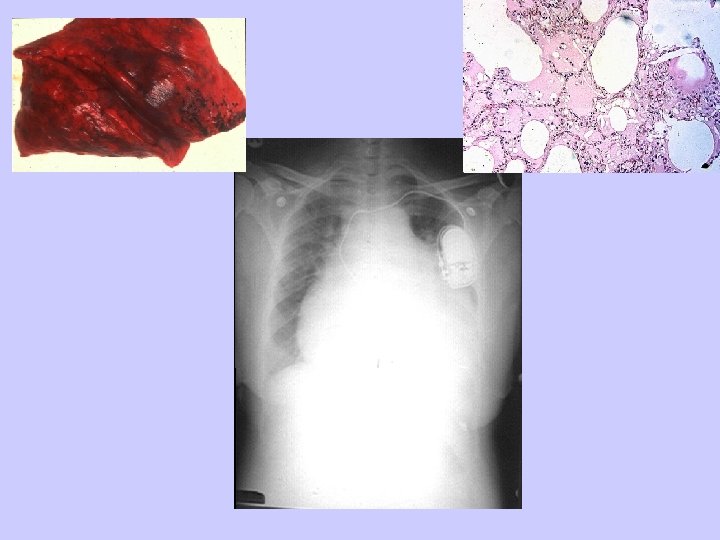
MAIN SYMPTOMES 1. CONGESTION - left-sided - DYSPNEA, LUNG EDEMA - right-sided - LOWER EXTREMITY EDEMAS, HEPATOMEGALY… 2. DECREASED CARDIAC OUTPUT WEAKNESS, FATIGUE, DECREASED ORGAN PERFUSION
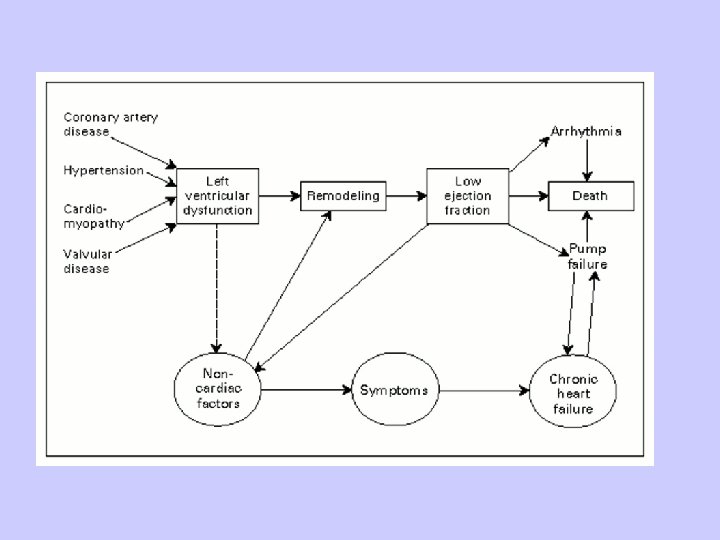
Causes of heart failure Myocardial failure - defect in myocardial contraction (ischemia, cardiomyopathy) - loss of myocardium (myocardial infarction) Excessive, long-term hemodynamic burden - increased pressure burden (systemic or lung hypertension) - increased volume burden (valvular abnormalities) - hyperkinetic cirkulation (increased CO) Most commonly it is the combination of CHD and arterial hypertension
In heart failure CO decreases Activation of compensatory mechanisms trying to increase CO back to normal values How are the distinct mechanism influencing the CO regulated ?
Cardiac output (CO) = heart rate (HR) × stroke volume (SV) 70 /min 70 ml 4 900 ml/min
Heart rate - vegetative nerves - (disturbances in) heart rhythm - has impact of heart cycle duration, mainly shortens diastole – when the heart is filling with blood Increases CO but high rates decrease the ventricle filling and heart is easier exhausted
Cardiac output (CO) = heart rate (HR) × stroke volume (SV) 70 /min 70 ml 4 900 ml/min
Stroke volume - preload - contractility - afterload * How is the heart filled before the systole * What is its „force“ of contraction * What is resistance against the pumping
Preload filling of the heart at the end of the diastole enddiastolic volume = EDV Frank-Starling mechanisms Volume in the ventricle corresponds to the pressure – enddiastolic pressure, EDP, filling pressure
Factors influencing preload - Venous return total blood volume blood distribution (position of the body, intrathoracic pressure, venous tonus…) - atrial systole - size of ventricle cavity - intrapericardial pressure Low preload is the cause of the decreased CO in case of syncope and shock In heart failure the preload is not decreased but it is increased as one of the compensatory mechanisms
The relation between the filling of the ventricle and the intraventricular pressure diastolic filling curve volume: EDV - enddiastolic volume pressure: EDP - enddiastolic pressure, filling pressure - amount of the blood in the ventricle - properties of the ventricle wall
J. Kofránek intraventricular pressure EDP ventricle volume
enddiastolic volume (EDV) preload changes in the ventricle geometry dilatation increased wall tension increased oxygen consumption failure of Frank-Starling mech. P V enddiastolic pressure (EDP) pressure “transfer” to the regions „precceding the heart“ ventricle wall properties (compliance) ischemia - impaired relaxation fibrosis hypertrophy/dilatation L - lung edema P - e. g. hepatomegaly
Iso vo lum ic ma xm is Intraventricular pressure Enddiastolic volume s ic ton m axi m Iso as i D Systolic volume ic l o t g n i l il f preload Systolic residual volume Ventricle volume J. Kofránek
J. Kofránek Iso vo lum ic ma xim s Intraventricular pressure s ic ton m axi m Izo g Systolic volume ic ol t s a n if lli Di Increased preload. . . Ventricle volume …increases cardiac output.
J. Kofránek Intraventricular pressure Syst. volume Systolic volume Decreased preload. . . Ventricle volume …decreases cardiac output.
Stroke volume - preload - contractility - afterload * How is the heart filled before the systole * What is its „force“ of contraction * What is resistance against the pumping
Contractility Increase: sympatic nerves, catecholamines Decreased ischemia, hypoxia, acidosis, proinflammatory cytokines, some drugs etc. Decreased contractility is often the causative mechanism of heart failure.
Catecholamines increase systolic volume Sh ifte di catecholamines sov olu Iso mic m vo lum axim s ic ma xim s Intraventricular pressure Systolic volume …without increasing preload Ventricle volume J. Kofránek
Stroke volume - preload - contractility - afterload * How is the heart filled before the systole * What is its „force“ of contraction * What is resistance against the pumping
Afterload the force against which it contracts, the tension or stress developed in the ventricular wall during ejection - arterial pressure - systemic vascular resistence - blood viskosity - geometry of the ventricle (Laplace law) T=P×r/d Increased volume of the ventricle and thiner wall (i. e. dilatation) increase afterload contribute to the decrease of CO increase requirements for oxygen
Izo vo lum ic ma xim x Intraventricular pressure Increase of „afterload“ does not chane SV s ic ton m axi m Iso Syst. volume Systolic volume ic Di ol t s a ng i l l fi . . . but preload is increased Ventricular volume J. Kofránek
Types of heart failure According to the ventricle - left-sided Acc. to the intensity - right-sided (cor pulmonale due to lung diseases, lung embolism etc. ) - both-sided According to the course - acute According to the CO - chronic (development of the compensatory mechanisms): - high-output (hyperkinetic cirkulation) compensated decompensated - low-output (most)
whether the principal abnormality is - the inability to contract normally and expel sufficient blood (systolic failure) - or to relax and fill normally (diastolic failure) Systolic failure Blood ejection from the ventricle is disturbed Stroke volume might be maintaind at the costs of increased EDV (and EDP)
Ejection fraction EF = SV / EDV the ratio of stroke volume to end-diastolic volume normal value = 67 ± 8 percent SV = 70 ml, EDV = 120 ml EF = 70 / 120 = 58 %
Normal heart stimulated by the sympatic nerves - EF increases, SV increases (contractility increased) Heart with noncompensated systolic failure - EF low, SV low Heart with compensated systolic failure and increased preload - EF low, SV might be normal (EDV is increased)
EDV 1 End of diastole 1
SV 1 End of systole 1 EF 1 = SV 1/EDV 1
SV 2 EF 2 = SV 2/EDV 2 EF 2 > EF 1 End of systole 2
EDV 2 End of diastole 2
SV 3 End of systole 3 EF 3 = SV 3/EDV 3
SV 1 EF 1 = SV 1/EDV 1 EF 1 > EF 3 SV 1 = SV 3 End of systole 1
Diastolic failure usually the decrease in compliance of heart wall EDP increases - CHD - Hypertension with hypertrophy - Some cardiomyopathies etc.
J. Kofránek intraentricular pressure filling pressure ventricular volume
EDP measurement heart catheterization as a pulmonary wedge pressure
Evaluation / monitoring of hemodynamic heart function - EF (ultrasound) - cardiac output (ultrasound or catheterization) - EDP (catheterization) - Heart rate (HR) - Blood pressure (BP)
Symptoms of heart failure from the hemodynamic point of view Low CO Weakness, fatigue, decreased organ perfusion incl. kidneys, muscles - redistribution of CO FORWARD Blood congestion in organs from which blood is collected to the failing ventricle Edemas etc. BACKWARD
Apart from hemodynamic changes heart failure is characterized by important involvement of compensatory mechanisms, mainly neurohumoral, which can, however, if persisting, lead to further progression of failure. Another changes involve the heart itself. Compensatory mechanisms – can in short-term have a positive role, in long-term persistence contribute to the worsening of the failure.
Main compensatory mechanisms in heart failure They lead to incrase (maintain) CO 1. 2. 3. 4. Sympatic activity Increase of preload Salt and water retention Myocardium changes Short-term effective, long-term have deletirious effects themselves and contribute to the symptoms and progression of HF Vitious circle
Sympatic activity in heart failure Heart rate Contractility Venous return CO
Negative consequences Tachycardia: Increase in oxygen consumption shortening of the diastole (impairment of diastolic filling and myocardial blood flow) Increased risk for arrhytmias
Norepinephrine cardiotoxicity (increase of calcium in myocardium) Periphery vasoconstriction increase of afterload CO/blood flow redistribution Metabolic action hyperlipidemia, hyperglycemia During the heart failure the receptors in myocardium are down-regulated
Low doses of betablockers are nowadays used to treat and improve the moderately severe heart failure. Extremely activated sympatic activity is in SHOCK
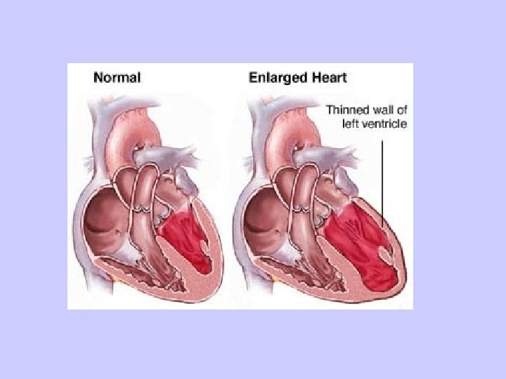
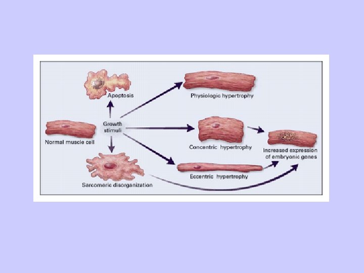
Heart changes Reaction to biomechanical stress (tension in the wall) and to neurohumoral stimuli REMODELATION important for further outcome of heart failure
* Dilatation primary du to volume burden thin wall – increased tension in the wall (higher r, lower h) secondary from previous hypertrophy (excentric hypertrophy) * Hypertrophy concentric in hypertension excentric secund. in increased volume burden and increased preload
Molecular and cellular changes Angiotensin II endotelin IGF-I growth factors cytokins IL-6 cardiotropin 1 etc. Distension leads to gene expression, e. g. of the genes for natriuretic peptides and fetal genes
Heart changes – cellular level - dysregulated myogenesis (abnormal, „embryonal “ growth) - apoptosis Further worsening of heart function
Consequences of heart changes - increased wall tension in dilatation - increase in afterload and oxygen consumption - impaired oxygen delivery in hypertrophy - decrease of compliance - diastolic failure - overstretched dilatation impairs contraction and leads to relative valvular insuficiency - arrhytmias - prognostic factor
Water and salt retention increase in preload Negative consequences: - heart dilatation - congestion, edemas - changes in water/mineral equilibrium, sodium retention and potassium depletion – contributes to electrical nestability of the myocardium
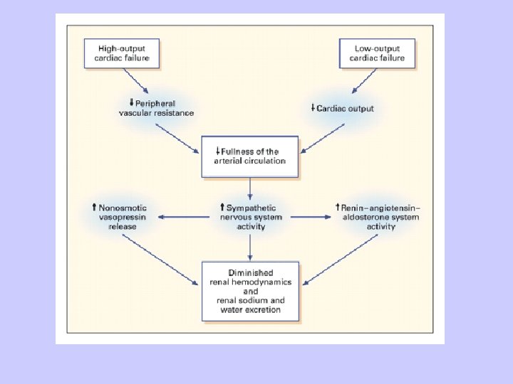
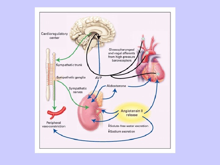
cardiac output - decrease in effective plasmatic volume tissue perfusion sympaticus ADH glomerular filtration renin angiotensin II periphery vasoconstriction BP afterload aldosterone sec. hyperaldosteronisms ECT expansion preload
Neurohumoral adjustments influence vasoconstriction, fluid retention, myocardium - angiotensin II - aldosterone - natriuretic peptids - norepinephrin - ADH - endotelin - prostaglandins keeping the renal perfusion
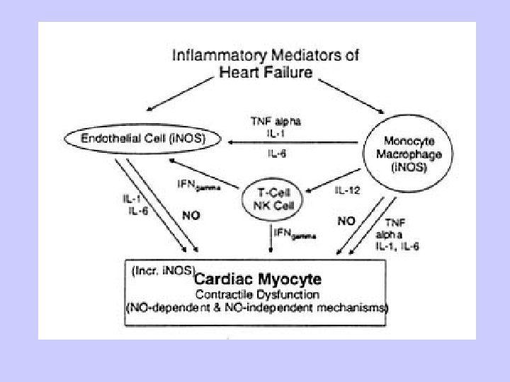
Cytokines in heart function / heart failure Action: negative inotropic proapoptotic fibroplastic arrhytmogenic etc. Mainly proinflammatory cytokines: TNF , IL-1, IL-6 Originate in systemic inflammatory reaction (inflammation, tumor) locally in heart failure as a response to hemodynamic changes
Right-sided failure BACKWARD FORWARD decreased ejection from RV decreased flow from RV to the lungs EDV, EDP in RV decreased flow to the left atrium pressure in R atrium cardiac output volume and pressure in large veins fluid retention symptomes of decreased CO capillary pressure volume in distensible organs (hepatosplenomegaly) edemas, transsudation (ascites, hydrothorax)
Left-sided failure BACKWARD FORWARD decreased ejection from LV EDV, EDP in LV cardiac output kidney perfusion tissue perfusion pressure in L atrium symptomes of decreased CO volume and pressure in pulmonary veins fluid retention capillary pressure fluid volume in the lung fluid transsudation to the alveoli pulmonary edema, dyspnea fatigue, weakness pale skin impairment of organ functions
Left-sided failure Right-sided failure BACKWARD decreased ejection from LV EDV, EDP in LV pressure in L atrium volume and pressure in pulmonary veins capillary pressure fluid volume in the lung fluid transsudation to the alveoli pulmonary edema, dyspnea postcapillary pulmonary hypertension
left-sided failure increase of filling pressure + fluid retention pulmonary edema hypoxemia myocardial hypoxia impairment of myocardial contractility dyspnea - increase burden for respiratory muscles
Heart failure classification NYHA -New York Heart Association: according to the dyspnea ·Class I: patients with no limitation of activities; they suffer no symptoms from ordinary activities. ·Class II: patients with slight, mild limitation of activity; they are comfortable with rest or with mild exertion. ·Class III: patients with marked limitation of activity; they are comfortable only at rest. ·Class IV: patients who should be at complete rest, confined to bed or chair; any physical activity brings on discomfort and symptoms occur at rest.
Principles of the treatment (acc. to Harrison’s Principles of Internal Medicine) - reduction of cardiac work load - control of excessive fluid retention - diuretics - vasodilator therapy - improves (decreases) afterload - enhancement of myocardial contractility - digitalis - sympathomimetic amines - dopamine… - betablockers
The End
Sympathetic nerves catecholamins Izovolumic maxims Intraventricular pressure Failing heart as i D J. Kofránek ic l o t g n i l il f Ventricle volume
Intraventricular pressure Izotonic maxims s ic ton m axi m Iso as i D J. Kofránek ic l o t g n i l il f Ventricle volume
Iso tion trac con nic oto s ic ton m axi m Iso Aux Isovolumic contraction Intraventricular pressure vo lum ic ma xm is Denoting the condition in which a contracting muscle shortens against an increasing load. Relating to a muscle contracting to accommodate an increasing load. as i D ic l o t g n i l il f Isotonic contraction J. Kofránek Ventricle volume
Cardiac output insufficiency increase of syst. volume decrease of „Starling curve“ decrease of syst. volume increased preload J. Kofránek Enddiastolic pressure
catecholamins increase of diastolic compliance Cardiac output insufficiency increase of „Starling curve“ increase of syst. volume decrease of „Starling curve“ increased preload J. Kofránek Enddiastolic pressure