HEART FAILURE Heart failure HF occurs when the
 occurs when the heart is unable to pump enough")


 and left ventricular end diastolic pressure")


 & ACEI(↓TPRafter load & ↓plasma volume –preload). -Diuretics(↓ plasma volume).")


. -↓CO→fatigue. Diagnostic tools")


- Slides: 58
HEART FAILURE Heart failure (HF) occurs when the heart is unable to pump enough blood out to meet the O 2 and nutrient demands of the body. HF can result from either diastolic or systolic dysfunction.
HEART FAILURE Diastolic heart failure may develop alone or in conjunction with systolic heart failure. It often follows prolonged hypertension. When the ventricle must pump continually against a very high afterload, muscle cells hypertrophy and become stiff.
HEART FAILURE Stiffness of the muscle cells causes a reduction in ventricular compliance , leading to decreased filling, abnormal diastolic relaxation , and decreased SV.
HEART FAILURE Left ventricular end diastolic volume (LVEDV) and left ventricular end diastolic pressure (LVEDP) are elevated and reflected back into the pulmonary circulation , causing pulmonary hypertension.
HEART FAILURE Because SV and hence BP fall, baroreceptor reflexes are activated. Systolic dysfunction as a cause of HF results from injury to the ventricle, usually from a MI.
HEART FAILURE The damaged muscle is unable to contract forcefully , and again, SV falls. Decreased SV leads to a decrease in BP , quickly followed by the initiation of reflex responses geared to reversed the trend.
HEART FAILURE Because the damaged ventricle is unable to bring SV back up, the reflexes continue. Sympathetic stimulation of cardiac β 1 receptors becomes chronic, chronically activated sympathetic response reduces Ca++ levels in , and release of Ca++from, the myocardial cells sarcoplasmic reticulum.
HEART FAILURE Reduction in myocardial Ca++ causes defective excitation-contraction coupling, leading to diminished myocardial force production, dysarhythmia, and eventual contractile dysfunction &cardiac muscle cell remodeling.
HEART FAILURE Also worsening of the HF is the effect the progressive increase in EDV has no stretching the cardiac muscle cell causing less tension to be produced as the ventricle become more distended with blood. HF becomes a worsening cycle:
HEART FAILURE The more overfilled the ventricle becomes, the less blood it can pump out, leading to further accumulation of blood and additional stretch of the muscle fibers. As a result, SV, C. O and BP all remain low.
HEART FAILURE The body’s reflex responses initiated in response to the fall in pressure continue and significantly worsen the situation.
Reflexes initiated during heart failure ↓BP is sensed by the baroreceptors. Most reflex responses initiated by baroreceptor activation significantly advance HF progression.
Reflexes initiated during heart failure
Reflexes initiated during heart failure This occurs because the reflex responses either further ↑ventricular filling (preload) or further reduce SV by ↑ the afterload against which the ventricle must pump. ↑ preload and afterload serve to ↑ the workload and O 2 demand to the heart.
Reflexes initiated during heart failure If the ↑ O 2 demand cannot be met, the muscle fibers become ↑ hypoxic and contractility worsens. The downward spiral of heart failure continues. As each of these reflexes further fill and stretch the heart andor ↑ after load, BP continues to be below normal,
Reflexes initiated during heart failure Causing those same reflexes to be maintained. Chronically activated sympathetic response leads to diminished intracellular Ca++ release from the sarcoplasmic reticulum, and ultimately to contractile dysfunction. HF continues unless the cycle of overfill, decreased SV and decreased BP is broken.
Reflexes initiated during heart failure One reflex response that is advantageous during heart failure is that which occurs from atrial over filling. As blood is poorly pumped out of the ventricle, it soon begins to accumulate in the atria.
Reflexes initiated during heart failure Expansion of atria leads to stretching of atrial baroreceptors and the release of the ANP which work on the kidney to increase the excretion of Na+& causing VD. BNP is released from an over stretched ventricle.
Reflexes initiated during heart failure -BNP is significantly correlated with HF severity and is excellent markers of disease progression.
Causes of heart failure HF may result from : -No cardiac causes such as long standing systemic pulmonary hypertension or, disorders such as kidney failure or water intoxication , which increase plasma volume to such a degree that ventricular fibers are stretched beyond their optimum length.
Causes of heart failure Cardiac causes of heart failure include MI, cardiac myopathy, valvular defects, and congenital malformation. Pathways leading to HF following MI and chronic hypertension.
Progression of heart failure HF can begin on either the left or right side of the heart. For instance longstanding systemic hypertension would cause the right ventricle to hypertrophy and fail , the site of a MI would determine which side of the heart is first affected following a heart attack.
Progression of heart failure Because a failing left ventricle would cause blood to back up in the right atrium , it is apparent that left HF can eventually lead to right HF. As blood is poorly pumped out of the right side of the heart, it begins to pool in the peripheral venous system.
Clinical manifestations Forward effects of left HF -↓ABP. -Fatigue. -↑ HR. -↓ urine output. - Plasma volume expansion.
Clinical manifestations Backward effects of left HF -↑pulmonary congestion especially when lying down. -Dyspnea. ↓ Forward effects of right HF -↓pulmonary blood flow. -↓blood oxygenation -Fatigue. -↓ABP
Clinical manifestations Backward effects of right HF -↑venous pooling of blood, edema of the ankles &feet -Jugular venous distension. -Hepatomegaly&splenomegaly.
Diagnostic tools -3 ed heart sound may be present. -Radiological identification of pulmonary congestion and ventricular enlargement may indicate HF. -MRI (ventricular enlargement)
Diagnostic tools -Measurement of ventricular EDP with a catheter inserted into the pulmonary artery (reflecting left ventricular pressure) or into the vena cava (reflecting right ventricular pressure) can diagnose HF. Left ventricular pressure usually reflects left ventricular volume.
Diagnostic tools -Echocardiography can demonstrate abnormal dilatation of the cardiac chambers and abnormalities in contractility. -Measurement of serum BNP &ANP provides information on disease severity.
TREATMENT -Beta blockers (↓HR→↑SV) & ACEI(↓TPRafter load & ↓plasma volume –preload). -Diuretics(↓ plasma volume). -O 2 therapy -Nitrates (↓pre &after load) -Digoxin.
Mitral valve stenosis Usually occurs due to a buildup of scar tissue following rheumatic fever or another cardiac infection. It may also result from a congenital defect in valve structure.
Mitral valve stenosis In order to pump blood through the narrowed orifice , the left atrium must contract more forcefully. If the left atrium is unable to pump through the orifice, blood will pool in the atrium and back up into the lungs and right side of the heart.
Mitral valve stenosis Right HF can result, especially if flow through the valve is so restricted that SV &CO are too low to maintain normal range systemic BP. If this occurs , baroreceptor reflexes initiating sympathetic &abnormal responses will be activated →↑plasma volume &↑TPR& ↑ABP
Mitral valve stenosis Clinical manifestations: -Pulmonary congestion, dyspnea &pulmonary hypertension. -↑HR. Diagnostic tools: -Low-pitched diastolic murmur -Echocardiography may be used to diagnose abnormal valve structure&motion.
Mitral valve stenosis Complications -Left atrial hypertrophy may cause atrial dysrhythmia or right HF. Treatment -Treatment for CHF. -Valve replacement or surgical correction.
Aortic valve stenosis Aortic stenosis follows RF or is a congenital malformation. The left ventricle must pump more forcefully to expel blood through the narrow orifice. This causes ventricular hypertrophy and eventually reduces compliance.
Aortic valve stenosis Clinical manifestations -Pulmonary congestion , with sign of dypnea &pulmonary hypertension. -Fatigue may occur due to ↓CO& ↓SV&↑HR. Diagnostic tools -A systolic murmur may be heard. -Echo. Complications -Left ventricular hypertrophy may develop , leading to CHF
Pulmonary valve stenosis Pulmonary stenosis occurs due to a congenital defect, in this case the right ventricle must pump more forcefully to expel blood. This can lead to right ventricular hypertrophy , backing up into the right atrium and causing dilatation of the vena cava &blood accumulation in the systemic veins.
Pulmonary valve stenosis Blood flow into the lungs and left side of the heart will be reduced if the stenosis is sever , leading to a decrease in blood pressure. Right HF may develop.
Pulmonary valve stenosis Clinical manifestations -↓pulmonary flow causes poor oxygenation of the blood and feelings of weakness and fatigue. -Venous distention &swelling of the ankles and feet are common. Diagnostic tools: Echo Complications Right heart failure.
Mitral valve regurgitation Clinical manifestations -Pulmonary congestion (dyspnea & pulmonary hypertension). -↓CO→fatigue. Diagnostic tools -A systolic heart murmur may be heard. Complications -CHF
Aortic valve regurgitation Clinical manifestations -A wide pulse pressure. -HF. Diagnostic tools -A high- pitched diastolic heart murmur.
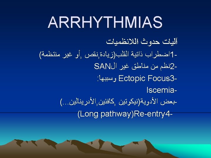
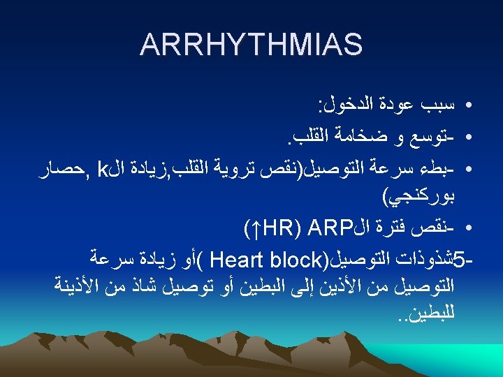
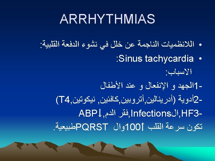
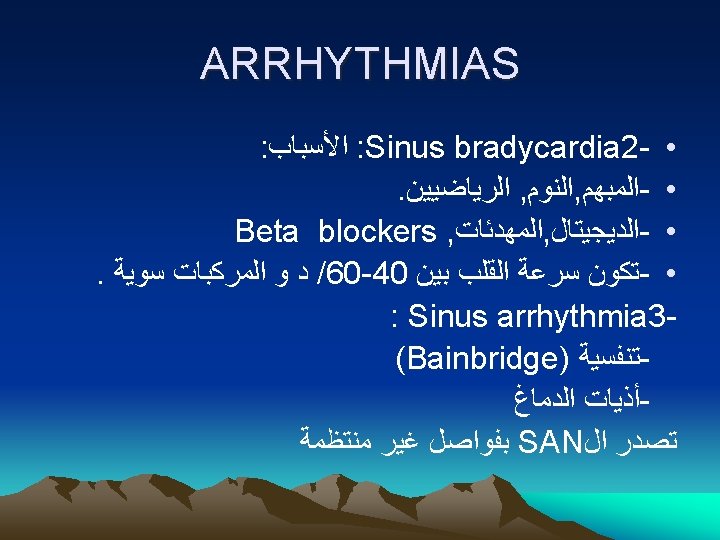
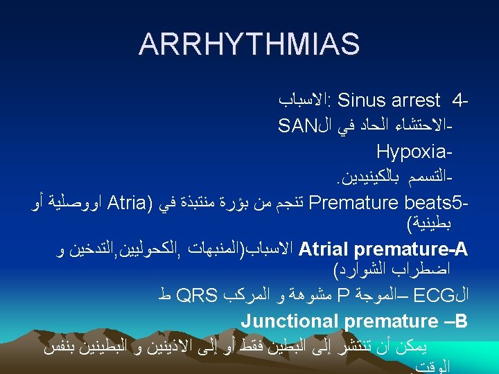
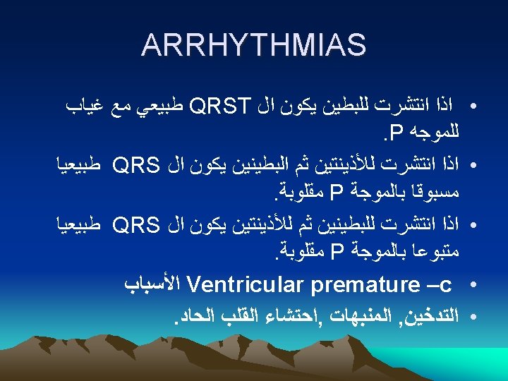
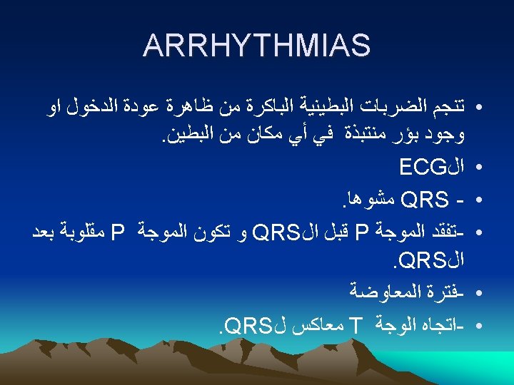
ARRHYTHMIAS