Heart Dissection Prepare for dissection Put on goggles


.")


- Slides: 26
Heart Dissection
Prepare for dissection Put on goggles and gloves. Obtain a deer heart and immediately place it in your tray so the blood does not drip all over.
Wash the heart to remove excess blood.
Remove the pericardium if it is still present, show others if they don’t have one
These students discovered that the pericardium was attached to the diaphragm.
External Anatomy Place the heart in the dissecting tray so that the anterior side is toward you. The major blood vessels will be located superiorly and the apex is inferior. Locate the • Interventricular sulcus • Apex Answer questions 1 -4 on the lab.
Locate the chambers of the heart. Three of the chambers are shown in the picture. Answer questions 5 -8.
Major Blood Vessels Find the pulmonary artery first. Insert the dull probe into the pulmonary artery. Answer question 9.
Next find the aorta. It is on the superior side of the heart and is the largest blood vessel in the body. It has thick walls. Answer question 10.
Next you will locate the superior vena cava and the inferior vena cava on the posterior side. Since they both lead into the right atrium, your probe (or finger) can connect the two. Answer questions 11 -12.
Find the pulmonary veins and you’re all set! Anterior view Posterior view
Time to cut it open. First cut. Cut into the pulmonary artery and parallel to the interventricular sulcus
• With your fingers, push open the heart at the cut to examine the internal structure. If there is dried blood inside the chambers, rinse out the heart. • Locate the right atrium. Notice thinner muscular wall of this receiving chamber. • Find where the inferior and superior vena cava enter this chamber and notice the lack of valves.
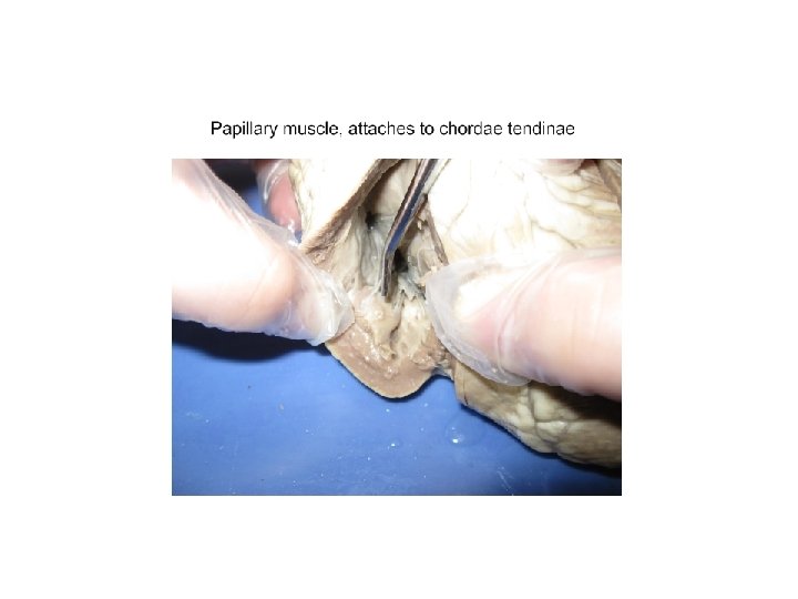
Locate the valve that is between the right atrium and right ventricle. This is called the tricuspid valve. The valve consists of three leaflets and has long fibers of connective tissue called chordae tendinae that attach the valve to papillary muscles of the heart. Answer question 13.
Use your fingers to feel the thickness of the right ventricle and its smooth lining. Also note the network of irregular muscular cords on the inner wall of this chamber.
Find the septum on the right side of the right ventricle. This thick muscular wall separates the right and left pumping ventricles from each other.
Locate the pulmonary valve between the right ventricle and the pulmonary artery. The pulmonary valve is called a semi-lunar valve because of its half moon shape. Answer question 14.
Cut 2 Cut from the left atrium into the left ventricle all the way to the apex.
Examine the left atrium. Find the openings for the pulmonary veins. See if you can locate semi-lunar valves at the entrance to these veins. Answer question 15.
Inside this chamber, look for the valve that controls blood flow between the upper left atrium and lower left ventricle. This valve is called the bicuspid or mitral valve. This valve consists of two leaflets shaped like a headpiece called a miter that you may see bishops wear. Answer question 16.
Examine the left ventricle. Notice thickness of the ventricular wall. Compare the wall of the left ventricle to the wall of the right ventricle. Answer question 17.
Using your scissors, cut across the left ventricle toward the aorta. (cut 3).
Find the aortic valve. Answer questions 18 -19.
Using scissors, cut through the aorta and examine the inside. Find the hole or coronary sinus in the wall of this major artery. This leads into the coronary artery which carries blood to the heart muscle to nourishment it. Answer question 20.
Dispose of your heart When you have finished dissecting the heart, wrap it in paper towel and put in the waste can Clean and dry all the dissecting equipment, including the tray. Wash your hands.