
Heart and Circulatory System I Daphne T Hsu


- Slides: 41
Heart and Circulatory System I Daphne T. Hsu, MD Professor of Clinical Pediatrics dh 17@columbia. edu
Outline • • • Vasculogenesis Embryonic Folding Formation of the Primary Heart Tube Looping Atrial Septation Primitive Ventricular Septum Atrioventricular Canal/Endocardial Cushions Conotruncal Septation Ventricular septation Congenital Heart Defects
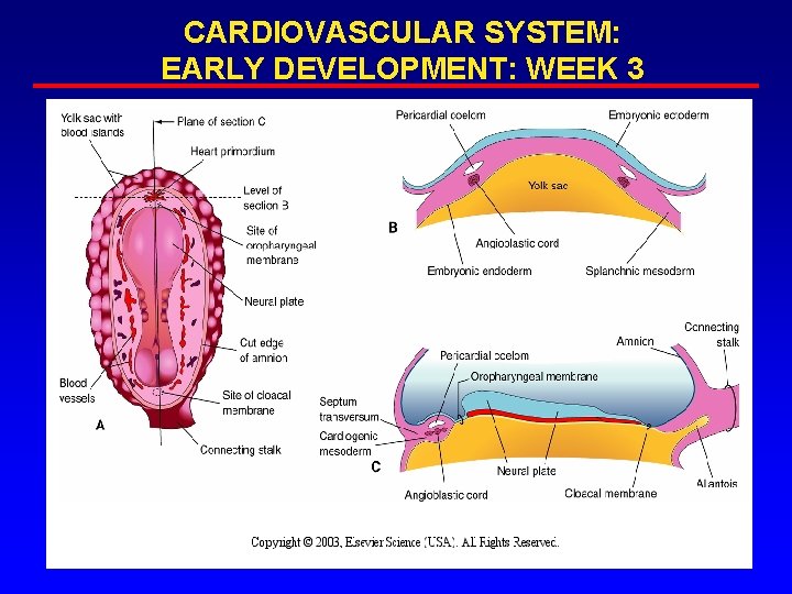
CARDIOVASCULAR SYSTEM: EARLY DEVELOPMENT: WEEK 3
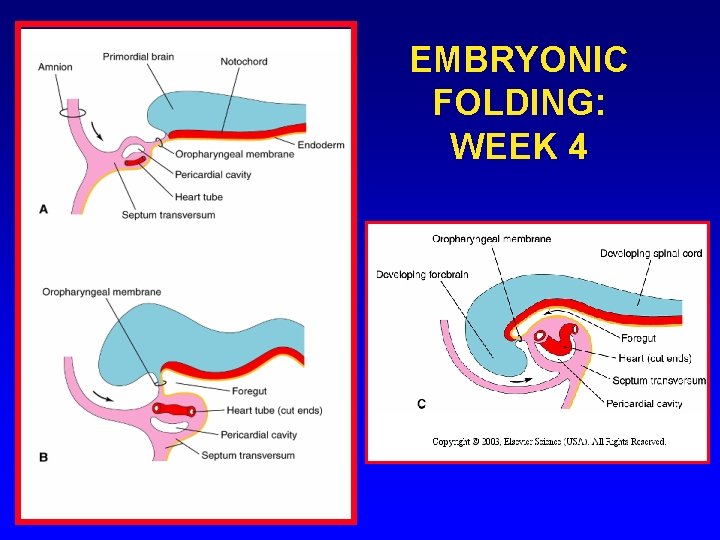
EMBRYONIC FOLDING: WEEK 4
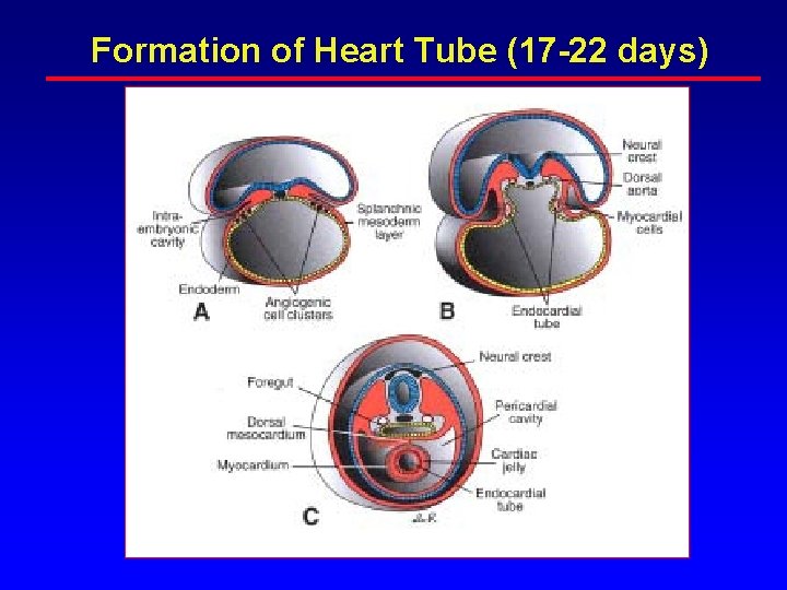
Formation of Heart Tube (17 -22 days)
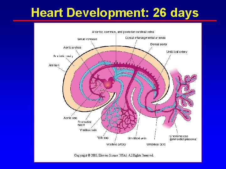
Heart Development: 26 days
PRIMITIVE HEART TUBE: WEEK 4
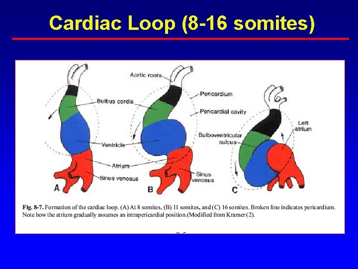
Cardiac Loop (8 -16 somites)
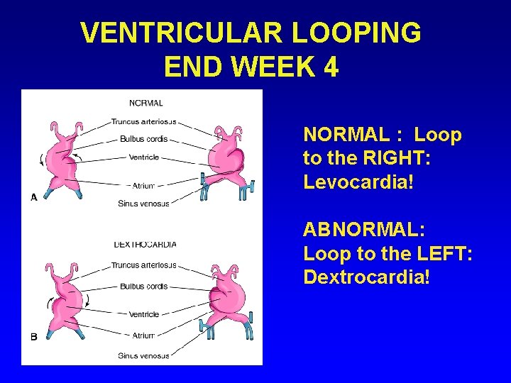
VENTRICULAR LOOPING END WEEK 4 NORMAL : Loop to the RIGHT: Levocardia! ABNORMAL: Loop to the LEFT: Dextrocardia!
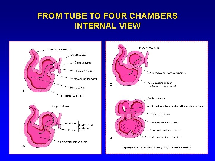
FROM TUBE TO FOUR CHAMBERS INTERNAL VIEW
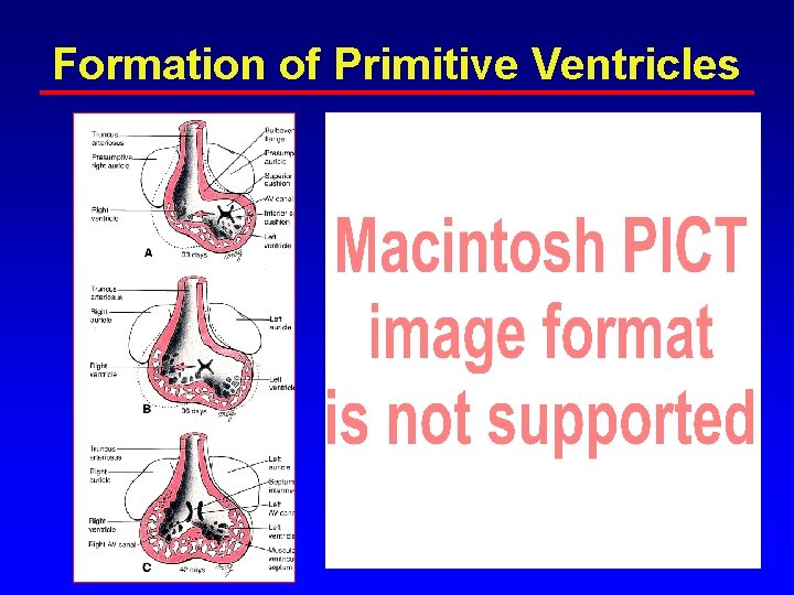
Formation of Primitive Ventricles
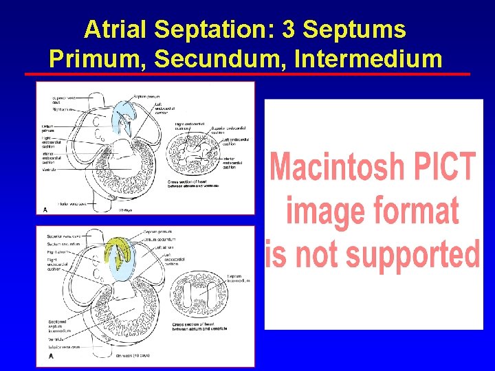
Atrial Septation: 3 Septums Primum, Secundum, Intermedium
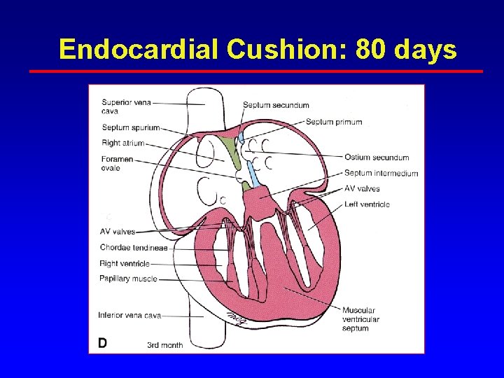
Endocardial Cushion: 80 days
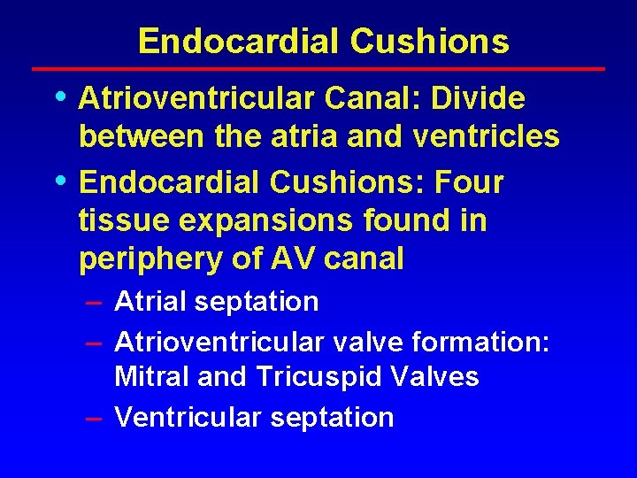
Endocardial Cushions • Atrioventricular Canal: Divide • between the atria and ventricles Endocardial Cushions: Four tissue expansions found in periphery of AV canal – Atrial septation – Atrioventricular valve formation: Mitral and Tricuspid Valves – Ventricular septation
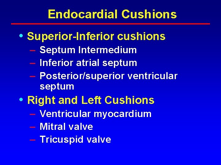
Endocardial Cushions • Superior-Inferior cushions – Septum Intermedium – Inferior atrial septum – Posterior/superior ventricular septum • Right and Left Cushions – Ventricular myocardium – Mitral valve – Tricuspid valve
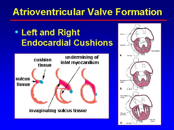
Atrioventricular Valve Formation • Left and Right Endocardial Cushions
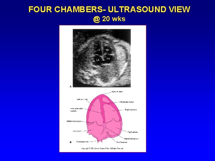
FOUR CHAMBERS- ULTRASOUND VIEW @ 20 wks
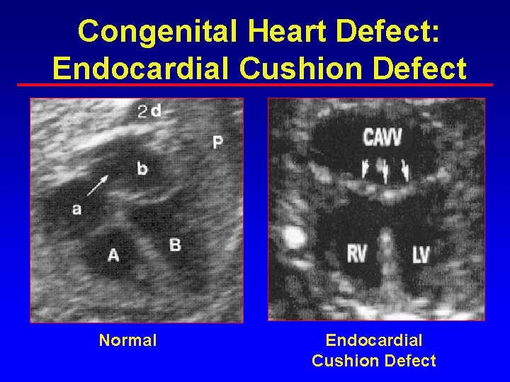
Congenital Heart Defect: Endocardial Cushion Defect Normal Endocardial Cushion Defect
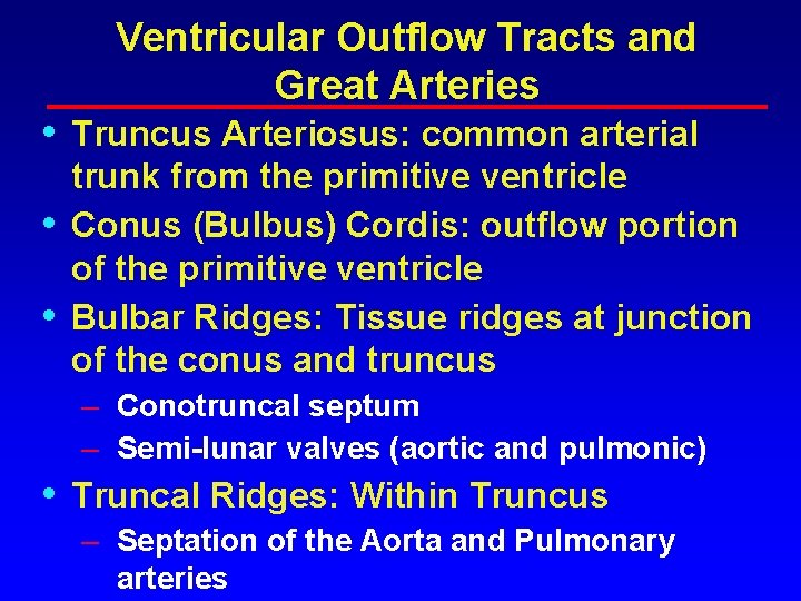
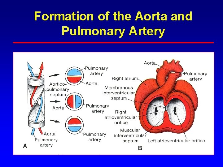
• • • Ventricular Outflow Tracts and Great Arteries Truncus Arteriosus: common arterial trunk from the primitive ventricle Conus (Bulbus) Cordis: outflow portion of the primitive ventricle Bulbar Ridges: Tissue ridges at junction of the conus and truncus – Conotruncal septum – Semi-lunar valves (aortic and pulmonic) • Truncal Ridges: Within Truncus – Septation of the Aorta and Pulmonary arteries
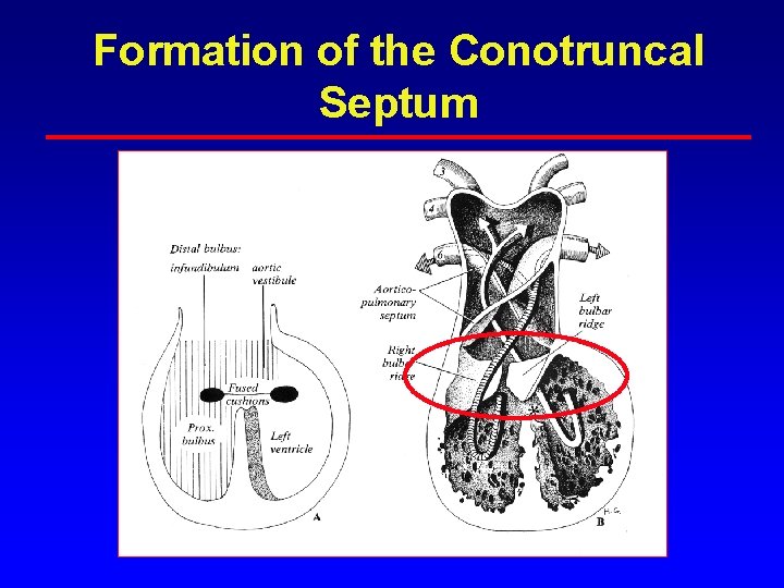
Formation of the Conotruncal Septum
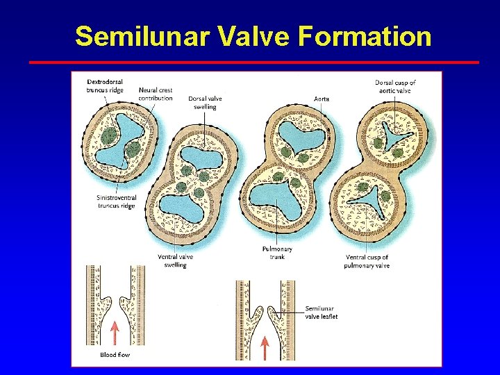
Semilunar Valve Formation
Formation of the Aorta and Pulmonary Artery
Conotruncal Formation
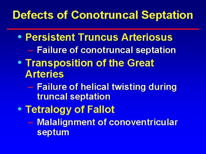
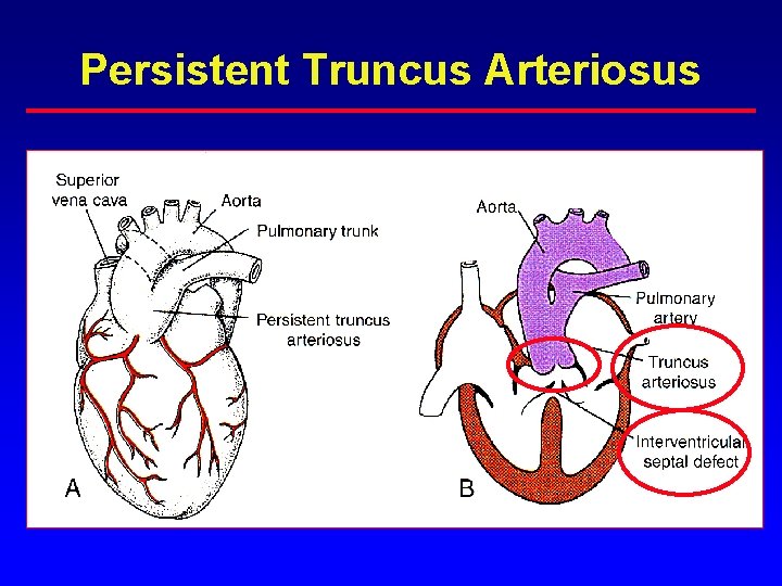
Defects of Conotruncal Septation • Persistent Truncus Arteriosus – Failure of conotruncal septation • Transposition of the Great Arteries – Failure of helical twisting during truncal septation • Tetralogy of Fallot – Malalignment of conoventricular septum
Persistent Truncus Arteriosus
Transposition of the Great Arteries • • Failure of helical twisting during truncal septation Aorta arises from RV Pulmonary artery arises from LV VSD in 20% of cases
Transposition of the Great Arteries
Tetralogy of Fallot • Malalignment of conoventricular septum 1. Ventricular septal defect 2. Aortic valve override 3. Pulmonary stenosis 4. Right ventricular hypertrophy
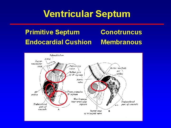
Ventricular Septum Primitive Septum Conotruncus Endocardial Cushion Membranous
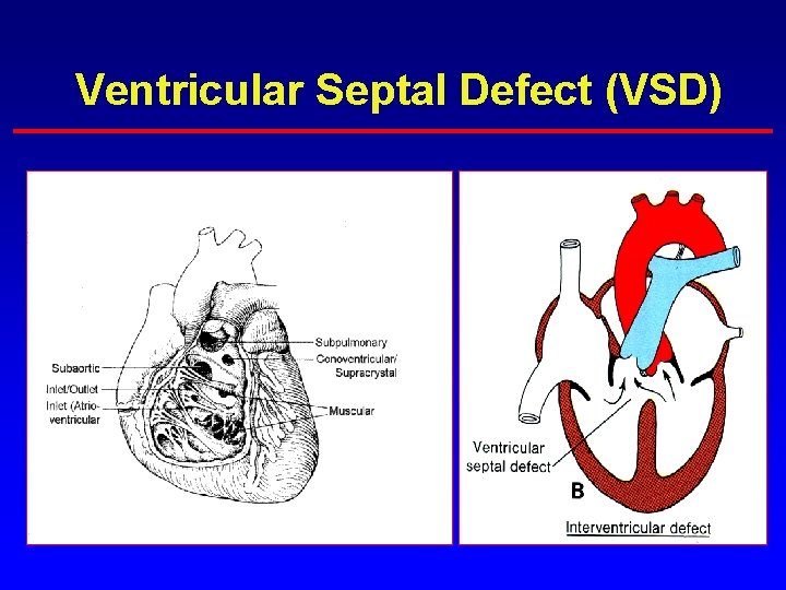
Ventricular Septal Defect (VSD)
Muscular VSD
Endocardial Cushion (Inlet VSD)
Supracristal VSD
Membranous VSD
Echocardiogram: Membranous VSD
Angiogram: VSD
Heart Formation • Heart structures form completely • by week 12 Week 3 -4: Vitelline Stage – Heart tube forms – “Looping” occurs – Venous system starts to form • Week 5 – Placenta provides nutrients and liver takes of hematopoeis
Heart Formation con’t • Weeks 5 -8 – – SVC, IVC form Right and Left Atrium divide Ventricles start to form Aorta and pulmonary arteries • Weeks 8 -12 – Ventricles and mitral and tricuspid valves – Aorta and pulmonary artery, aortic arch • Week 12: Fetal Circulation begins
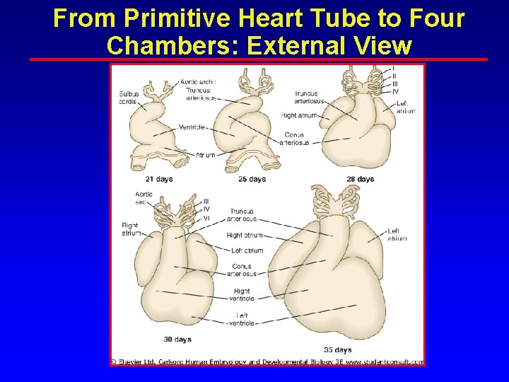
From Primitive Heart Tube to Four Chambers: External View
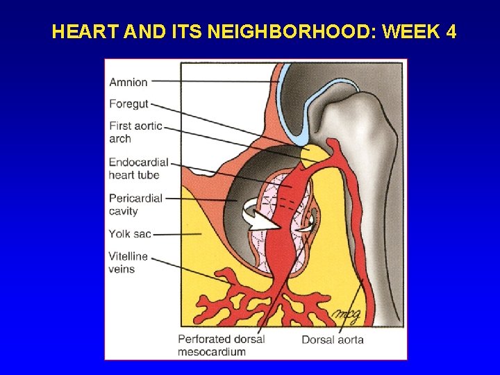
HEART AND ITS NEIGHBORHOOD: WEEK 4
Multiple Defects: Bilateral Left-Sidedness • • • Systemic Veins – Interrupted IVC – Bilateral SVC Common Atrium Common Ventricle – VSD: endocardial cushion Pulmonary veins: – Ipsilateral Pulmonary Stenosis