Health Science Theory Lab Cardiovascular System Review Objective

Health Science Theory & Lab Cardiovascular System Review

Objective Upon completion of this lesson, students will be able to: • Explain the structure and function of the heart • Explain the movement of blood through the heart and circulatory system • Describe common cardiovascular diseases • Describe common clinical procedures to assess cardiac function

Essential Question • How is the structure of the heart related to its function? • How does blood move through the heart? • How does the heart beat? • What are the signs of poor circulation? • What tests can be done to identify cardiac problems?

The Heart: “The Body’s Workhorse • Your heart beats 100, 000 times per day • 35 million times per year • Pumps 1, 585 gallons of blood per day!

Introduction • • • Located in the thoracic cavity Behind the sternum Inside mediastinum (cavity) Top of the heart is the base Bottom is the apex

Layers of the Heart The heart has 3 layers • Epicardium • Myocardium • Endocardium • Pericardium is a sack that surrounds the heart

Epicardium • Outermost layer of the heart. • Coronary arteries run along this layer.
 Pure muscle Contractile muscle Damaged during a")
Myocardium • • Middle layer (thickest layer) Pure muscle Contractile muscle Damaged during a heart attack
 Lining of the heart chambers Folds to")
Endocardium • • • Innermost layer (thin) Lining of the heart chambers Folds to form the heart valves Watertight Conduction system housed here

Pericardium • A double-walled sack that encloses the heart • Anchors the heart to the diaphragm and great vessels • Pericardial fluid minimizes friction during heartbeat

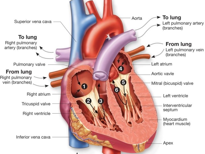
Heart Chambers • • Right atrium Right ventricle Left atrium Left ventricle

Roles of the Chambers • Atria deliver blood to the ventricles that lie beneath them • Ventricles are high pressure pumping chambers that move blood to the body and the lungs

Right Atrium • Receiving chamber for deoxygenated blood returning from the body • Blood is dark maroon with little oxygen • Blood has a high CO 2 content • Right atrium delivers it’s blood to the right ventricle

Right Ventricle • Receives blood from the right atrium • Pumps deoxygenated blood to the lungs • Thinner muscle layer

Left Atrium • Receiving chamber for blood returning from the lungs • 100% O 2 saturation • Blood is bright red

Left Ventricle • Pumps blood to the entire body • The major powerful pumping chamber! • Blood is bright red

Heart Valves • “Doors” between chambers that prevent the backflow of blood • There are 4 valves: 2 semilunar and 2 atrioventricular valves • Valves open and close based on pressure changes • Valves open ONLY in the direction of blood flow. • Heart sounds caused by closing of valves

Semilunar Valves • Seminlunar means “half moon. ” • Pulmonic valve: between RV and pulmonary artery • Aortic valves: between the left ventricle and aorta

Atrioventricular Valves • Located between atria and ventricles • Supported by chordae tendineae to prevent backflow • Tricuspid Valve – located between RA and RV (3 cusps) • Mitral Valve (Bicuspid) – located betwwen LA and LV (2 cusps)

Check on Learning • Which layer of the heart is the thickest? • Why is the endocardium layer water tight? • What is the percent of O 2 saturation of blood returning to the left atrium? • The inferior-most tip of the heart is called the ______ • Which chambers delivers blood to the ventricles?

Great Vessels of the Heart • 5 great vessels • Attached at the base of the heart – – – Superior Vena Cava (SVC) Inferior Vena Cava (IVC) Pulmonary Artery Pulmonary Veins Aorta
 • Returns deoxygenated blood to the right atrium from the")
Superior Vena Cava (SVC) • Returns deoxygenated blood to the right atrium from the head, neck, upper chest and arms
 • Returns deoxygenated blood to the right atrium from the")
Inferior Vena Cava (IVC) • Returns deoxygenated blood to the right atrium from the lower chest, abdomen and legs

Pulmonary Artery • Delivers deoxygenated blood from the RV to the lungs to pick up a load of oxygen and unload CO 2

Aorta • Largest artery in the body • Takes oxygenated blood from the LV to the body to feed all organs

Blood Flow Through The Heart Let’s track a single blood cell as it travels through the heart

“The Roller Coaster Ride” • • Blood cell enters the heart via one of the vena cava Enters the right atrium Travels through the tricuspid valve in to the right ventricle Passes through the pulmonic valve into the pulmonary artery then goes to the lungs • Sent back through the pulmonary veins into the left ventricle • Goes through the mitral valve into the left ventricle • Passes through the aortic valve into the aorta back to the body

The Cardiac Cycle • The mechanical events that occur to pump blood • Diastole • Systole

Diastole • Phase of blood flow where the ventricles relax and fill with blood • 3 phases: Rapid filling, Diastasis and Atrial Kick • Blood moves from atria to ventricles • S 1 sound heard at the end of diastole when the AV valves slam shut

Systole • Ventricles contract to push blood out of the heart toward the lungs and the body • 4 phases: isovolumetric contraction, ventricular ejection, protodiastole, isovolumetric relaxation.

Coronary Arteries • As a muscle, the heart itself needs oxygen • Coronary arteries deliver oxygen to the heart itself • Two major coronary arteries: Left Main Coronary Artery Right Coronary Artery
 • Feeds the anterior and lateral walls of the")
Left Main Coronary Artery (LMCA) • Feeds the anterior and lateral walls of the left ventricle
 • Feeds the right ventricle and inferior wall of the")
Right Coronary Artery (RCA) • Feeds the right ventricle and inferior wall of the left ventricle

What Makes the Heart Beat?

Heart Cells • Contractile cells: cause the heart muscle to contract resulting in heartbeat • Conduction system cells: create and conduct electrical signals to tell the heart when to beat.

Heart Cells • Sinoatrial Node: a small body of specialized muscle tissue in the wall of the right atrium that acts as a pacemaker by producing a signal at regular intervals • Atrioventricular Node: serves as an electrical relay station, slowing the electrical current sent by the SA node and passes down through to the ventricles

Heart Cells • Bundle of His: transmits impulses from the AV node to the ventricles of the heart. • Purkinje Fibers: carry the contraction impulse from both the left and right bundle branch to the myocardium of the ventricles

Check on Learning • Which blood vessel returns blood to the heart from the head? • Which blood vessel is the largest artery in the body? • Which blood vessels provide oxygenated blood to the heart muscle itself?
- Slides: 40