Healing and Repair Lecture 2 Dr Bushra AlTarawneh

Healing and Repair, Lecture 2 Dr. Bushra Al-Tarawneh, MD

Healing of skin wounds can be classified into healing by first intention (primary union), referring to epithelial regeneration with minimal scarring, as in well-apposed surgical incisions. Healing by second intention (secondary union), referring to larger wounds that heal by a combination of regeneration and scarring.

Mechanisms of Tissue Regeneration The importance of regeneration in the replacement of injured tissues varies in different types of tissues and with the severity of injury. • In epithelia of the intestinal tract and skin, injured cells are rapidly replaced by proliferation of residual cells and differentiation of cells derived from tissue stem cells, providing the underlying basement membrane is intact. • Tissue regeneration can occur in parenchymal organs whose cells are capable of proliferation, but with the exception of the liver, this is usually a limited process. Pancreas, adrenal, thyroid, and lung have some regenerative capacity.

Restoration of normal tissue architecture can occur only if the residual tissue is structurally intact, for example after partial surgical resection of the liver. By contrast, if the entire tissue is damaged by infection or inflammation, regeneration is incomplete and is accompanied by scarring. For example, extensive destruction of the liver with collapse of the reticulin framework, as occurs in a liver abscess, leads to scar formation even though the remaining liver cells have the capacity to regenerate.

LIVER REGENERATION The human liver has a remarkable capacity to regenerate, as demonstrated by its growth after partial hepatectomy, which may be performed for tumor resection or for living donor hepatic transplantation. Regeneration of the liver occurs by two major mechanisms: . Proliferation of remaining hepatocytes and repopulation from progenitor cells. Which mechanism plays the dominant role depends on the nature of the injury. • Proliferation of hepatocytes following partial hepatectomy. In humans, resection of up to 90% of the liver can be corrected by proliferation of the residual hepatocytes. This process is driven by cytokines such as IL-6 produced by Kupffer cells, and by growth factors such as hepatocyte growth factor (HGF) produced by many cell types.

REPAIR BY SCARRING If repair cannot be accomplished by regeneration alone, it occurs by replacement of the injured cells with connective tissue, leading to the formation of a scar, or by a combination of regeneration of some residual cells and scar formation. Scarring may happen if: -The tissue injury is severe or chronic and results in damage to parenchymal cells and epithelia as well as to the connective tissue framework, -Nondividing cells are injured.

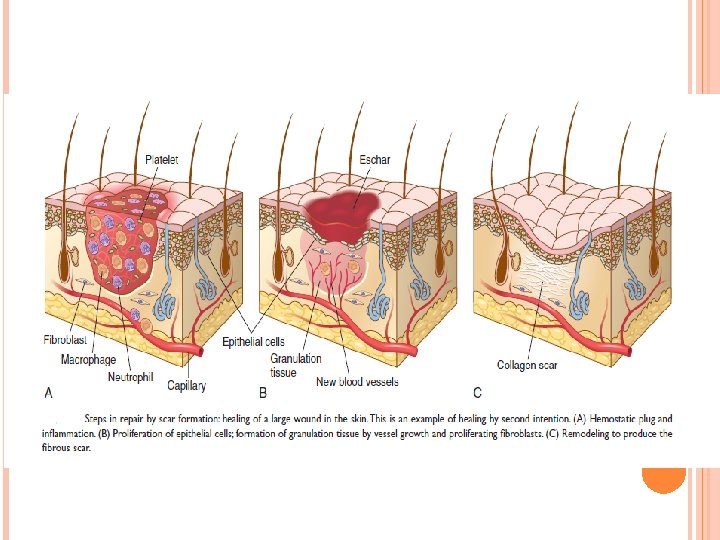
STEPS IN SCAR FORMATION Repair by connective tissue deposition consists of a series of sequential steps that follow tissue injury: 1 - Within minutes after injury, a hemostatic plug comprised of platelets is formed, which stops bleeding and provides a scaffold for infiltrating inflammatory cells. 2 -Inflammation. 3 - Cell proliferation. 4 - Activation of Fibroblasts and Deposition of Connective Tissue. 5 - Remodeling of Connective Tissue.

STEPS IN SCAR FORMATION …. CONTINUE 2 -INFLAMMATION. This step is comprised of the typical acute and chronic inflammatory responses-------to recruit neutrophils and then monocytes during the next 6 to 48 hours. Macrophages are the central cellular players in the repair process: - M 1 macrophages clear microbes and necrotic tissue and promote inflammation in a positive feedback loop - M 2 macrophages produce growth factors that stimulate the proliferation of many cell types in the next stage of repair. As the injurious agents and necrotic cells are cleared, the inflammation resolves.

STEPS IN SCAR FORMATION …. CONTINUE 3 - CELL PROLIFERATION. In the next stage, which takes up to 10 days, several cell types, including epithelial cells, endothelial and other vascular cells, and fibroblasts, proliferate and migrate to close the now-clean wound. Each cell type serves unique functions: • Epithelial cells respond to locally produced growth factors and migrate over the wound to cover it. • Endothelial and other vascular cells proliferate to form new blood vessels, a process known as angiogenesis. • Fibroblasts proliferate and migrate into the site of injury and lay down collagen fibers that form the scar.

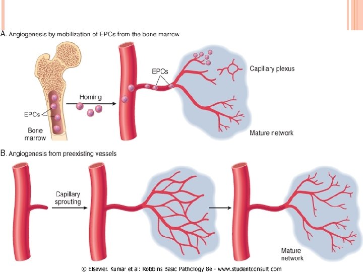
STEPS IN SCAR FORMATION …. CONTINUE ANGIOGENESIS Angiogenesis is the process of new blood vessel development from existing vessels. - It is critical in healing at sites of injury, in the development of collateral circulations at sites of ischemia, and in allowing tumors to increase in size beyond the constraints of their original blood supply. - Angiogenesis involves sprouting of new vessels from existing ones.

THE PROCESS OF ANGIOGENESIS • Vasodilation in response to NO and increased permeability induced by VEGF. • Separation of pericytes from the abluminal surface and breakdown of the basement membrane to allow formation of a vessel sprout. • Migration of endothelial cells toward the area of tissue Injury. • Proliferation of endothelial cells just behind the leading front (“tip”) of migrating cells. • Remodeling into capillary tubes. • Recruitment of periendothelial cells (pericytes for small capillaries and smooth muscle cells for larger vessels) to form the mature vessel. • Suppression of endothelial proliferation and migration and deposition of the basement membrane.

THE PROCESS OF ANGIOGENESIS Angiogenesis consists of the following steps: • Vasodilation in response to NO and increased permeability induced by VEGF. • Separation of pericytes from the abluminal surface and breakdown of the basement membrane to allow formation of a vessel sprout. • Migration of endothelial cells toward the area of tissue injury. • Proliferation of endothelial cells just behind the leading front (“tip”) of migrating cells. • Remodeling into capillary tubes. • Recruitment of periendothelial cells (pericytes for small capillaries and smooth muscle cells for larger vessels) to form the mature vessel. • Suppression of endothelial proliferation and migration and deposition of the basement membrane.
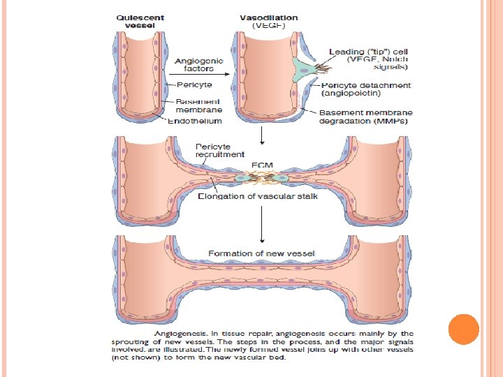

THE PROCESS OF ANGIOGENESIS INVOLVES SEVERAL SIGNALING PATHWAYS, CELL–CELL INTERACTIONS, ECM PROTEINS, AND TISSUE ENZYMES. • Growth factors. VEGFs, mainly VEGF-A stimulates both migration and proliferation of endothelial cells, thus initiating the process of capillary sprouting in angiogenesis. It promotes vasodilation by stimulating the production of NO and contributes to the formation of the vascular lumen. Fibroblast growth factors (FGFs), mainly FGF-2, stimulate the proliferation of endothelial cells. They also promote the migration of macrophages and fibroblasts to the damaged area, and stimulate epithelial cell migration to cover epidermal wounds. Newly formed vessels need to be stabilized by the recruitment of pericytes and smooth muscle cells and by the deposition of connective tissue. PDGF and TGF-β, likely participate in the stabilization process: PDGF recruits smooth muscle cells and TGF-β suppresses endothelial proliferation and migration, and enhances the production of ECM proteins.

THE PROCESS OF ANGIOGENESIS …. CONTINUE Notch signaling. Through “cross talk” with VEGF, the Notch signaling pathway regulates the sprouting and branching of new vessels and thus ensures that the new vessels that are formed have the proper spacing to effectively supply the healing tissue with blood. ECM proteins participate in the process of vessel sprouting in angiogenesis, largely through interactions with integrin receptors of endothelial cells and by providing the scaffold for vessel growth. Enzymes in the ECM, notably the matrix metalloproteinases (MMPs), degrade the ECM to permit remodeling and extension of the vascular tube.

THE PROCESS OF ANGIOGENESIS …. CONTINUE Newly formed vessels are leaky because of incomplete interendothelial junctions and because VEGF, the growth factor that drives angiogenesis, increases vascular permeability. This leakiness accounts in part for the edema that may persist in healing wounds long after the acute inflammatory response has resolved.

STEPS IN SCAR FORMATION …. CONTINUE 3 - CELL PROLIFERATION. The combination of proliferating fibroblasts, loose connective tissue, new blood vessels and scattered chronic inflammatory cells, forms a type of tissue that is unique to healing wounds and is called granulation tissue. This term derives from its pink, soft, granular gross appearance, such as that seen beneath the scab of a skin wound.

GRANULATION TISSUE : SPECIALIZED TYPE OF TISSUE THAT IS CHARACTERISTIC OF HEALING PROCESS. THE TERM GRANULATION TISSUE DERIVES FROM: PINK, SOFT, GRANULAR GROSS APPEARANCE.
")
Histologic appearance of grnulation tissue: 1. Proliferating fibroblasts. 2. Formation of new capillaries. (Angiogenesis) 3. Loose ECM formation.

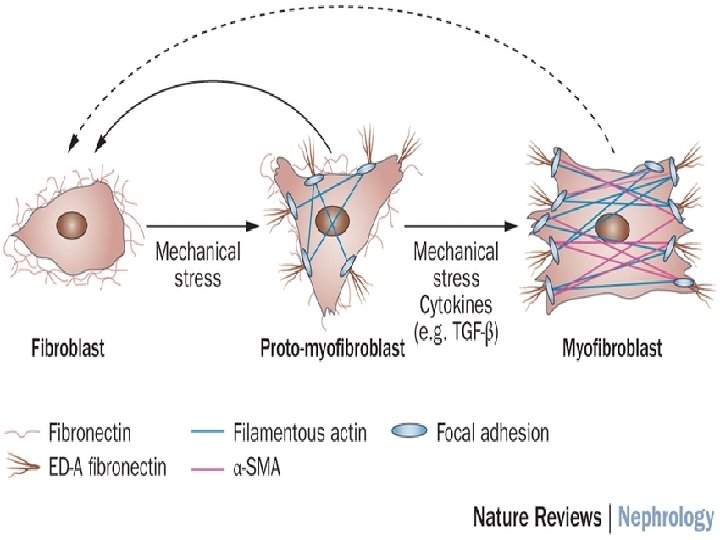
STEPS IN SCAR FORMATION …. CONTINUE 4 - ACTIVATION OF FIBROBLASTS AND DEPOSITION OF CONNECTIVE TISSUE The laying down of connective tissue occurs in two steps: (1) migration and proliferation of fibroblasts into the site of injury. (2) deposition of ECM proteins produced by these cells. These processes are orchestrated by locally produced cytokines and growth factors, including PDGF, FGF-2, and TGF-β. In response to cytokines and growth factors, fibroblasts enter the wound from the edges and migrate toward the center. Some of these cells may differentiate into cells called myofibroblasts, which contain smooth muscle actin and have increased contractile activity, and serve to close the wound by pulling its margins toward the center. Activated fibroblasts and myofibroblasts also increase their synthetic activity and produce connective tissue proteins, mainly collagen, which is the major component of the fully developed scar.

TGF-β is the most important cytokine for the synthesis and deposition of connective tissue proteins. It is produced by most of the cells in granulation tissue, including alternatively activated macrophages. TGF-β stimulates fibroblast migration and proliferation, increases the synthesis of collagen and fibronectin, and decreases the degradation of ECM by inhibiting metalloproteinases. TGF-β is involved not only in scar formation after injury but also in the development of fibrosis in lung, liver, and kidneys that follows chronic inflammation. TGF-β also has anti-inflammatory effects that serve to limit and terminate inflammatory responses. It does this by inhibiting lymphocyte proliferation and the activity of other leukocytes.

STEPS IN SCAR FORMATION …. CONTINUE 5 - REMODELING. The connective tissue that has been deposited by fibroblasts is reorganized to produce the stable fibrous scar. This process begins 2 to 3 weeks after injury and may continue for months or years.

STEPS IN SCAR FORMATION …. CONTINUE 5 -REMODELING OF CONNECTIVE TISSUE After the scar is formed, it is remodeled to increase its strength and contract it. Wound strength increases because of cross-linking of collagen and increased size of collagen fibers. In addition, there is a shift of the type of collagen deposited, from type III collagen early in repair to more resilient type I collagen. In well-sutured skin wounds, strength may recover to 70% to 80% of normal skin by 3 months. Wound contraction is initially caused by myofibroblasts and later by cross-linking of collagen fibers.

The degradation of collagens and other ECM components is accomplished by a family of matrix metalloproteinases (MMPs. are produced by a variety of cell types), and their synthesis and secretion are regulated by medioters. They include : q Interstitial collagenases, which cleave fibrillar collagen (MMP-1, -2, and -3) q Gelatinases (MMP-2 and 9), which degrade amorphous collagen and fibronectin. q Stromelysins (MMP-3, -10, and -11), which degrade a variety of ECM constituents. o Neutrophil elastase, cathepsin G, plasmin, and other serine proteinases can also degrade ECM but are less important in wound remodeling In addition, activated collagenases can be rapidly inhibited by specific tissue inhibitors of metalloproteinases (TIMPs), produced by most mesenchymal cells. Thus, a balance of MMPs and TIMPs regulates the size and nature of the scar.

As the scar matures, there is progressive vascular regression, which eventually transforms the highly vascularized granulation tissue into a pale, largely avascular scar.

THANK YOU
- Slides: 29