HEAD INJURY Done By Yara khaled yaseen Definition

HEAD INJURY Done By: Yara khaled yaseen

Definition • A head injury, as defined by the National Advisory Neurological Diseases and Stroke Council, is • a morbid state resulting from gross or subtle structural changes in the scalp, skull, and/or the contents of the skull, which is produced by mechanical forces. These injuries can range from a minor bump on the skull to serious brain injury. •

Epidemiology • Third most common cause of death in the US. • Third most common cause of death in Jordan. • Number One Killer in Trauma. • More than 50% of all trauma deaths. • 50% of all deaths from motor vehicle accidents. • 200, 000 people in the world live with the disability caused by these injuries.

• Traumatic brain injuries are more common in young patients, and men account for the majority (75%) of cases.

Etiology § 32% of head injuries are caused by falls , more common among the elderly people (>65 years) and in the very young (0 -4 years). § 17% by motor vehicle accidents, more common among adolescents and young adults (15 -24) years. § 16. 5% by being struck or against something. § 10% by assaults. § 21% by other ways.
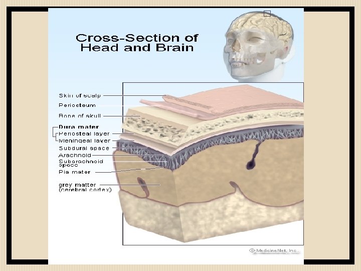

Classification • Head injuries can be classified broadly into: 1. Scalp injury. 2. Skull injury. 3. Brain injury.

Scalp anatomy • The scalp has 5 layers: 1. Skin. 2. Connective tissue. 3. Aponeurosis. 4. Loose areolar connective tissue. 5. Pericranium.

Scalp injury • It can manifest as: 1. Abrasion. 2. Bruising. 3. Laceration. 4. Subgaleal hematoma (subaponeuritic hematoma). 5. Subperiosteal hematoma (cephalhematoma).

Subgaleal hematoma • Subgaleal hemorrhage or hematoma is bleeding into the potential space between the skull periosteum and the scalp galea aponeurosis. • Majority (90%) result from vacuum applied to the head at delivery (Ventouse assisted delivery). • Results from bleeding from the emissary veins into the loose connective tissue layer.

Subgaleal hematoma • It has a high frequency of occurrence with associated head trauma (40%), such as intracranial hemorrhage or skull fracture.

Subgaleal hematoma • The diagnosis is generally clinical with a fluctuant boggy mass developing over the scalp (especially over the occiput) with superficial skin bruising. • The swelling develops gradually 12– 72 hours after delivery, although it may be noted immediately after delivery in severe cases.

Subgaleal hematoma • Patients with subgaleal hematoma may present with hemorrhagic shock. • The swelling may obscure the fontanel and cross suture lines (distinguishing it from cephalohematoma).

Subgaleal hematoma

Subgaleal hematoma

Subperiosteal hematoma • Hemorrhage of blood between the skull and the periosteum secondary to rupture of a blood vessel crossing the periosteum. • Less extensive than subgaleal hematoma. • In 80% of cases treatement is conservative.

Subperiosteal hematoma • Its firmer than the subgaleal hematoma. • Scalp moves freely over the mass. • Mostly doesn’t cross the midline. • Observe, give good analgesia and avoid aspiration in newborns.

Subperiosteal hematoma

Subperiosteal hematoma

Comparison

Skull Injury

Skull fractures • A skull fracture is a break in one or more of the eight bones that form the cranial portion of the skull, usually occurring as a result of blunt force trauma. • If the force of the impact is excessive, the bone may fracture at or near the site of the impact and cause damage to the underlying physical structures contained within the skull such as the membranes, blood vessels, and brain, even in the absence of a fracture.

Skull fractures • There are two major types of skull fractures: 1. Linear fractures which are the most common. 2. Depressed fractures.
. • Usually straight and")
Linear skull fractures • Most common of skull fractures (90%). • Usually straight and involve no displacement of the bone. • The common method of injury is blunt force trauma. • Are usually of little clinical significance unless they parallel in close proximity or transverse a suture, or they involve a venous sinus groove or vascular channel. • Usually require no intervention.

Linear skull fractures

Linear skull fractures • According to their site, linear skull fractures may be called: 1. Basilar skull fractures. 2. Diastatic skull fractures.

THANK YOU Hope you enjoy it !

HEAD INJURIES AHMAD AL ABBADI

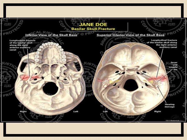
Basilar fractures • Are basically linear fractures that occur in the skull base. • May occur separately or as an extension of fractures of the vault. • They require more force to cause than other areas of the neurocranium; thus they are rare (4%).

Basilar fractures • Usual locations are: § Petrous part of temporal bone. § Orbital surface of frontal bone. § Basioocciput

Basilar fractures • They have characteristic signs: § hemotympanum § CSF leaking from the nose (rhinorrhea) or ears (otorrhea). § Periorbital ecchymosis often called raccoon eyes. § Retroauricular ecchymosis known as Battle's sign (bruising over the mastoid process).

Basilar fractures • § § - Treatment: Conservative : Admit the patient. stabilize airway, ventilation, and circulatory issues is the priority. cervical spine immobilization Neurological assessment every 2 hrs. Antibiotics. Surgery: if there’s Traumatic aneurysms. Posttraumatic carotid cavernous fistula. Meningitis or cerebral abscess. Facial palsy. Cosmetic deformities.

Battle’s sign

Racoon eyes

Diastatic fractures • Occur when the fracture line transverses one or more sutures of the skull causing a widening of the suture. • While this type of fracture is usually seen in infants and young children as the sutures are not yet fused it can also occur in adults. • When a diastatic fracture occurs in adults it usually affects the lambdoidal suture as this suture does not fully fuse in adults until about the age of 60 years.

Diastatic fractures

Depressed fractures • Depression beneath the vault. • Can be open or closed. • 2 types: 1. One piece fracture. 2. Comminuted fracture (multiple pieces).

Depressed fractures • Treatment could be: 1. Conservative. 2. Elevation. 3. Craniectomy with immediate/delayed cranioplasty.
: 1. Fracture is")
Depressed fractures • Criteria to elevate a depressed skull (surgical management): 1. Fracture is more than the thickness of the skull. 2. CSF leak. 3. Seizures. 4. Compound wounds. 5. Neurological signs. 6. Cosmetic. 7. Overlying an eloquent area of the brain.

Depressed fractures

Brain injury
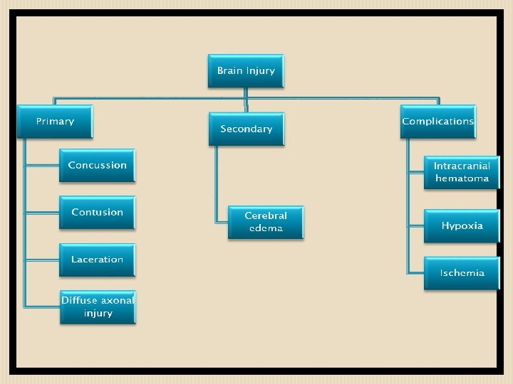

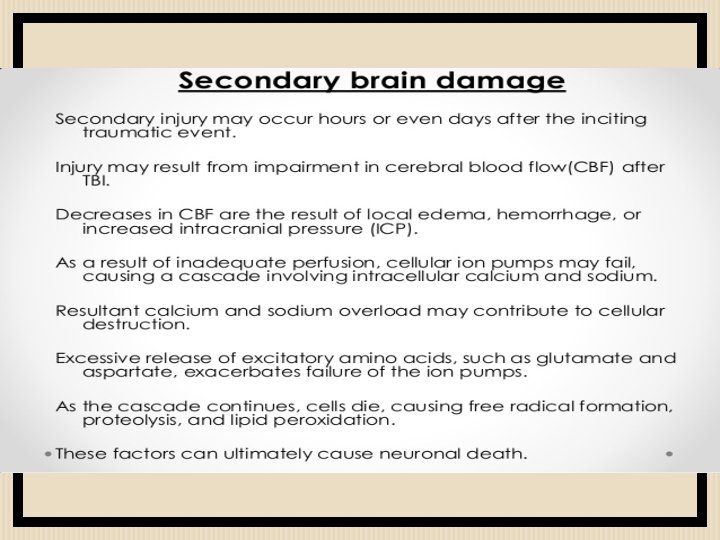
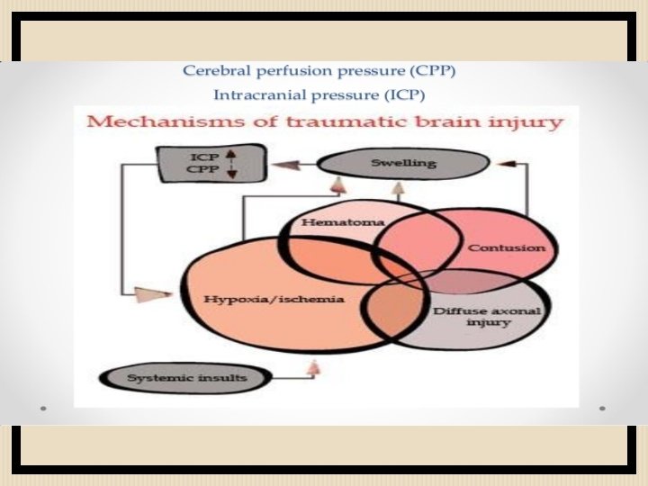
Primary brain injury • Primary brain injury occurs at the time of trauma. • It results from external mechanical forces transferred to intracranial contents. • The damage that results includes a combination of focal contusions and hematomas, as well as shearing of white matter tracts (diffuse axonal injury) along with cerebral edema and swelling.

Primary brain injury • Common mechanisms include: 1. Direct trauma. 2. Acceleration/ deceleration injuries. 3. Shearing forces.

Direct trauma • Can be classified into: 1. Closed Head Injury: no penetration of the skull (blunt). 2. Open head Injury: bullet, knife, or fracture (penetrating). • Here we have direct injury to the brain under the site of the impact.

Acceleration/ deceleration injuries • The skull has a minimal space for the brain to move within. • Here the brain is thrust against the skull opposite to the blow site due to the sudden movement of the brain inside the skull. • Also called coup-contrecoup injuries. • In old age patients, if the brain is atrophied, the insult is more severe, because of more space for movement.

coup and contrecoup injury • Damage may occur directly under the site of impact, or it may occur on the side opposite the impact (coup and contrecoup injury, respectively). When a moving object impacts the stationary head, coup injuries are typical, while contrecoup injuries are usually produced when the moving head strikes a stationary object

coup and contrecoup injury

subdural epidural

Thank you
- Slides: 53