Head Injury Done By Mohammad Badwan Abdelrahman Bdeir

Head Injury Done By: Mohammad Badwan Abdelrahman Bdeir

Definition • A head injury, as defined by the National Advisory Neurological Diseases and Stroke Council, is a morbid state resulting from gross or subtle structural changes in the scalp, skull, and/or the contents of the skull, which is produced by mechanical forces. • These injuries can range from a minor bump on the skull to serious brain injury.

Epidemiology • Third most common cause of death in the US. • Third most common cause of death in Jordan. • Number One Killer in Trauma. • More than 50% of all trauma deaths. • 50% of all deaths from motor vehicle accidents. • 200, 000 people in the world live with the disability caused by these injuries.

• Traumatic brain injuries are more common in young patients, and men account for the majority (75%) of cases.

Etiology § 32% of head injuries are caused by falls , more common among the elderly people (>65 years) and in the very young (0 -4 years). § 17% by motor vehicle accidents, more common among adolescents and young adults (15 -24) years. § 16. 5% by being struck or against something. § 10% by assaults. § 21% by other ways.

Classification • Head injuries can be classified broadly into: 1. Scalp injury. 2. Skull injury. 3. Brain injury.

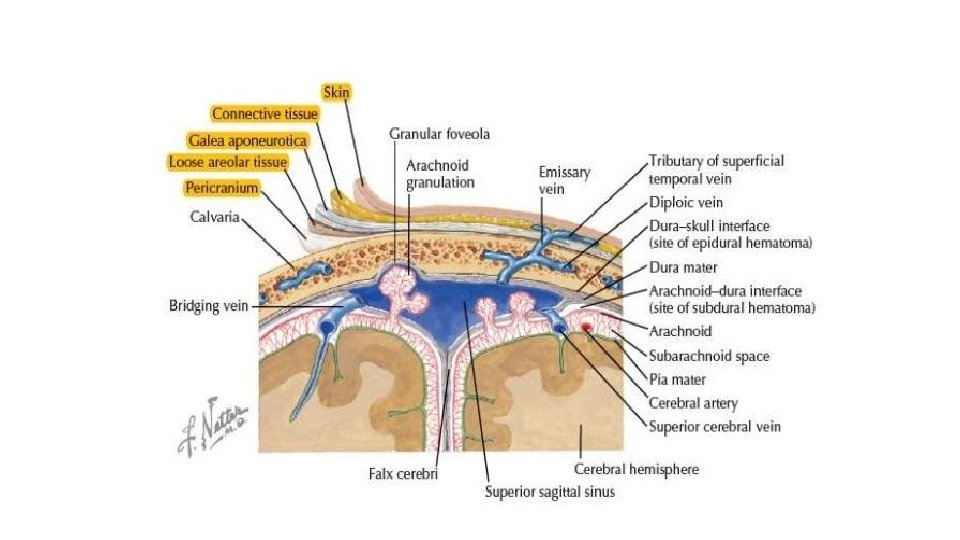
Scalp anatomy • The scalp has 5 layers: 1. Skin. 2. Connective tissue. 3. Aponeurosis. 4. Loose areolar connective tissue. 5. Pericranium.

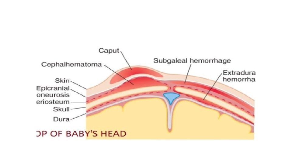
Scalp injury • It can manifest as: 1. 2. 3. 4. 5. Abrasion. Bruising. Laceration. Subgaleal hematoma (subaponeuritic hematoma). Subperiosteal hematoma (cephalhematoma).
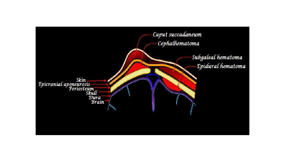

Subgaleal hematoma • Subgaleal hemorrhage or hematoma is bleeding into the potential space between the skull periosteum and the scalp galea aponeurosis. • Majority (90%) result from vacuum applied to the head at delivery (Ventouse assisted delivery). • Results from bleeding from the emissary veins into the loose connective tissue layer.

Subgaleal hematoma • It has a high frequency of occurrence with associated head trauma (40%), such as intracranial hemorrhage or skull fracture. • The occurrence of these features does not correlate significantly with the severity of subgaleal hemorrhage.

Subgaleal hematoma • The diagnosis is generally clinical with a fluctuant boggy mass developing over the scalp (especially over the occiput) with superficial skin bruising. • The swelling develops gradually 12– 72 hours after delivery, although it may be noted immediately after delivery in severe cases.

Subgaleal hematoma • Patients with subgaleal hematoma may present with hemorrhagic shock. • The swelling may obscure the fontanel and cross suture lines (distinguishing it from cephalohematoma).

Subgaleal hematoma

Subgaleal hematoma

Subperiosteal hematoma • Hemorrhage of blood between the skull and the periosteum secondary to rupture of a blood vessel crossing the periosteum. • Less extensive than subgaleal hematoma. • In 80% of cases treatement is conservative.

Subperiosteal hematoma • Its firmer than the subgaleal hematoma. • Scalp moves freely over the mass. • Mostly doesn’t cross the midline.

Subperiosteal hematoma

Comparison

Skull Injury

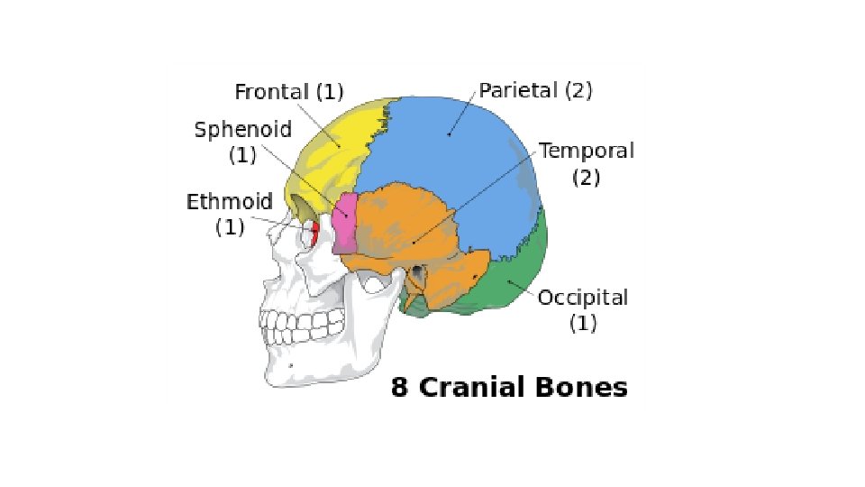
Skull fractures • A skull fracture is a break in one or more of the eight bones that form the cranial portion of the skull, usually occurring as a result of blunt force trauma. • If the force of the impact is excessive, the bone may fracture at or near the site of the impact and cause damage to the underlying physical structures contained within the skull such as the membranes, blood vessels, and brain, even in the absence of a fracture.

Skull fractures • There are two major types of skull fractures: 1. Linear fractures which are the most common. 2. Depressed fractures.
. • Usually straight and")
Linear skull fractures • Most common of skull fractures (90%). • Usually straight and involve no displacement of the bone. • The common method of injury is blunt force trauma. • Are usually of little clinical significance unless they parallel in close proximity or transverse a suture, or they involve a venous sinus groove or vascular channel. • Usually require no intervention.

Linear skull fractures

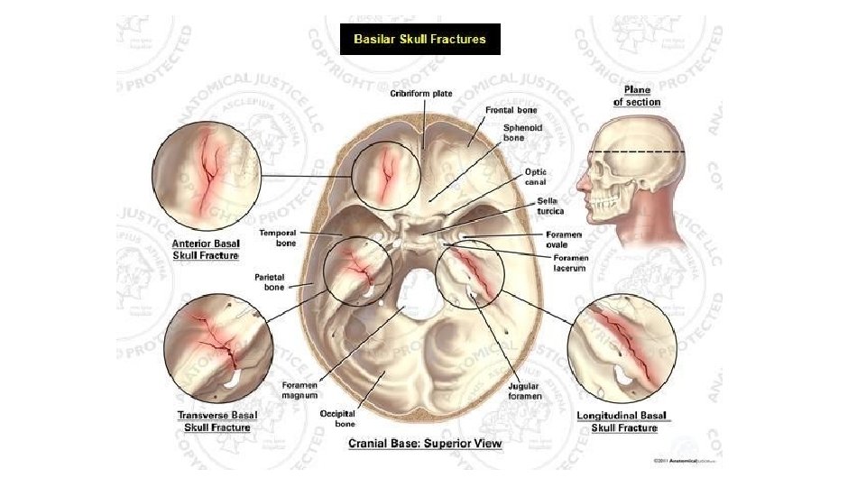
Linear skull fractures • According to their site, linear skull fractures may be called: 1. Basilar skull fractures. 2. Diastatic skull fractures.

THE END

SKULL FRACTURES • LINEAR break in the continuity of bone without alteration of relationship of parts cause- Low velocity injuries • DEPRESSED Inward indentation of skull cause- powerful blow • Comminuted multiple linear fractures with fragmentation of bones into pieces • Compound Depressed skull fractures and scalp laceration communicating intracranial cavity

ACCORDING TO LOCATION • Frontal fracture • Temporal fracture : Boggy temporal muscle because extravasation of blood , battle sign & Otorrhoea • Parietal fracture : Deafness, otorrhoea , Bulging of tympanic membrane by blood or CSF & Facial paralysis • Posterior fossa fracture • Orbital fracture : Periorbital ecchymosis(RACCOON EYES) & Optic nerve injury • Basilar skull fracture

Basilar fractures • Are basically linear fractures that occur in the skull base. • They require more force to cause than other areas of the neurocranium; thus they are rare (4%). Usual locations are: §Petrous part of temporal bone. §Orbital surface of frontal bone. §Basioocciput
")
Basilar skull fracture They have characteristic signs: • CSF leaking from the nose (rhinorrhea) or ears (otorrhea). • Periorbital ecchymosis often called raccoon eyes. • Retroauricular ecchymosis known as Battle's sign (bruising over the mastoid process). • Bulging of tympanic membrane • Facial paralysis • Tinnittis , vertigo

Raccoon eyes Battel sign

Treatment • Conservative : -Admit the patient. - stabilize airway, ventilation, and circulatory issues is the priority. -cervical spine immobilization -Neurological assessment every 2 hrs. -Antibiotics. • Surgery: if there’s -Traumatic aneurysms. -Posttraumatic carotid cavernous fistula. -Meningitis or cerebral abscess. -Facial palsy. -Cosmetic deformities.

Diastatic fracture • Occur when the fracture line transverses one ormore sutures of the skull causing a widening of the suture. • While this type of fracture is usually seen in infants and young children as the sutures are not yet fused it can also occur in adults. • When a diastatic fracture occurs in adults it usually affects the lambdoidal suture as this suture does not fully fuse in adults until about the age of 60 years.

Depressed skull fracture • Depression beneath the vault. • Can be open or closed. • 2 types: 1. One piece fracture. 2. Comminuted fracture (multiple pieces).

Treatment 1. Conservative. 2. Elevation. 3. Craniectomy with immediate/delayed cranioplasty. Criteria to elevate a depressed skull (surgical management): 1. Fracture is more than the thickness of the skull. 2. CSF leak. 3. Seizures. 4. Compound wounds. 5. Neurological signs. 6. Cosmetic. 7. Overlying an eloquent area of the brain.

Test to determine CSF leakage Method 1 : • Check for presence of glucose • Dextrostrip/ Tes-Tape strip • If blood is present in the fluid • The test become unreliable • Go for 2 nd method Method 2( halo ring sign) : • Allow leaking fluid drip onto a white pad/towel • Observes the drainage • Within a few minutes the blood coalesces into center and a yellowish ring encircles the blood

Brain injury • CONCUSSION : A sudden transient mechanical head injury with disruption of neuronal activity and a change in the LOC , It occurs When the brain suddenly shifts inside the skull and knocks against the skulls bony surface • CONTUSION : It is the bruising of the brain tissue within a focal area , It is usually associated with a closed head injury • LACERATIONS It involve actual tearing of brain tissue and often occur in association with depressed , open fractures and penetrating injuries • Secondary brain injury occur after hours to days after trauma impairment in CBF edema increase ICB.

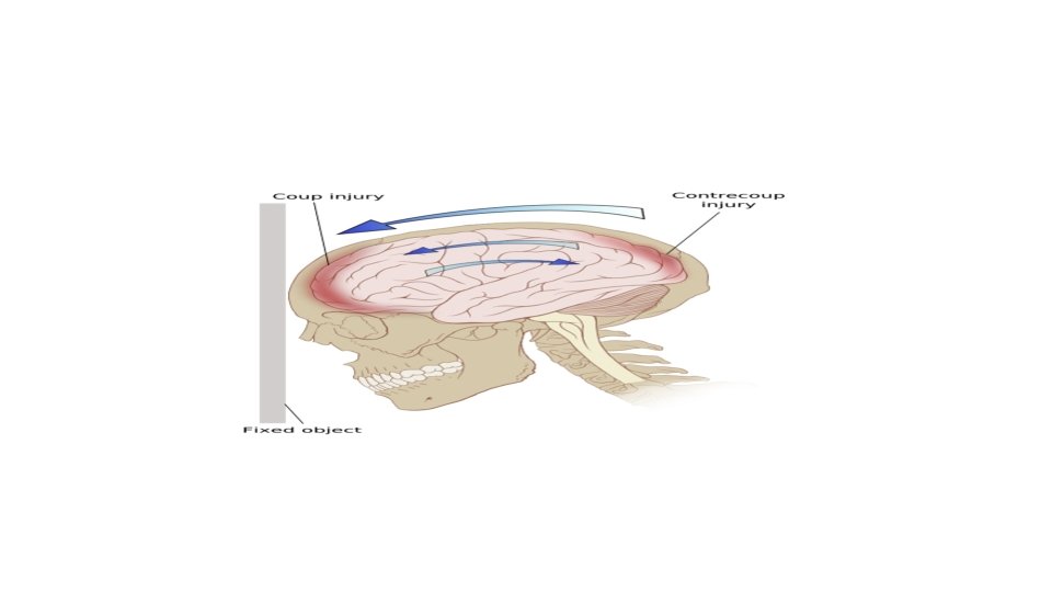
Acceleration deceleration injures Here the brain is thrust against the skull opposite to the blow site due to the sudden movement of the brain inside the skull. Also called coup-contrecoup injuries, Damage may occur directly under the site of impact, or it may occur on the side opposite the impact (coup and contrecoup injury, respectively). When a moving object impacts the stationary head, coup injuries are typical, while contrecoup injuries are usually produced when the moving head strikes a stationary object In old age patients, if the brain is atrophied, the insult is more severe, because of more space for movement.
 EPIDURAL HEMORRHAGE • A neurologic emergency • Most")
COMPLICATIONS • INTRACRANIAL HAEMORRHAGES : a) EPIDURAL HEMORRHAGE • A neurologic emergency • Most common type of intracranial hemorrhage • bleeding between the dura and the inner surface of the skull • Blow to the temporal, parietal bone • Commonly bleeding by arterial origin breakage to middle meningeal artery • On head CT the clot is bright, biconvex shaped clot and has a welldefined border that usually respects cranial suture lines
 SUB-DURAL HEMATOMA • Subdural hematoma occurs from bleeding between the dura matter and")
b) SUB-DURAL HEMATOMA • Subdural hematoma occurs from bleeding between the dura matter and the arachnoid layer of the meninges • Types 1. acute 2. subacute 3. Chronic • SDH usually results from venous bleeding, usually from tearing of a bridging vein running from the cerebral cortex to the dural sinuses. • Hematoma may be slower to develop • On head CT scan, the clot is bright or mixed-density, crescent-shaped (lunate), may have a less distinct border
 SUB ARACHNOID HEMORRHAGE • Bleeding occurs between the arachnoid and pia mater CAUSES")
c) SUB ARACHNOID HEMORRHAGE • Bleeding occurs between the arachnoid and pia mater CAUSES • Rupture of Berry aneurism • Trauma (fracture at the base of the skull leading to internal carotid aneurysm) • CT scan Increased attenuation is seen in the CSF Spaces over the cerebral hemisphere
 Intracerebral Hemorrhage (ICH) • Intraaxial hemorrhage is hemorrhage that occurs within the brain")
d) Intracerebral Hemorrhage (ICH) • Intraaxial hemorrhage is hemorrhage that occurs within the brain tissue itself Two main types: 1) Intraparencymal hemorrahge- ICH extending into brain parenchyma; 2) Intra-ventricular hemorrhage- ICH extending into ventricles; • CAUSES: Hypertensive vasculopathy (70 -80%) Ruptured Aneurism Trauma- 16%

Taking a history in head injury • Mechanism of injury • Loss of consciousness or amnesia • Level of consciousness at scene and on transfer • Evidence of seizures • History of vomiting • Pre-existing medical conditions • Medications (especially anticoagulants) • Illicit drugs and alcohol

Physical examination • Glasgow Coma Score • Pupil size and response • Signs of skull fracture Bilateral periorbital edema (raccoon eyes) Battle’s sign (bruising over mastoid) Cerebrospinal fluid rhinorrhoea or otorrhoea Haemotympanum or bleeding from ear • Full neurological examination: tone, power, sensation, reflexes

CT scan is considered the best diagnostic test to evaluate for craniocerebral trauma because it allows rapid diagnosis and intervention in the setting -NICE guidelines for (CT) in head injury : • Glasgow Coma Score (GCS) < 13 at any point • GCS 13 or 14 at 2 hours • Focal neurological deficit • Suspected open, depressed or basal skull fracture • Seizure • Vomiting > one episode -Urgent CT head scan if none of the above but: • Age > 65 • Coagulopathy (e. g. on warfarin) • Antegrade amnesia > 30 min (CT within 8 hours)

Management • The patient should be positioned with the head up 30 degree • It is important to ensure that the cervical immobilisation collar does not obstruct venous return from the head • Maintain Airway and ventilation • Maintain Circulation and cerebral perfusion pressure • Maintain Control of intracranial pressure -MEDICATIONS • Osmotic diuretics MANNITOL • Anticonvulsants PHENYTOIN where it may inhibit spread of seizure activity in motor cortex • Barbiturates PENTOBARBITAL It will reduce the brain metabolic rate and helps reduce ICP • Calcium Channel Blockers

Surgical management No surgical intervention if collection <10 ml Indication of surgical decompression: • The GCS score decreases by 2 or more points between the time of injury and hospital evaluation • The patient presents with fixed and dilated pupils • The intracranial pressure (ICP) exceeds 20 mm Hg Types: • Burr-holeopening into cranium with a drill • Craniotomy bone flap is temporarily removed from the skull to access the brain for evacuation • Craniectomy – Excision into the cranium to cut away a bone flap • Cranioplasty surgical repair of a defect or deformity of a skull

Done by : Abdelrahman bdeir Mohammad badwan
- Slides: 53