
Head CT Basics Trauma Micelle Haydel MD LSUNew
Head CT Basics : Trauma Micelle Haydel, MD LSU-New Orleans
First, a little test…
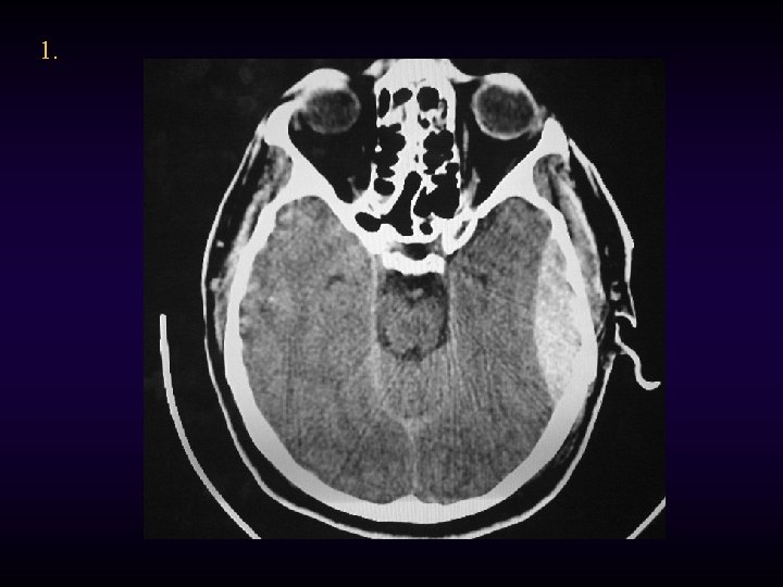
1.
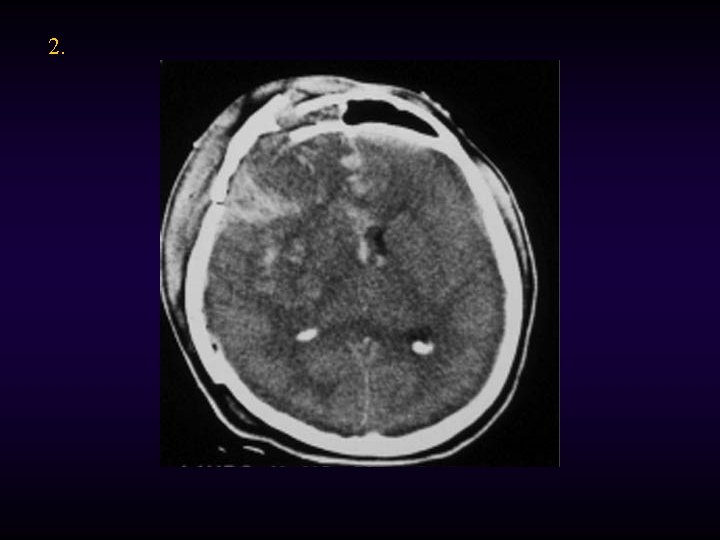
2.
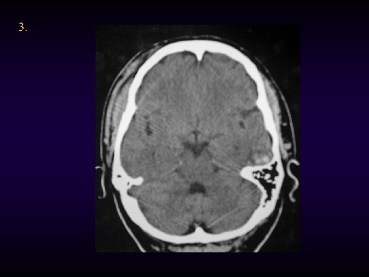
3.
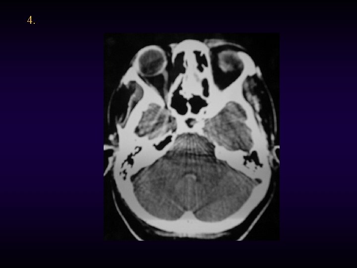
4.
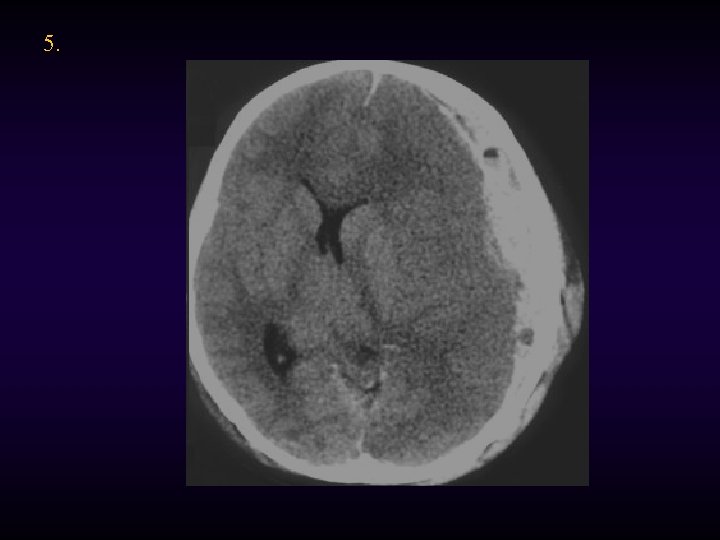
5.
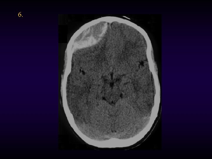
6.
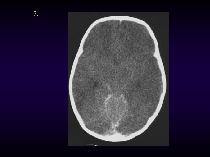
7.
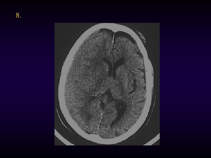
8.
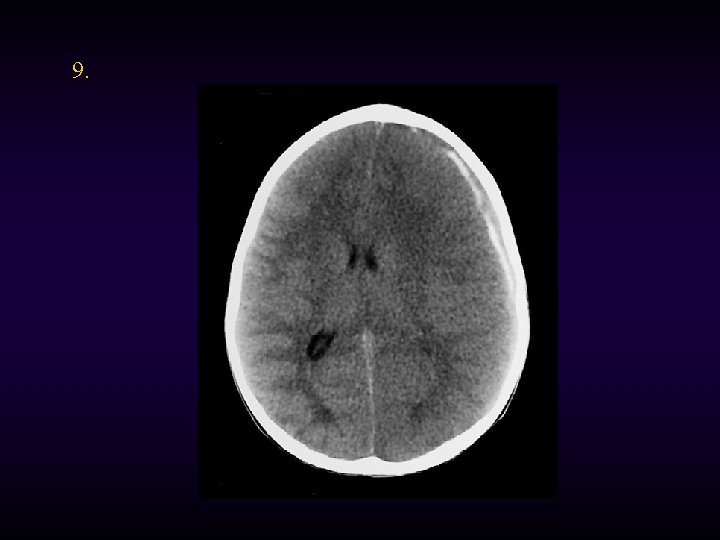
9.
Head CT Interpretation the basics
A Little History. . . • In the early 1970 s, Hounsfield developed a way of computerizing Xrays to select certain densities for viewing. – Plain films: black, white or a few shades of grey – CT: thousands of shades of grey based on the density of the tissue (Hounsfield Units)
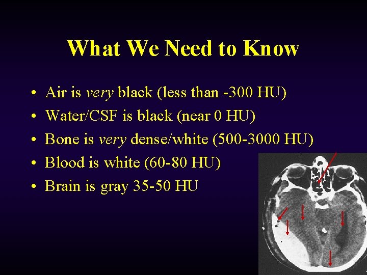
What We Need to Know • • • Air is very black (less than -300 HU) Water/CSF is black (near 0 HU) Bone is very dense/white (500 -3000 HU) Blood is white (60 -80 HU) Brain is gray 35 -50 HU
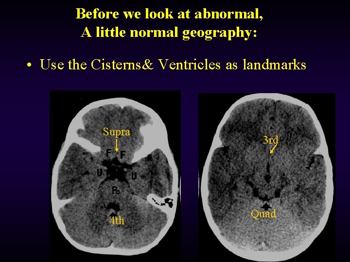
Before we look at abnormal, A little normal geography: • Use the Cisterns& Ventricles as landmarks Supra 4 th 3 rd Quad
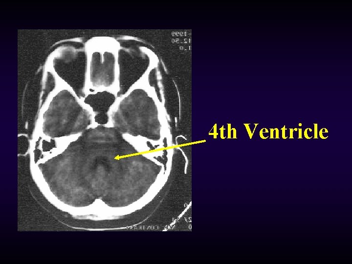
4 th Ventricle
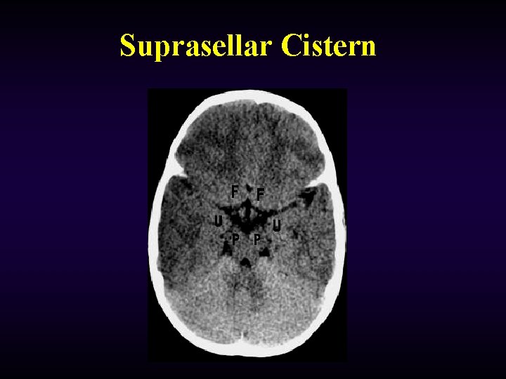
Suprasellar Cistern
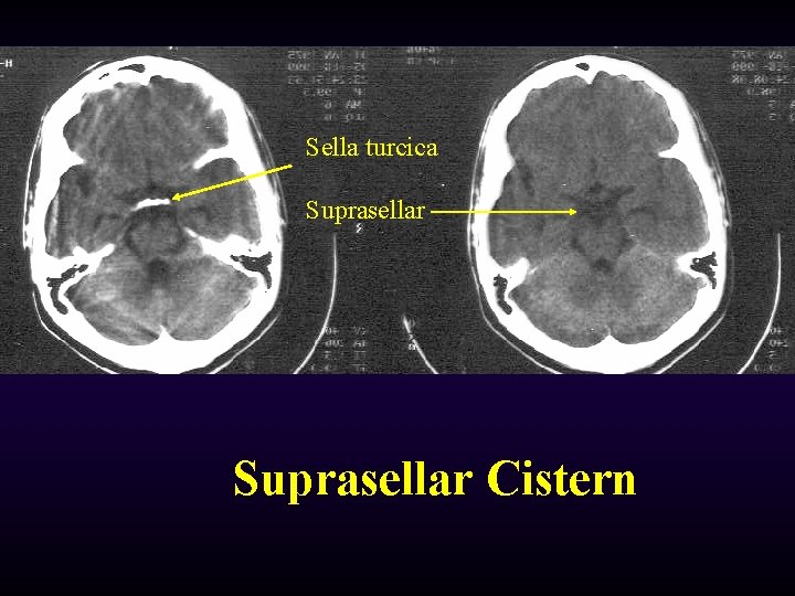
Sella turcica Suprasellar Cistern
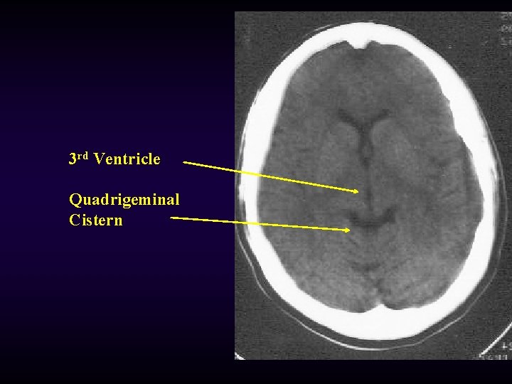
3 rd Ventricle Quadrigeminal Cistern
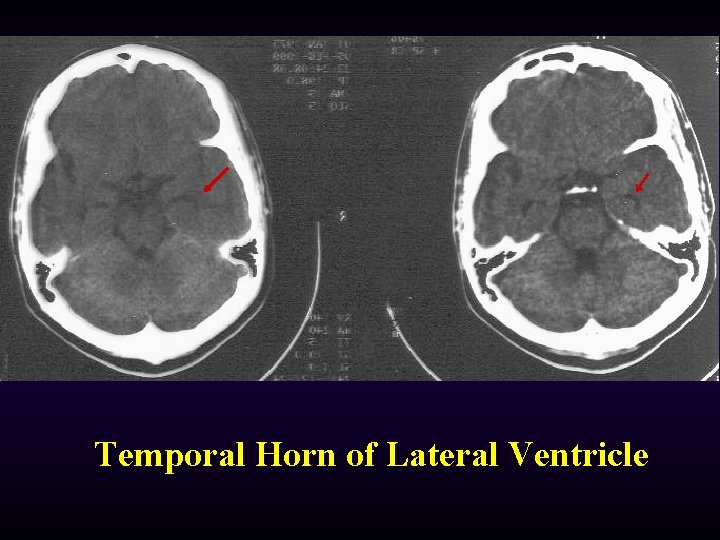
Temporal Horn of Lateral Ventricle
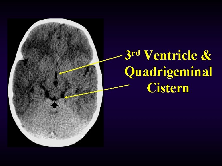
3 rd Ventricle & Quadrigeminal Cistern
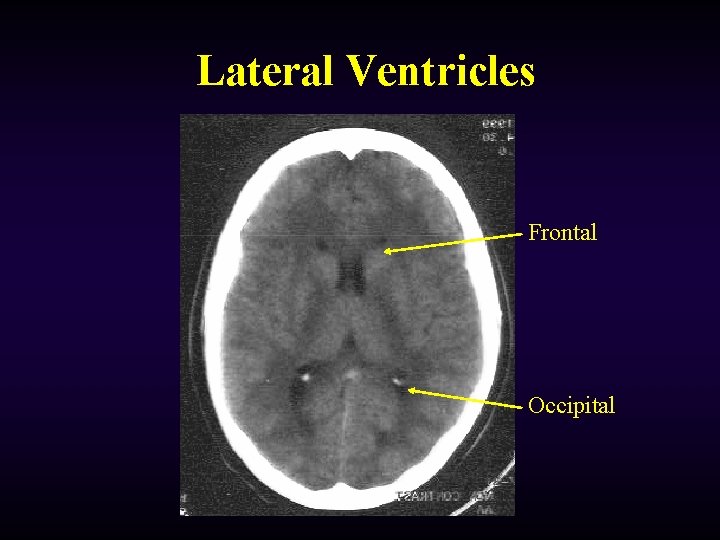
Lateral Ventricles Frontal Occipital
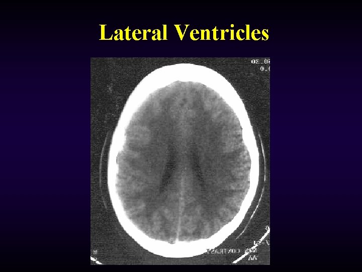
Lateral Ventricles
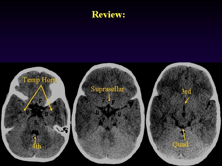
Review: Temp Horn Suprasellar 4 th 3 rd Quad
Normal Head
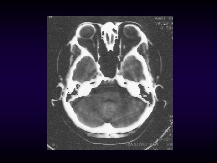
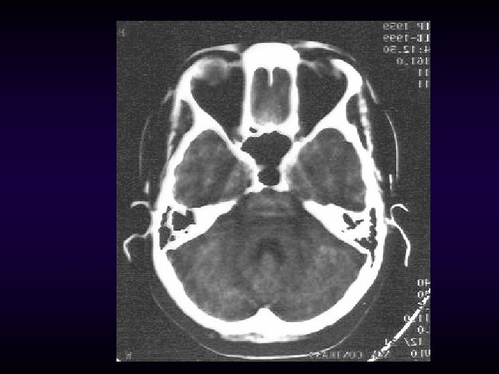
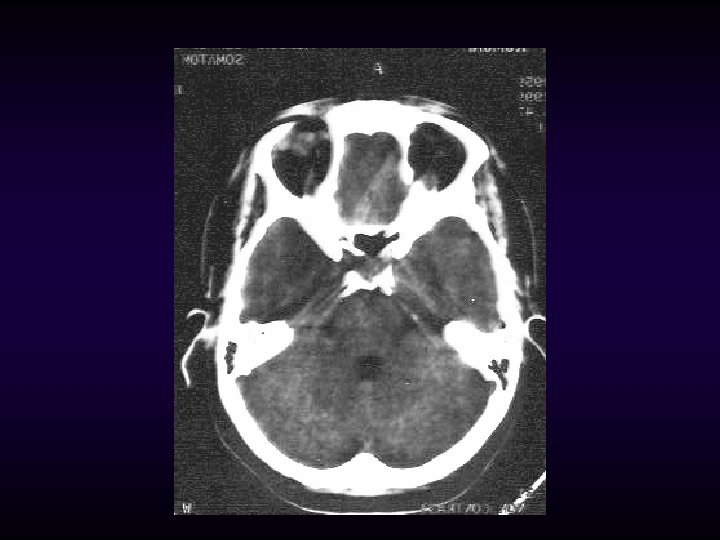
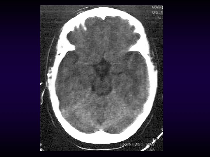
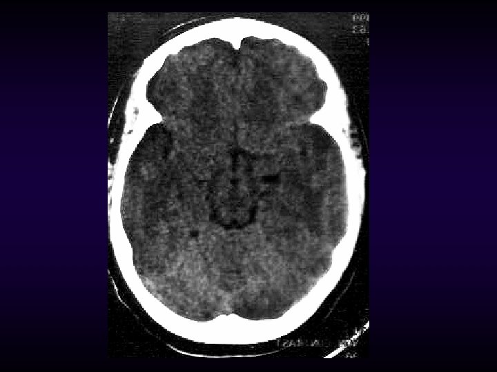
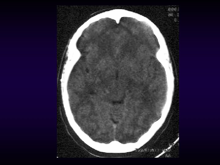
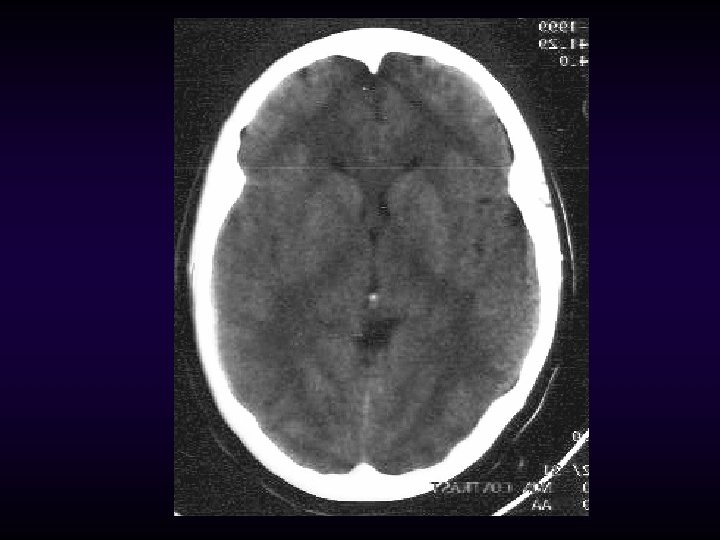
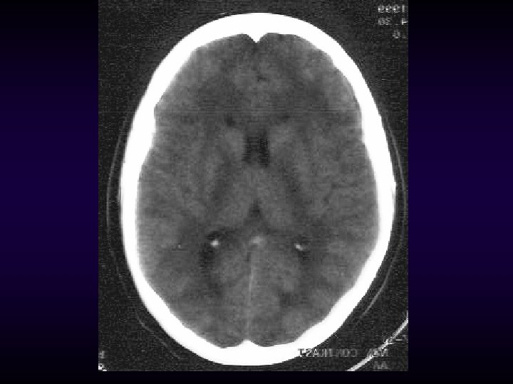
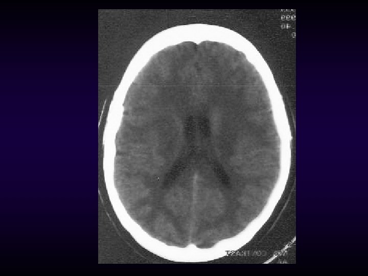
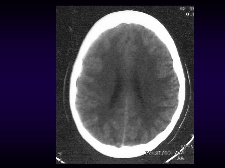
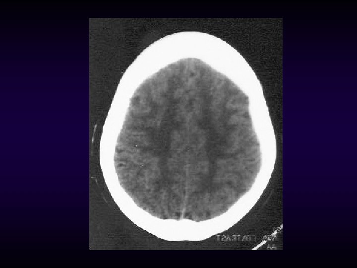
ABNORMAL CTs
On each brain slice look for: § Symmetry, symmetry • sulci • cisterns and ventricles • grey-white differentiation
Identify What Doesn’t Belong • Hyperdensities (whiter) extra-axial hematomas (SDH, EDH) • ICB or contusion • SAH in sulci, cisterns or ventricles • • Hypodensities (darker) pneumocephaly (air is darker than CSF) • infarction •
Identify What Doesn’t Belong • Localized or diffuse edema effacement of sulci or cisterns • distortion of gray-white matter interface • enlarged ventricles, temporal horn • • Fractures • soft tissue swelling • fluid (blood) in sinuses or mastoid air cells • in children, look for widened sutures • Always look at bone windows
Blood: Acute blood is white (60 -80 HU) on CT, due to the density of hemoglobin. As hgb breaks down, the HU decrease (i. e. subacute and isodense hematomas)
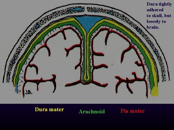
Dura tightly adhered to skull, but loosely to brain. Dura mater Arachnoid Pia mater
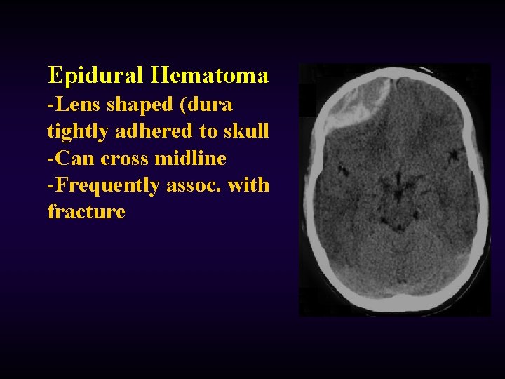
Epidural Hematoma -Lens shaped (dura tightly adhered to skull -Can cross midline -Frequently assoc. with fracture
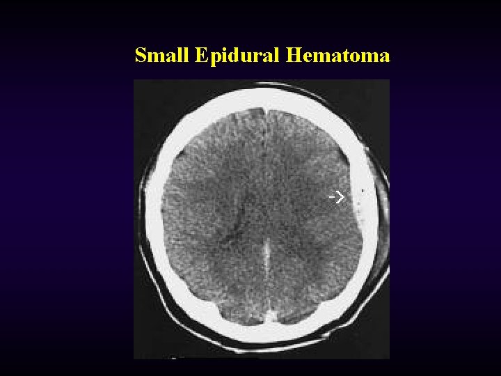
Small Epidural Hematoma
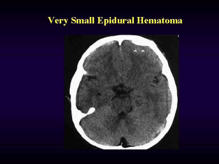
Very Small Epidural Hematoma
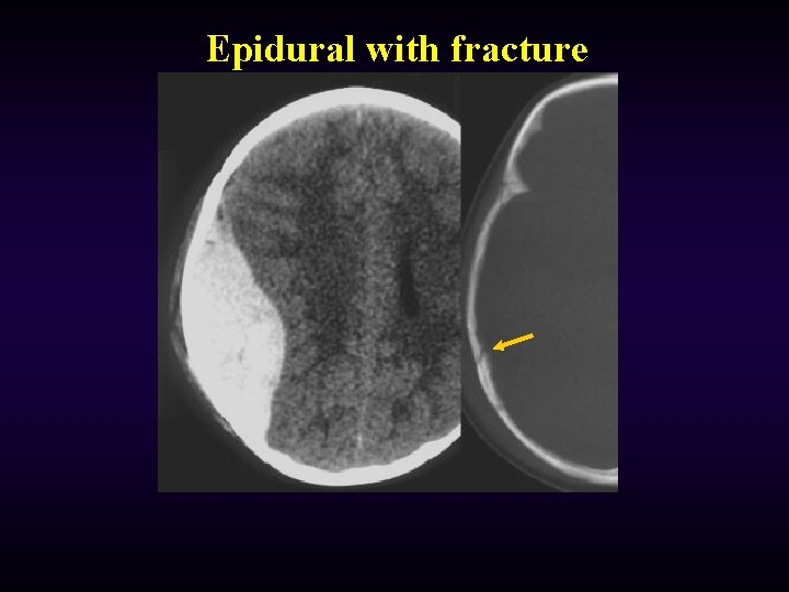
Epidural with fracture
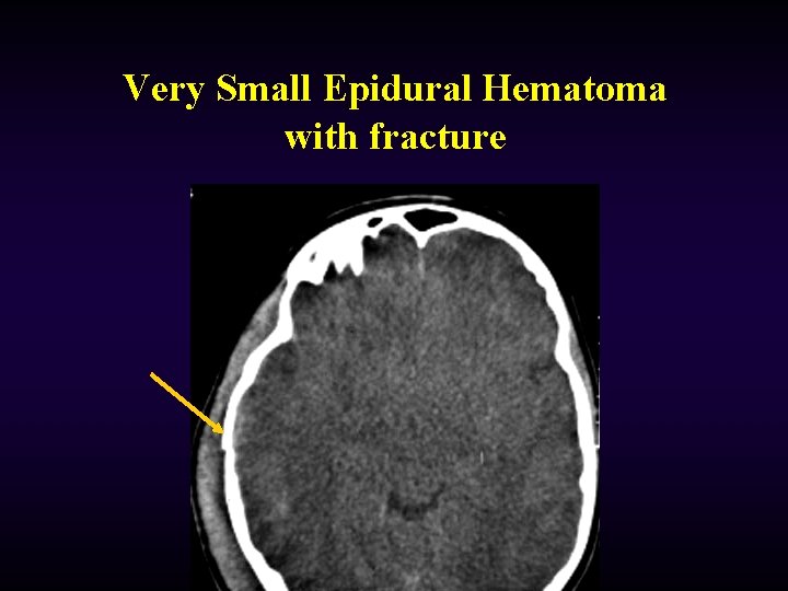
Very Small Epidural Hematoma with fracture
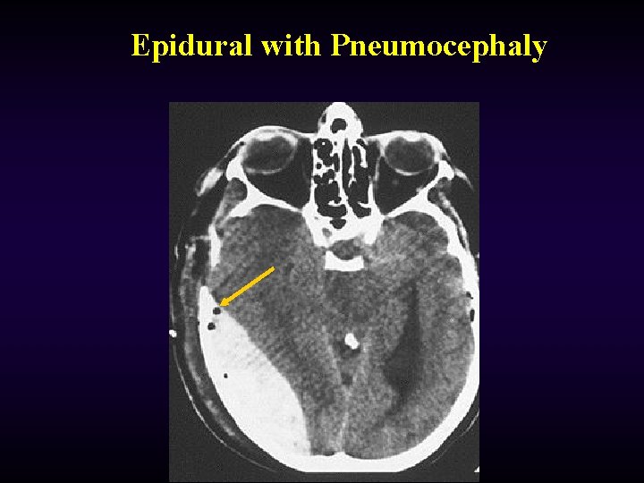
Epidural with Pneumocephaly
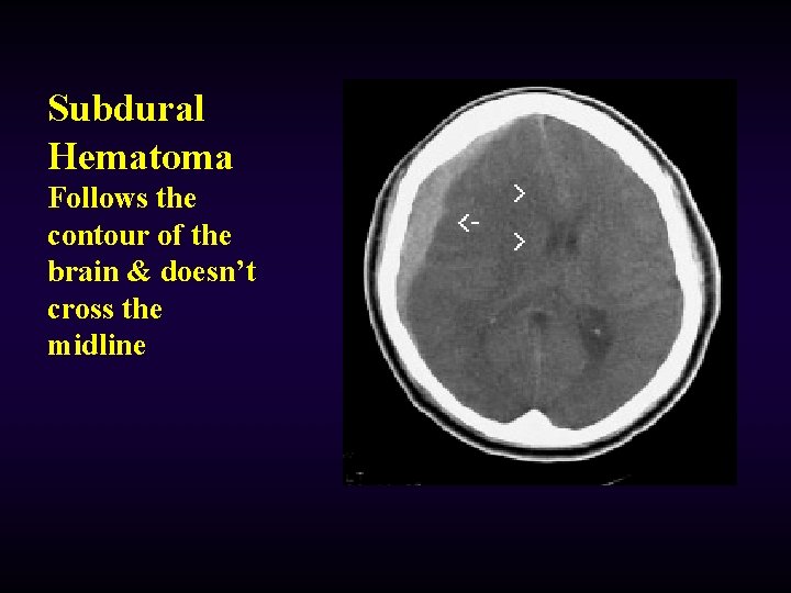
Subdural Hematoma Follows the contour of the brain & doesn’t cross the midline
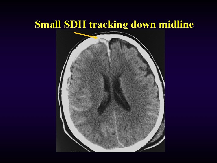
Small SDH tracking down midline
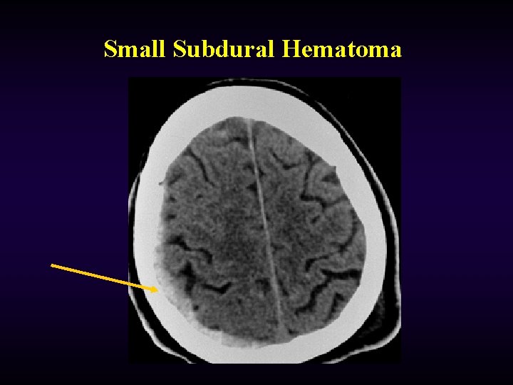
Small Subdural Hematoma
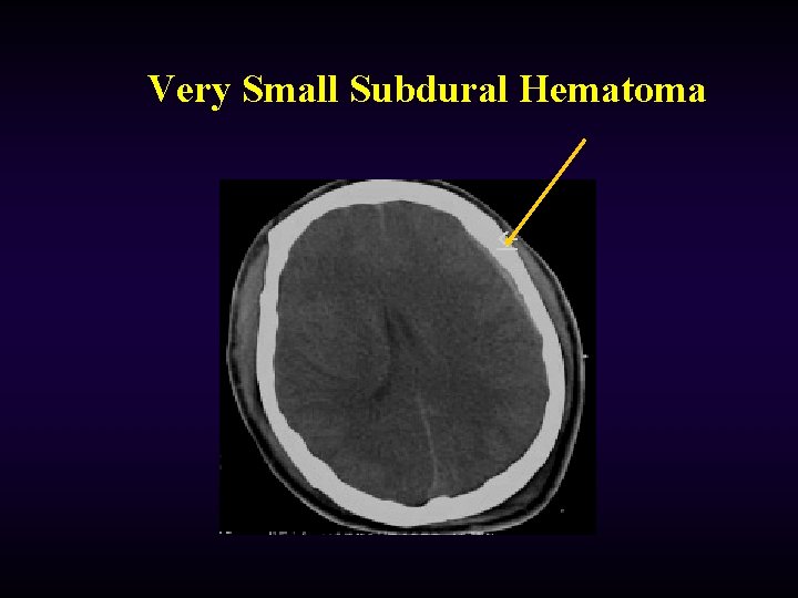
Very Small Subdural Hematoma
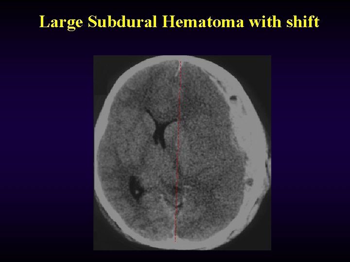
Large Subdural Hematoma with shift
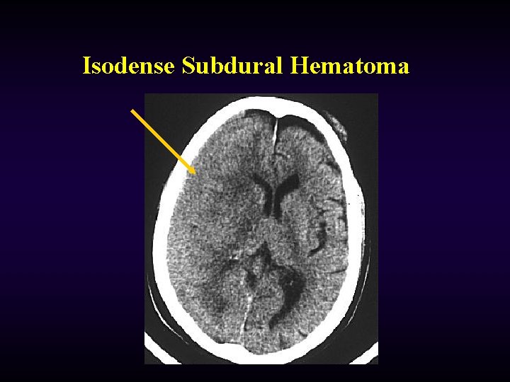
Isodense Subdural Hematoma
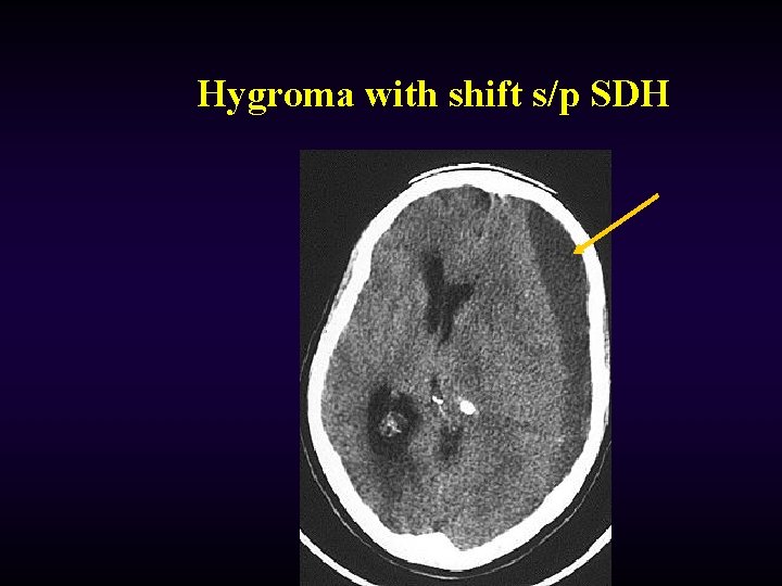
Hygroma with shift s/p SDH
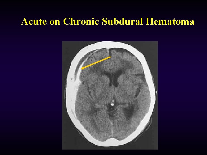
Acute on Chronic Subdural Hematoma
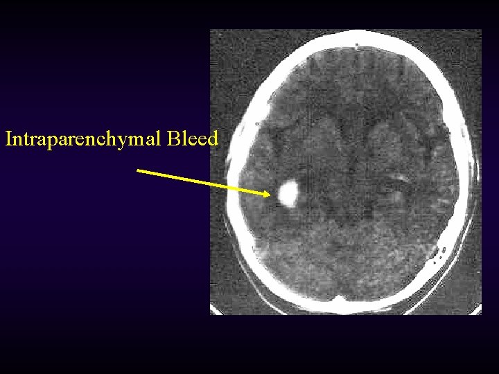
Intraparenchymal Bleed
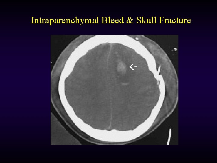
Intraparenchymal Bleed & Skull Fracture
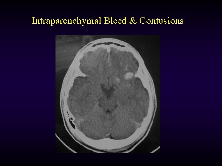
Intraparenchymal Bleed & Contusions
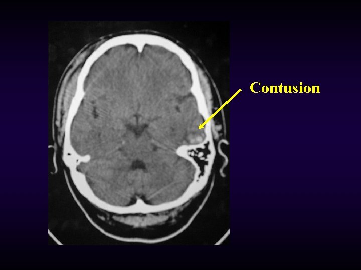
Contusion
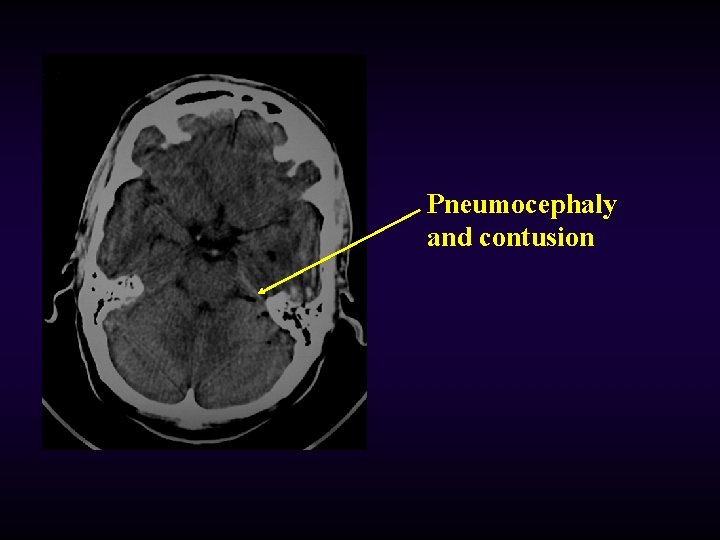
Pneumocephaly and contusion
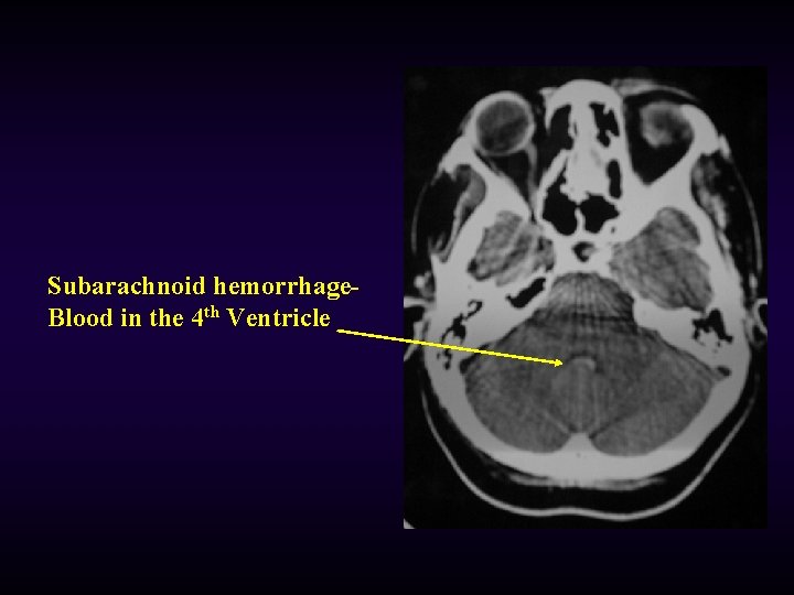
Subarachnoid hemorrhage. Blood in the 4 th Ventricle
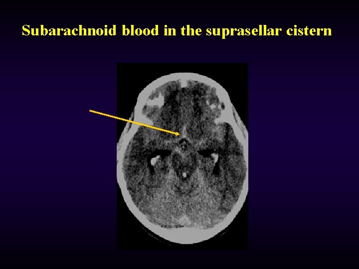
Subarachnoid blood in the suprasellar cistern
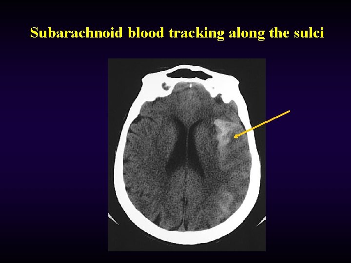
Subarachnoid blood tracking along the sulci
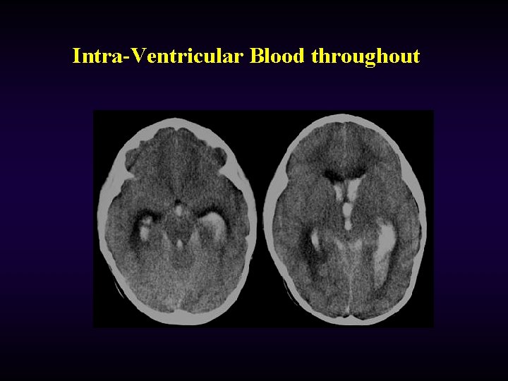
Intra-Ventricular Blood throughout
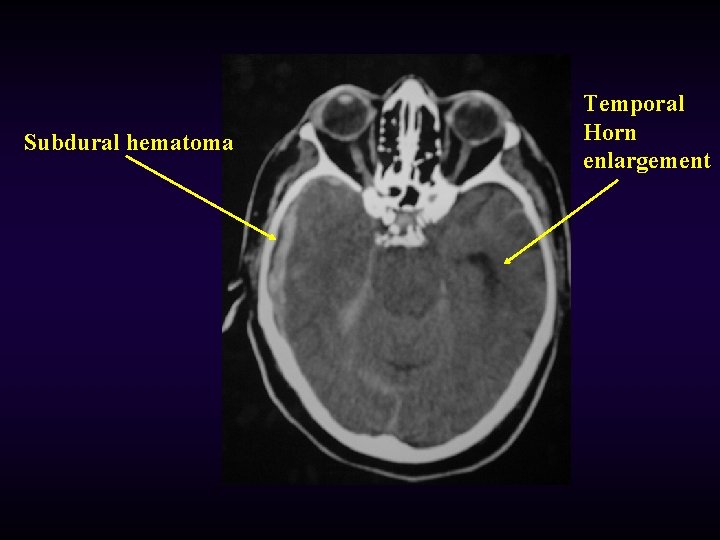
Subdural hematoma Temporal Horn enlargement
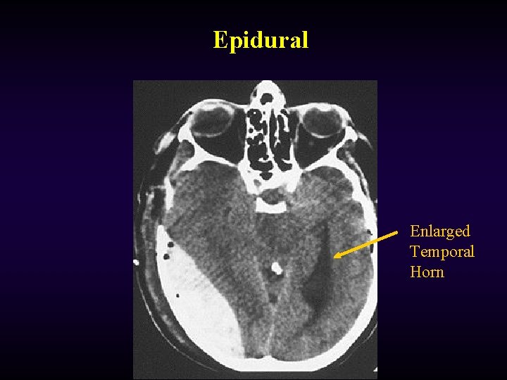
Epidural Enlarged Temporal Horn
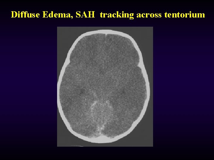
Diffuse Edema, SAH tracking across tentorium
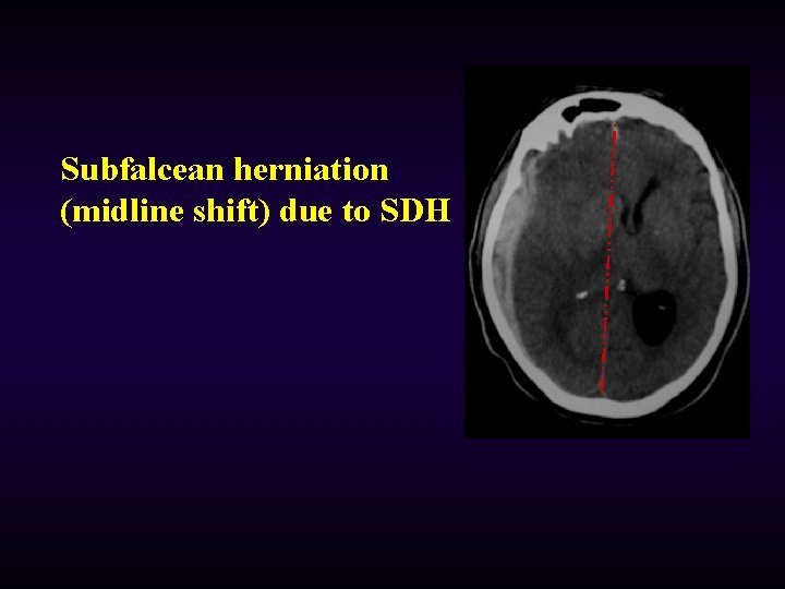
Subfalcean herniation (midline shift) due to SDH
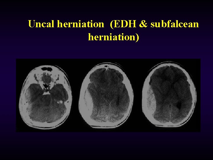
Uncal herniation (EDH & subfalcean herniation)
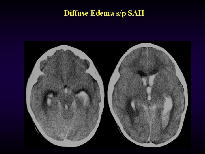
Diffuse Edema s/p SAH
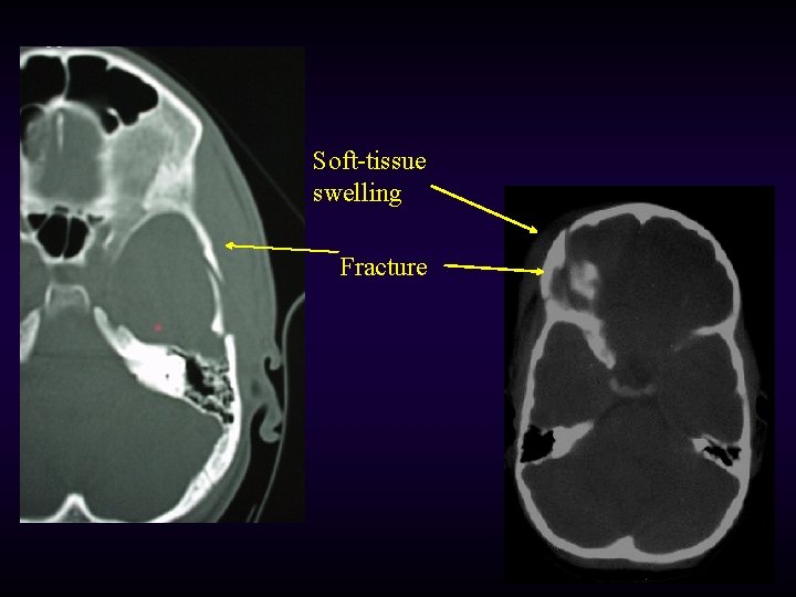
Soft-tissue swelling Fracture

…. the results
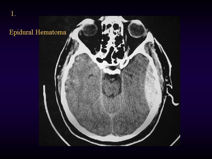
1. Epidural Hematoma
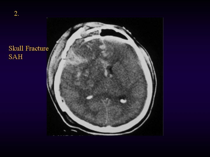
2. Skull Fracture SAH
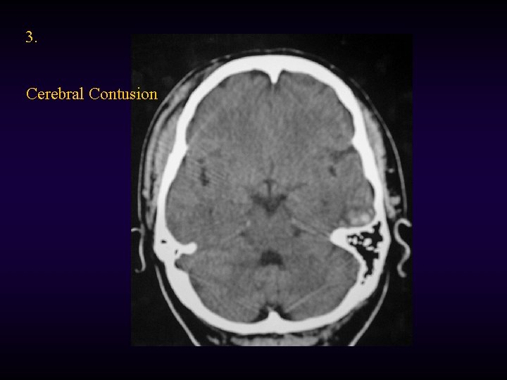
3. Cerebral Contusion
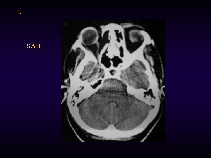
4. SAH
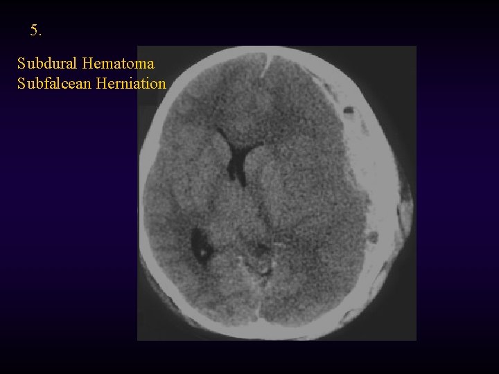
5. Subdural Hematoma Subfalcean Herniation
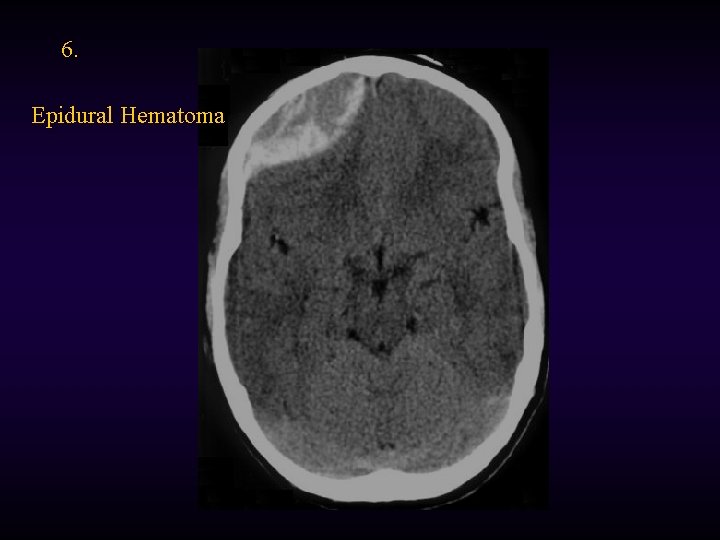
6. Epidural Hematoma
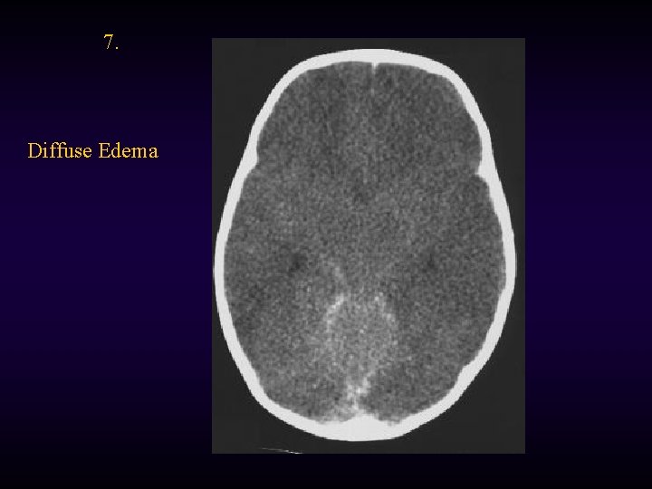
7. Diffuse Edema
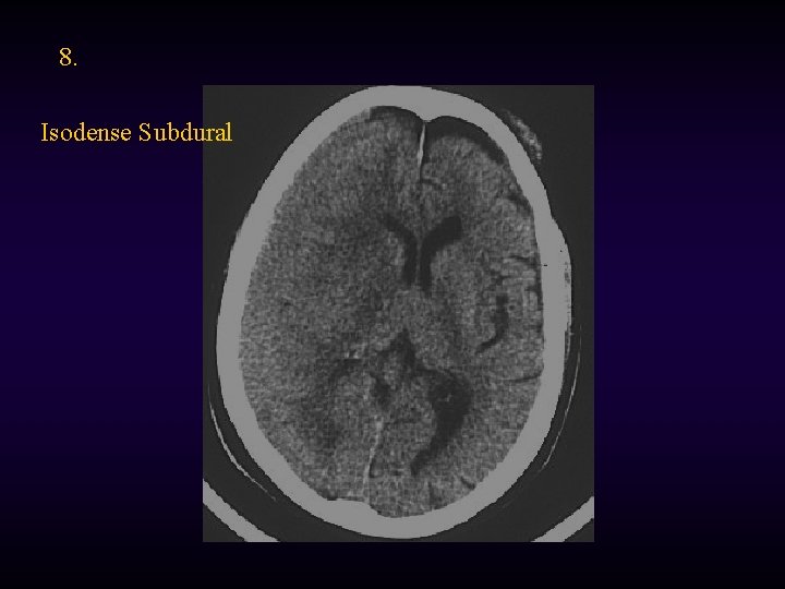
8. Isodense Subdural
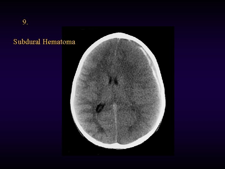
9. Subdural Hematoma

- Slides: 83