
Happy Year Awatif Jamal MD MSc FRCPC FIAC







- Slides: 46
Happy Year
Awatif Jamal, MD, MSc, FRCPC, FIAC Consultant & Associate Professor Department of Pathology King Abdulaziz University Hospital
Hemodynamic Disorders Thrombosis & Shock l Edema • Edema l Hyperemia and Congestion l Hemorrhage l Hemostasis & Thrombosis l Embolism l Infarction l Shock
INTRODUCTION The health of cells and tissues depend on; 1 -Intact circulation ; to deliver oxygen and remove wastes. 2 -Normal fluid homeostasis; which encompasses the following; A- maintenance of BV wall integrity. B- maintenance of intravascular pressure. C- maintenance of protein content or osmolarity within BV. D- maintenance of blood as a liquid until such time as injury necessitates clot formation.
EDEMA Fluid extravasations and accumulation in the interstitial spaces 60% of body weight is water, distributed as follow: Two thirds intracellular 5% intravascular The rest is interstitial
EDEMA Increased fluid in the interstitial tissue spaces Fluid may also accumulate in body cavities: 1. Hydrothorax 2. Hydropericardium 3. Hydroperitoneum is also called Ascites Massive generalized edema is called Anasarca
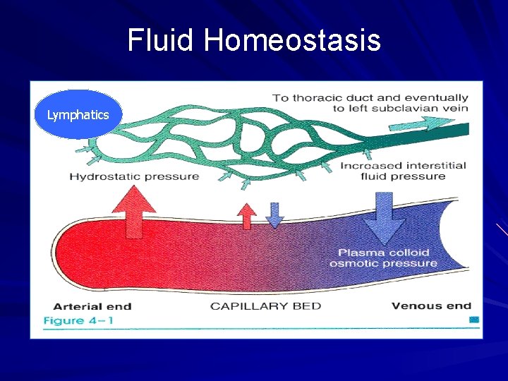
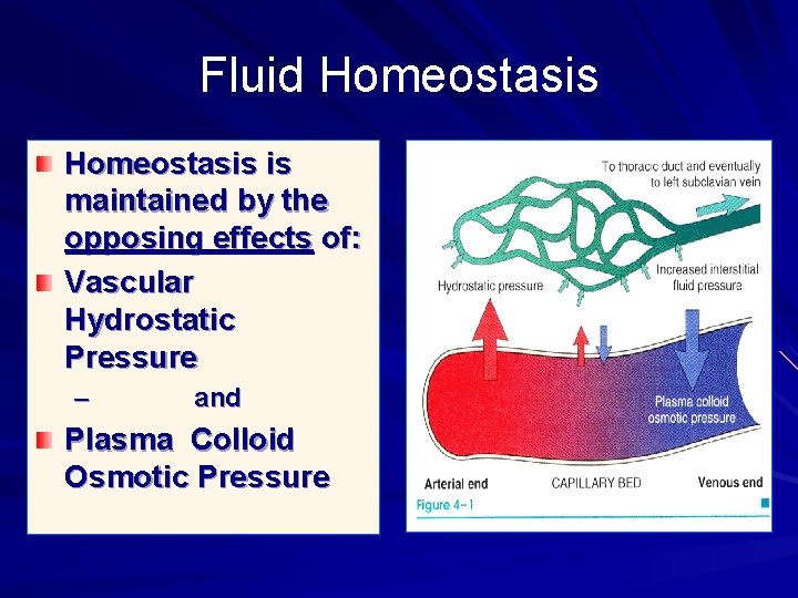
Pathogenesis The opposing effects of vascular hydrostatic pressure and plasma colloid osmotic pressure are the major factors that control the movement of fluid between vascular and interstitial tissues. Normally, the exit of fluid into the interstitium from the arteriolar end of microcirculation is nearly balanced by inflow of fluid at the venular end; a small residual amount of excess interstitial fluid is drained by the lymphatics
Fluid Homeostasis Lymphatics
Fluid Homeostasis is maintained by the opposing effects of: Vascular Hydrostatic Pressure – and Plasma Colloid Osmotic Pressure
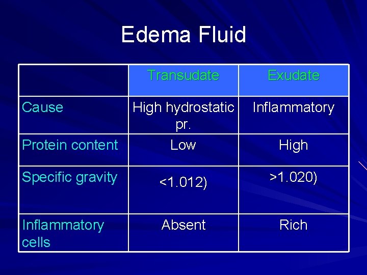
Edema Fluid = TRANSUDATE transudate is protein-poor (specific gravity <1. 012) An exudate is protein-rich (specific gravity >1. 020) = (inflammatory edema)
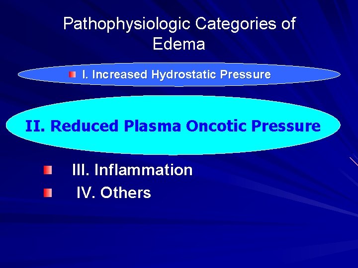
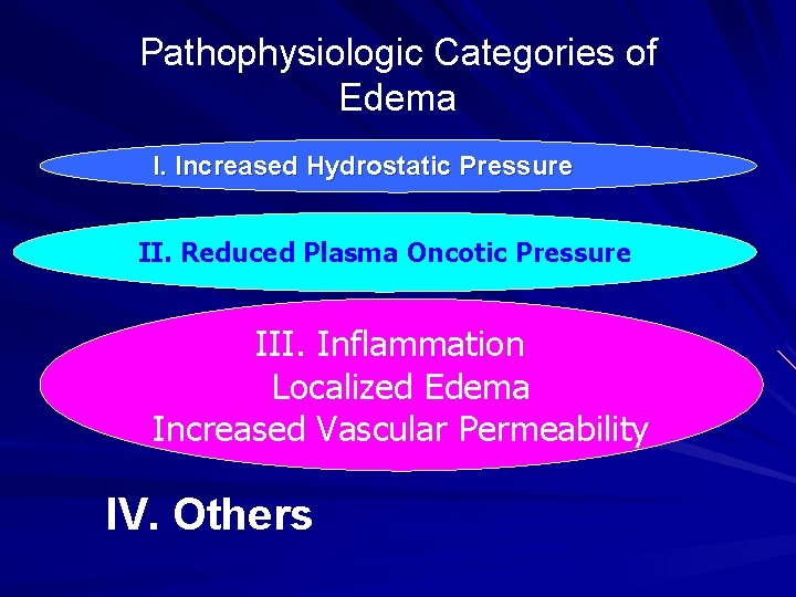
Pathophysiologic Categories of Edema I. Increased Hydrostatic Pressure II. Reduced Plasma Oncotic Pressure III. Inflammation –IV. Others
Patho-physiologic Categories of Edema Increased Hydrostatic Pressure Increased intravascular pressure may be due to 1 - Impaired venous return; A) Localized: Venous Thrombosis in lower extremities (local edema). B) Generalized: Congestive Heart Failure (generalized edema). 2 - Increased arteriolar dilatation; a) b) Heat Neurohumoral dysregulation
Increased Hydrostatic Pressure Congestive Heart Failure: Congestive Heart Failure is the most common cause of EDEMA due to Increased Hydrostatic Pressure “Generalized increased venous pressure, resulting in systemic edema, occur most commonly in CONGESTIVE HEART FAILURE”
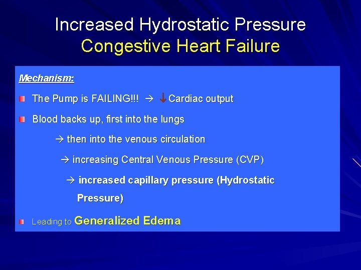
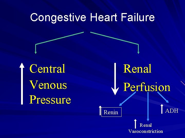
Increased Hydrostatic Pressure Congestive Heart Failure Mechanism: The Pump is FAILING!!! Cardiac output Blood backs up, first into the lungs then into the venous circulation increasing Central Venous Pressure (CVP) increased capillary pressure (Hydrostatic Pressure) Leading to Generalized Edema
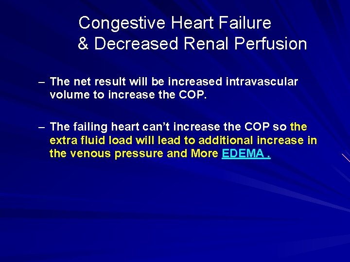
Congestive Heart Failure & Decreased Renal Perfusion Congestive heart failure Decreased Cardiac Output Decreased ARTERIAL blood volume “Less arterial blood…Less renal perfusion. . . The Kidney doesn’t see enough blood coming through …….
Congestive Heart Failure & Decreased Renal Perfusion activates the Renal Defense Mechanisms: 1. Renin-Angiotensin-Aldosterone axis Na & H 2 O retention 2. Renal Vasoconstriction 3. Increased Renal Anti-diuretic Hormone (ADH)
Congestive Heart Failure & Decreased Renal Perfusion – The net result will be increased intravascular volume to increase the COP. – The failing heart can’t increase the COP so the extra fluid load will lead to additional increase in the venous pressure and More EDEMA.
Congestive Heart Failure Central Venous Pressure Renal Perfusion ADH Renin Renal Vasoconstriction
Pathophysiologic Categories of Edema I. Increased Hydrostatic Pressure II. Reduced Plasma Oncotic Pressure III. Inflammation IV. Others
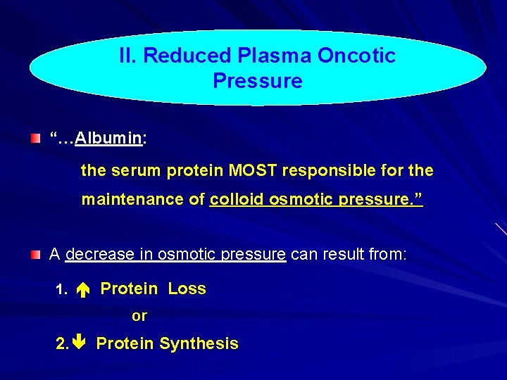
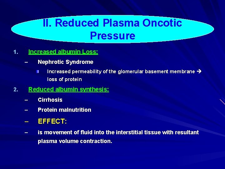
II. Reduced Plasma Oncotic Pressure “…Albumin: the serum protein MOST responsible for the maintenance of colloid osmotic pressure. ” A decrease in osmotic pressure can result from: 1. Protein Loss or 2. Protein Synthesis
II. Reduced Plasma Oncotic Pressure Increased albumin Loss: 1. – Nephrotic Syndrome Increased permeability of the glomerular basement membrane loss of protein Reduced albumin synthesis: 2. – Cirrhosis – Protein malnutrition – EFFECT: – is movement of fluid into the interstitial tissue with resultant plasma volume contraction.
Pathophysiologic Categories of Edema I. Increased Hydrostatic Pressure II. Reduced Plasma Oncotic Pressure III. Inflammation Localized Edema Increased Vascular Permeability IV. Others
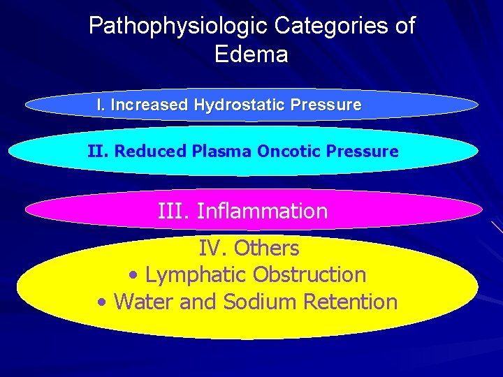
Pathophysiologic Categories of Edema I. Increased Hydrostatic Pressure II. Reduced Plasma Oncotic Pressure III. Inflammation IV. Others • Lymphatic Obstruction • Water and Sodium Retention
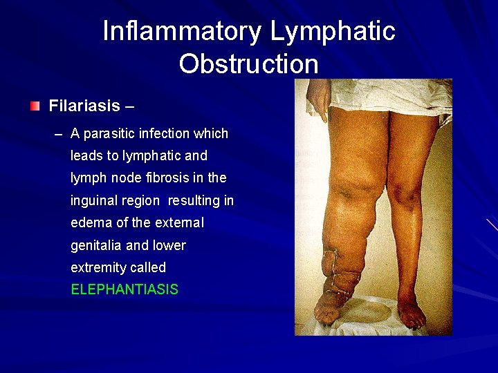
Lymphatic Obstruction Impaired lymphatic drainage with resultant lymphedema LOCALIZED EDEMA caused by : INFLAMMATION or NEOPLASTIC OBSTRUCTION
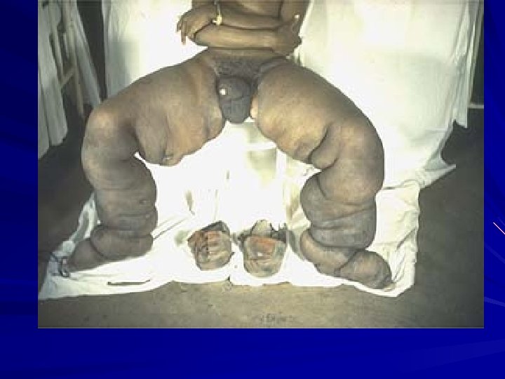
Inflammatory Lymphatic Obstruction Filariasis – – A parasitic infection which leads to lymphatic and lymph node fibrosis in the inguinal region resulting in edema of the external genitalia and lower extremity called ELEPHANTIASIS
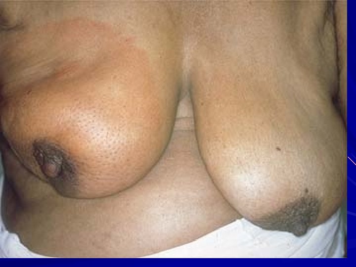
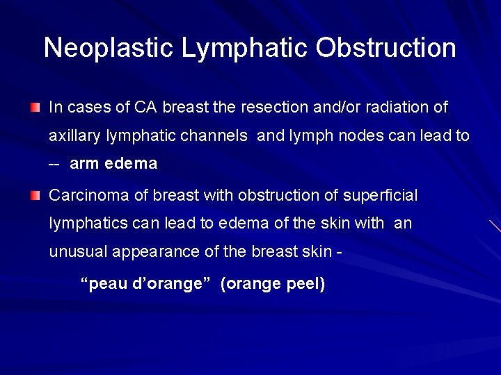
Neoplastic Lymphatic Obstruction In cases of CA breast the resection and/or radiation of axillary lymphatic channels and lymph nodes can lead to -- arm edema Carcinoma of breast with obstruction of superficial lymphatics can lead to edema of the skin with an unusual appearance of the breast skin “peau d’orange” (orange peel)
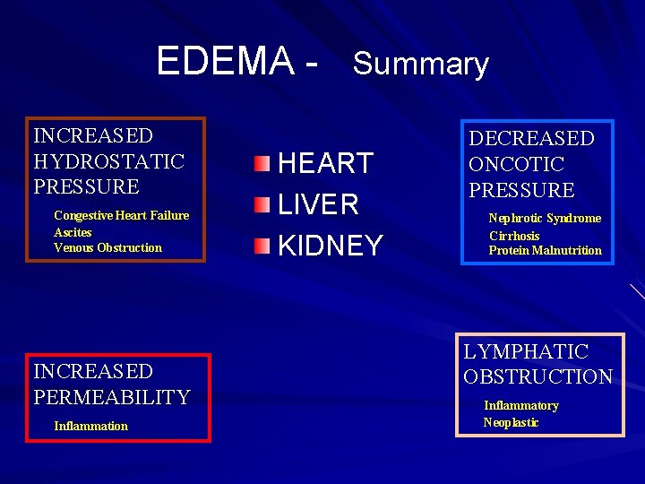
EDEMA - Summary INCREASED HYDROSTATIC PRESSURE Congestive Heart Failure Ascites Venous Obstruction INCREASED PERMEABILITY Inflammation HEART LIVER KIDNEY DECREASED ONCOTIC PRESSURE Nephrotic Syndrome Cirrhosis Protein Malnutrition LYMPHATIC OBSTRUCTION Inflammatory Neoplastic
Edema Fluid Transudate Cause High hydrostatic pr. Protein content Low Exudate Inflammatory High Specific gravity <1. 012) >1. 020) Inflammatory cells Absent Rich
GENERALIZED EDEMA HEART LIVER KIDNEY
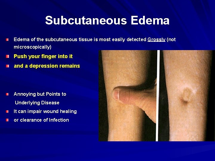
Subcutaneous Edema of the subcutaneous tissue is most easily detected Grossly (not microscopically) Push your finger into it and a depression remains Annoying but Points to Underlying Disease It can impair wound healing or clearance of Infection
Edema Dependent Edema is a prominent feature of Congestive Heart Failure; in legs if standing or sacrum in sleeping patient Periorbital edema is often the initial manifestation of Nephrotic Syndrome, while late cases will lead to generalized edema.
Pulmonary Edema is most frequently seen in Congestive Heart Failure – May also be present in renal failure, adult respiratory distress syndrome (ARDS), pulmonary infections and hypersensitivity reactions
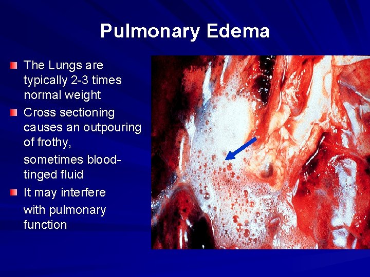
Pulmonary Edema The Lungs are typically 2 -3 times normal weight Cross sectioning causes an outpouring of frothy, sometimes bloodtinged fluid It may interfere with pulmonary function
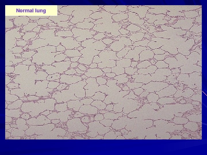
Normal lung
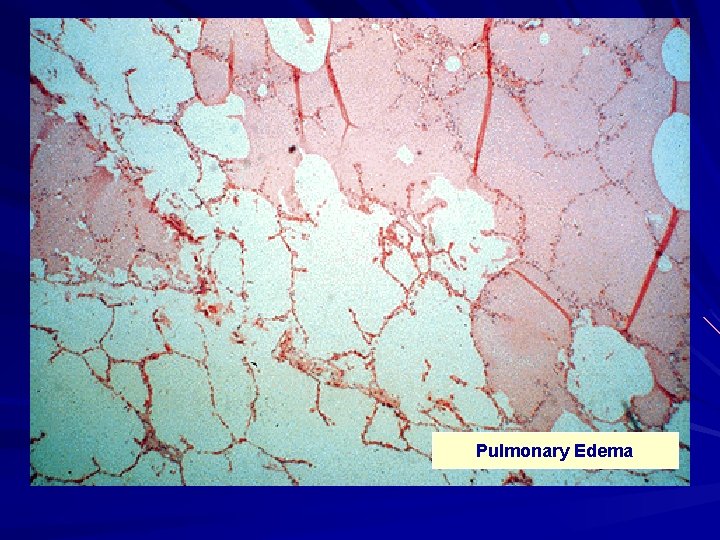
Pulmonary Edema
Pulmonary Edema Clinical Correlation May cause death by interfering with Oxygen and Carbon Dioxide exchange Creates a favorable environment for infection THINK it resembles “Culture Media”!!!
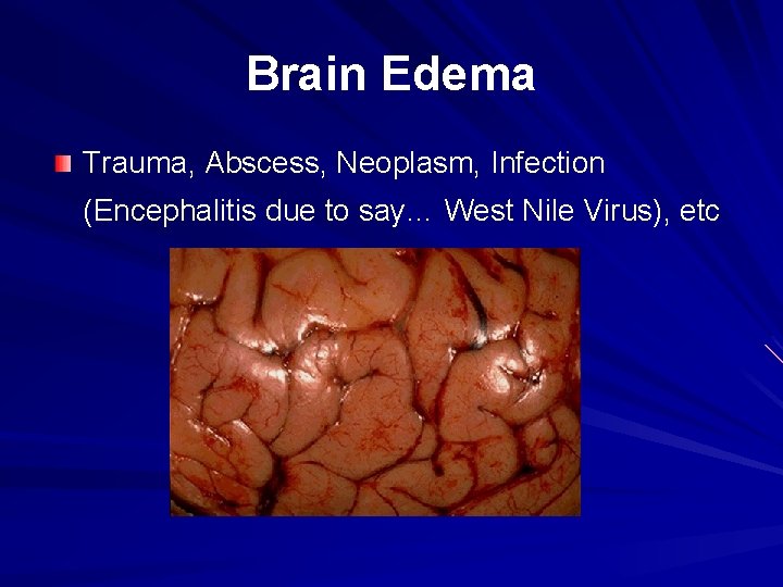
Brain Edema Trauma, Abscess, Neoplasm, Infection (Encephalitis due to say… West Nile Virus), etc
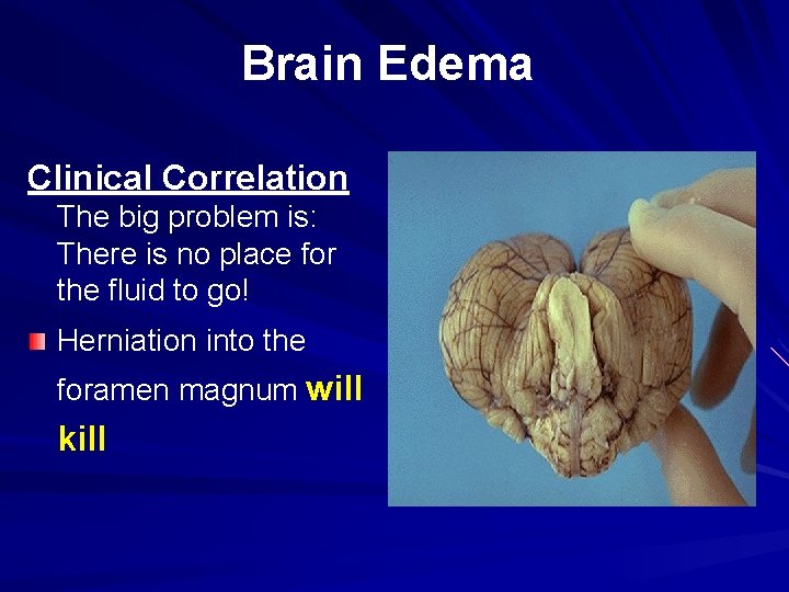
Brain Edema Clinical Correlation The big problem is: There is no place for the fluid to go! Herniation into the foramen magnum will kill
Clinical Correlation of Edema The effect of edema may be just annoying to fatal condition. It usually points to an underlying disease. However, it can impair wound healing or clearance of Infection. Creates a favorable environment for infection. THINK “Culture Media” May cause death by interfering with Oxygen and Carbon Dioxide exchange.
Thank you