Haneen bani younis Tasneem Migdadi Pulmonary Surfactant Action

Haneen bani younis Tasneem Migdadi

Pulmonary Surfactant: Action and Structure and Composition: Pulmonary surfactant is a mixture of lipoprotein: ▪ lipids (85% to 90%, predominantly phospholipids) and proteins (10% to 15%) The principal surface tension lowering agent in surfactant is dipalmitoylphosphatidyl choline (DPPC). � Source: Alveolar type II cells. Electron dense lamellar inclusion bodies. � Functions of Surfactant : Prevents lung collapse Promotes alveolar stability Helps to prevent edema in the lung decreases the work of breathing � Regulation of Synthesis and Secretion: Hormonal factors: Glucocorticoid, insulin, thyroxine Stretching of lungs � Surface tension between the fluid and the air is 7 -14 times more than that between surfactant and the air.

� Structure of surfactant : � Surfactant is lipid molecule , synthesized by alveolar cell type 2 , it is amphipathic molecule which means it has 2 positions : 1. hydrophilic 2. hydrophobic � The raw materials : glucose , fatty acids and more other which are necessary- to synthesis the surfactant- from the circulation. these raw materials are stored within a structure located also inside type 2 cells election dense lamilar bodies. � There are two theories about that : � 1. the raw materials stored and then used when need � 2. the surfactant is already synthesized and stored the (type 2 cells) � there are many stimuli that affect its synthesis like hormones , cortisone , insulin. . etc. which have an important role in controlling surfactant synthesis and the bodies mentioned above. � * surfactant is a lipoprotein =lipid +protein.

PULMONARY FUNCTION TESTS Pulmonary function tests � Are very useful tests to diagnose several lung diseases. � The most informative tests of lung function is Forced Expiratory Volume (FEV 1) It is the volume of gas exhaled in one second by a forced expiration following a full inspiration. � The total volume of the gas exhaled after a full inspiration represents the vital capacity. However, the total volume of the gas exhaled forcefully after a full inspiration represents forced vital capacity (FVC). � The normal ratio of the FEV 1 is 80 % of FVC.

� function of surfactant : � 1. Reduction of surface tension. how ? � since it is an amphipathic molecule, so has two portions , hydrophilic + � hydrophobic. The hydrophilic portions dissolves in watery , while the hydrophobic portions is oriental toward due alveolar gas or air. so , now there is a layer of surfactant between fluid and air. reduction in surface tension by 7 -14 times. (note : no complete elimination of surface tension occurs ). � 2. By reduction of surface tension , this prevents (lung collapse ) consequently. � *at low lung volume , surfactant is more action and so prevent lung collapse in this law volume during expiration to prevent the lung from biro totally collapsed. ﺣﻜﻲ ﻛﺜﻴﺮ ﺑﺲ ﺍﻟﻔﻜﺮﺓ ﻭﺣﺪﺓ � *(it is more efficient in smaller alveoli than in the larger one because it is more concentrated in the lower surface areas rather than larger ones )

� 3. prevent edema formation : � -surface tension does not just collapse the alveoli , it sucks the fluids from the alveoli to the capillaries. (more filtration ). proteins cannot leave the capillaries since they large , so they create a pressure which lead to the suction of fluids edema � *the process of exchange controlled by 2 pressures : � 1. hydro static pressures : filtration of the fluid. � 2. colloid osmotic pressure : by the presence of proteins , suck the fluid from the interstitial to the capillaries. � *But in the percent of surfactant , that does not happen. � 4. decreases the work of breathing cause we need more effort to re-inflate the lungs if surfactant is not present. (surfactant increases the compliance )

Interpretation of tests of forced expiration

�*pulmonary function test : �very important to measure the efficiency of RS , and to determine if there any lung disease, specifically , it determines or distinguish the presence or absence of restrictive and obstructive lung disease �*forced expiratory volume 1: the volume of gas expiration forcefully and rapidly , (because time is important )in the first second.

�*while the volume of air expired normally (not forclly after deep and maximum inspiration ) called vital capacity (VC) �*FVC : forced vital capacity , almost this volume is less than VC , because in vital capacity it is static volume , time is not considered like in FVC is considered. The portion of FVC which expired at the first second called (FEV 1) �*so the FVC is the maximum amount of air expired , and this air may go out of the lung completely during 4 seconds after forced inhalation. �*FEV 1/FVC=4/5=0. 8=80% Of air expired at the first second

�*but in the case of obstructive and restrictive disease this ratio changes : �FEV 1: in both cases reduction �FVC : in both cases reduction �*the ratio decreased in "obstructive case " �the ratio increase in "restriction case "or normal. ﺑﺘﻜﻮﻥ ﻣﺶ ﻃﺒﻴﻌﻴﺔ ﻣﺜﻼ FEV 1 ﻣﺮﺍﺕ ﺍﻟﻨﺴﺒﺔ ﺑﺘﻜﻮﻥ ﻧﻮﺭﻣﺎﻝ ﺑﺲ ﺍﻝ what is the obstructive disease ? (RV). (difficult in expiration (trapping * � the air inside ). due do resistant ﺑﺲ ﻛﻴﻒ ؟ﺑﻜﻮﻥ ﺍﻟﻬﻮﺍﺀ ﺍﻟﻠﻲ ﺩﺧﻞ ﺍﺻﻼ ﻗﻠﻴﻞ , � ﻭﻣﻤﻜﻦ ﺍﻟﻨﺴﺒﺔ ﺗﻜﻮﻥ ﻋﺎﻟﻴﺔ ﻭﺑﺎﻟﺘﺎﻟﻲ ﺍﻟﻠﻲ ﻓﺎﺕ ﻋﻠﻴﺔ ﺭﺡ ﻳﻄﻠﻊ ﻭﻫﻮ expiration ﻭﻣﺎ ﻋﻨﺪﻱ ﻣﺸﻜﻠﺔ ﺑﺎﻝ , restrictive disease ﺑﺘﻜﻮﻥ ﺍﻟﻨﺴﺒﺔ ﺗﻘﺮﻳﺒﺎ ﻋﺎﻟﻴﺔ ﻋﻨﺪ ﺍﻝ

Alveolar Ventilation � Important Terms: Ventilation: is a dynamic process that moves air in and out of the lungs. Minute ventilation: ▪ is the volume of air that enters the lung each minute. ▪ This is the product of tidal volume (500 ml) and respiratory rate (12 per minute). Expired minute ventilation: ▪ is the volume of air that leaves the lung each minute. ▪ amount of expired minute ventilation at rest is same as minute ventilation as the inspired and expired tidal volume is the same. Alveolar ventilation: The volume of the gas reaching the respiratory zone of the airways. However, not all of the Minute ventilation volume reaches the alveoli. 150 ml of the TV (500 ml) is left behind in the airways, which does not contain alveoli, therefore does not contribute the diffusion (Anatomic death space). Dead Space: ▪ Anatomic dead space ▪ Alveolar (functional), dead space
: the amount of enter and leaves the lung or the exchange")
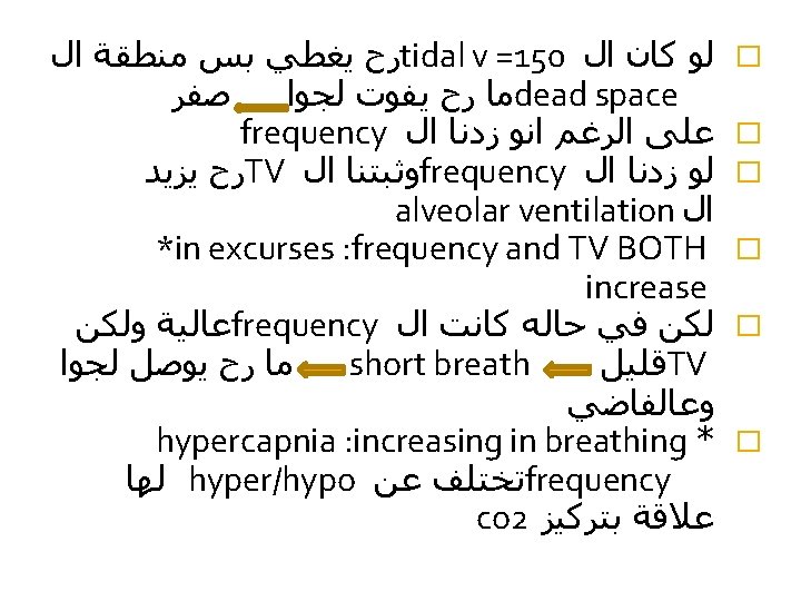
*Alveolar Ventilation -Ventilation(generally): the amount of enter and leaves the lung or the exchange of air between the lung and the environment. � * Minute Ventilation : exchange of air between the lung and the environment in one minute � - 500 X 12=600 ml= 6 Liter inspired and expired � � � * expired minute Ventilation : but just expired which is the same as inspired , but differs in the contact ( Presence of co 2). � *Now, The alveolar Ventilation: The Volume of air Reaching the alveoli or the Respiratory Zone. *The Tidal volume is 600 ml , just 2/3 reach to the R Zone (=350 ml). The remaining I 50 ml, Still in the Conducting Zone, does not have o 2 and does not do through gas exchange (1/3). which Called dead space (wasted air). � * Alveolar ventilation rate = Volume x R rate (at rest) 350 (fresh air � =350 X 12 =4. 2 Liter � � *Anatomical dead space : the volume of air remaining in the C Zone (150 ml ) From nose to the terminal alveoli � *alveolar dead space : in alveoli and not functional

* and it is a mismatch between Ventilation airation � and perfusion. occurs when Perfusion is little or not percent (no Ventilation)- non Functional alveolar dead ﻣﺜﻼ ﻭﻋﺎﺀ ﺩﻣﻮﻱ ﻣﺴﻜﺮ ﻋﻠﻰ ﺍﻻﺧﺮ ﻣﺎ ﺭﺡ ﻳﺼﻴﺮ ﻓﻴﻪ space. ﺗﺒﺎﺩﻝ ﻟﻠﻬﻮﺍﺀ ﺭﻏﻢ ﻭﺟﻮﺩ ﺍﻟﻬﻮﺍﺀ ﻭﻟﺬﻟﻚ ﻟﻌﺪﻡ ﻭﺻﻮﻝ ﺍﻟﺪﻡ B. V ﺍﻟﻰ ﻫﺬﻩ ﺍﻟﻤﻨﻄﻘﺔ ﺑﺤﻴﺚ ﺍﻧﻪ ﻳﺬﻫﺐ ﻟﻤﻨﻄﻘﺔ ﻓﻴﻬﺎ diluted. � *Physiological dead space = Anatomical t Alveolar ( called space ) � *in normal Person: Phy d. S = anatomical dead space (because this is no alveolar ds) � * So, by Comparing physiological ds with the TV we can estimate How much air wasted. � * Many ways to measure the phys ds , one of them by calculating the Fractional Concentration of Co 2 in the alveoli or the partial pressure of it( Co 2) in the expired air (tidal volume) : 1. alveolar volume 2. anatomical dead space).

Anatomic Dead Space Refers to the volume of air present in the conducting zone of the respiratory passage, i. e. from nose to terminal bronchioles. Since, the dead space is due to the anatomy of the airways, it is referred to as anatomical dead. � Due to the anatomical dead space, out of 500 ml of tidal inspiration, 350 ml of air enters the alveoli. Thus, the normal anatomical dead space volume is 150 ml. �

Alveolar Dead Space � Ventilated alveoli that do not participate in gas exchange � Mismatch of ventilation and perfusion � The sum of the anatomical dead space and alveolar dead space is called physiological dead space. � In normal persons, the physiologic dead space is nearly equal to the anatomic dead space � By comparing the physiologic dead space with the tidal volume, it is possible to estimate how much ventilation is “wasted. ”

Measurement of Physiological Dead Space For example, if the alveolar PCO 2 is 40 mm Hg and the mixed-expired PCO 2 is 28 mm Hg, then o Normally, VD/VT is 20% to 30% of minute ventilation. If VD= zero? ?
 of the tidal volume")
ALVEOLAR VENTILATION � Alveolar ventilation. Only the alveolar part (VA) of the tidal volume (VT) reaches the alveoli. The rest goes to dead space (VD). The volume of air that actually reaches the alveoli per minute is known as alveolar ventilation. Therefore, to know the volume of air that takes part in alveolar ventilation, first the dead space volume is subtracted from the tidal volume: VA = VT – VD (L). Multiplying. alveolar volume by the respiratory rate (f in min – 1) results in alveolar ventilation ( VA): Which is the volume of fresh air per minute actually reaching the alveoli. Alveolar ventilation is given by the following equation: If anatomic dead space volume is increased due to any cause, alveolar ventilation decreases.

� the amount of Co 2 In alveolar air= alveolar volume concentration of Co 2 * Co 2 in dead space = Volume of ds* its conc. in inspired � ﻭﻟﻜﻦ ﺗﺮﻛﻴﺰ ﺛﺎﻧﻲ ﺍﻛﺴﻴﺪ ﺍﻟﻜﺮﺑﻮﻥ air (no exchange in gases) inspired air ﺻﻔﺮ ﻓﻲ ﺍﻝ � the amount of Co 2 in expired =VT * its Conc. in inspired air � = VA*conc of co 2 in alveoli � =CVT-dead space V)*conc. of Co 2 in alveoli= � VT * Conc. of co 2 in expired air : � ﺍﻟﻤﻌﺎﺩﻟﺔ ﺍﻟﻨﻬﺎﺋﻴﺔ � dead space volume =TV *factor this factor represent dilution of co 2 � =alveolar conc. of co 2 - conc. of co 2 in expired air � alveolar conc. of co 2 ﻛﻴﻒ ؟ co 2 in alveoli. . . � *ﺍﻟﻤﺸﻜﻠﻪ ﻫﻮﻥ ﻣﺎ ﺑﻘﺪﺭ ﺍﻗﻴﺲ systemic artery ﻷﻨﺔ ﺑﺼﻴﺮ ﻋﻨﺪﻱ ﺍﺗﺰﺍﻥ � alveoli and � ﻳﻌﻨﻲ ﻟﻤﺎ ﻳﺼﻴﺮ ﻋﻨﺪﻱ ﺗﺒﺎﺩﻝ ﻏﺎﺯﺍﺕ ﺑﻴﻦ ﺍﻝ capillary equilibrium

*ﺑﺎﻟﺘﺎﻟﻲ ﻟﻤﺎ ﺍﺣﺴﺐ ﻋﻴﻨﺔ ﺩﻡ ﻭﺃﻘﻴﺲ ﻣﻨﻬﺎ ﺗﺮﻛﻴﺰ ﺛﺎﻧﻲ ﺍﻛﺴﻴﺪ alveoli ﺍﻟﻜﺮﺑﻮﻥ ﺑﻜﻮﻥ ﻧﻔﺴﻪ ﻓﻲ ﺍﻝ *tidal volume =500 *pco 2 in blood =40 *pco 32 =30 ﻋﺸﺎﻥ ﻫﻴﻚ dilution ﺑﺘﻜﻮﻥ expired ﻓﻲ co 2 * ﻗﻴﻤﺔ ﺗﺮﻛﻴﺰ ﺍﻝ C zone ﻷﻨﺔ ﺭﺡ ﻳﺨﺘﻠﻂ ﺑﺎﻟﻬﻮﺍﺀ ﺍﻟﻠﻲ ﺑﺎﻝ dilution factor , ﻓﻲ ﻑ ﺭﺡ ﻳﻘﻞ ﺗﺮﻛﻴﺰﻩ , *the co 2 in expired air is lower than in alveolar 500, 600 ؟ ﺣﺴﺐ 150 (ﻉ ﺍﺳﺎﺱ 40 -30)/40=125 ml *in another way : dilution factor =vd/vt =(40 -28)/40 =30% the portion of dead space is 30% of tidal volume (30/100)*500=150 ml *alveolar volume =tidal volume-dead space volume *alveolar ventilation =alveolar volume * breathing frequency ﺭﺡ ﺗﻘﻞ alveolar ventilation ds * ﺍﺫﺍ ﺻﺎﺭ ﻋﻨﺪﻱ ﺯﻳﺎﺩﺓ ﻓﻲ ﺍﻝ TV ﺍﺯﻳﺪ ﻭﺑﺎﻟﺘﺎﻟﻲ ﺍﻟﻄﺮﻳﻘﺔ ﺍﻟﻮﺣﻴﺪﺓ ﺣﺘﻰ ﺍﺯﻳﺪﺓ deep breath

ALVEOLAR VENTILATION Physiological Significance hypercapnea hypocapnea
- Slides: 21