HAND Palm of Hand Superficial fascia it contains


 • Boundaries: • Laterally: intermediate palmar septum •")

 • Boundaries: -Laterally: lateral palmar septum - Medially:")


- Slides: 34
HAND
Palm of Hand • Superficial fascia: -it contains the following structures: 1. Palmer cutaneous branch of ulnar nerve 2. Palmer cutaneous branch of median nerve 3. Palmaris brevis muscle
Palmaris Brevis Palmaris brevis is a thin, quadrilateral muscle, placed beneath the integument of the ulnar side of the hand. It acts to fold the skin of the hypothenar eminence transversally. Origin: Flexor retinaculum (medial) and palmar aponeurosis Nerve: Superficial branch of ulnar nerve Actions: Pulls on skin over hypothenar eminence, deepening the cup of the palm and so improving grip
• Deep fascia: -Thin on hypothenar and eminences but thick in the central part to form palmar aponeurosis -Palmer aponeurosis: is a triangular sheet of deep fascia on the central area of palm with its apex receives insertion of palmaris longus muscle
Fascial spaces of the palm • lateral and medial palmer septa divides palm into 3 fascial compartments: 1. Lateral compartment: contains thenar muscles 2. Medial compartment: contains hypothenar muscles 3. Intermediate compartment: -Lies deep to palmar aponeurosis between lateral and medial palmar septa -It is divided by intermediate palmar septum into: • Medial central palmar space (midpalmar space) • Lateral central palmar space (thenar space) • intermediate septum: It extends from the deep surface of palmar aponeurosis to 3 rd metacarpal bone
Medial central palmar space (midpalmar space) • Boundaries: • Laterally: intermediate palmar septum • Medially: medial palmar septum • Anteriorly: Palmar aponeurosis • Posteriorly: 3 rd, 4 th, 5 th metacarpal bones and intervenning interosseous muscles • Contents: - Tendons of flexor digitorum superficialis and profundus to 3 rd, 4 th, 5 th fingers - 2 nd, 3 rd, 4 th lumbrical muscles -Digital vessels and nerves to medial 3 fingers
• Communications of midpalmar space: • Distally: with webs between roots of medial 4 fingers • Proximally: the front of forearm.
Lateral central palmar space (thenar space) • Boundaries: -Laterally: lateral palmar septum - Medially: intermediate palmar septum -Anteriorly: Palmar aponeurosis -Posteriorly: Adductor pollicis and 1 st dorsal interosseous muscle • Contents: -Long flexor tendons to index finger -Tendon of flexor pollicis longus -1 st lumbrical muscle -Digital nerves and vessels to lateral 2 fingers
• Communications of thenar space: • Distally: with web between thumb and index fingers • Proximally: Extends behind flexor retinaculum
• Web space: -Subcutaneous space lies in interdigital clefts - It extends from free margin of web to transverse metacarpal ligament - Filled with loose areolar tissue, lumbrical tendon and digital nerve and vessels
• Pulp Space: -It is the space lies over palmar surface of distal ¾ of terminal phalanx -Boundaries: Anteriorly: deep fascia Posteriorly: terminal phalanx -It is traversed by many fibrous septa from skin to bone dividing the space into a number of loculi filled with subcutaneous fat - It is traversed by digital arteries to distal end of terminal phalanx
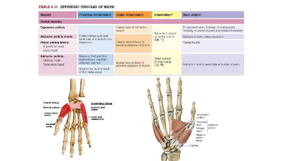
Muscles of the hand Thenar muscles Opponens pollicis Flexor pollicis brevis Abductor pollicis brevis
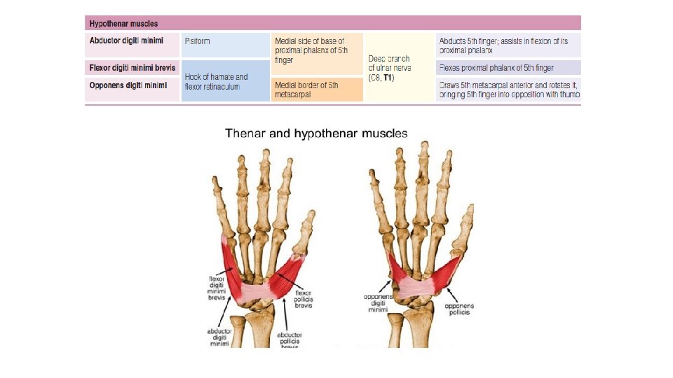
Hypothenar muscles Opponens digiti minimi Flexor digiti minimi Abductor digiti minimi
Adductor pollicis
Adductor pollicis
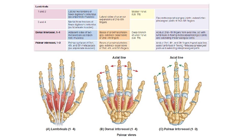
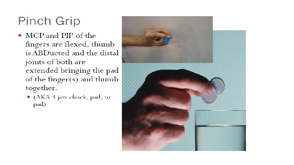
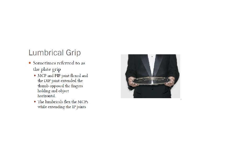
Lumbrical Muscles • They are 4 slender muscles arise from tendons of flexor digitorum profundus • They put the hand in writing position: - Flexion of metacarpo-phalangeal joints - Extension of interphalngeal joints
Interossei Muscles • Palmer interossei: • Dorsal interossei: • 4 unipennate muscles • 4 bipennate muscles • They adduct fingers to middle • They abduct fingers away from middle line of middle finger
• Both palmer and dorsal interossei put hand in writing position by flexion of metacarpo-phalangeal joints and extension of interphalangeal joints
Nerve Supply of Muscles of Hand Thenar Muscles Hypothenar Muscles Lumbrical muscles Abductor pollicis brevis Median Nerve Flexor pollicis brevis Median Nerve Opponens pollicis Median Nerve Adductor pollicis Deep branch of ulnar nerve Abductor digiti minimi Deep branch of ulnar nerve Flexor digiti minimi Deep branch of ulnar nerve Opponens digiti minimi Deep branch of ulnar nerve Lateral two (1 st and 2 nd ) Median Nerve Medial two (3 rd and 4 th ) Deep branch of ulnar nerve Palmer interossei Deep branch of ulnar nerve Dorsal interossei Deep branch of ulnar nerve
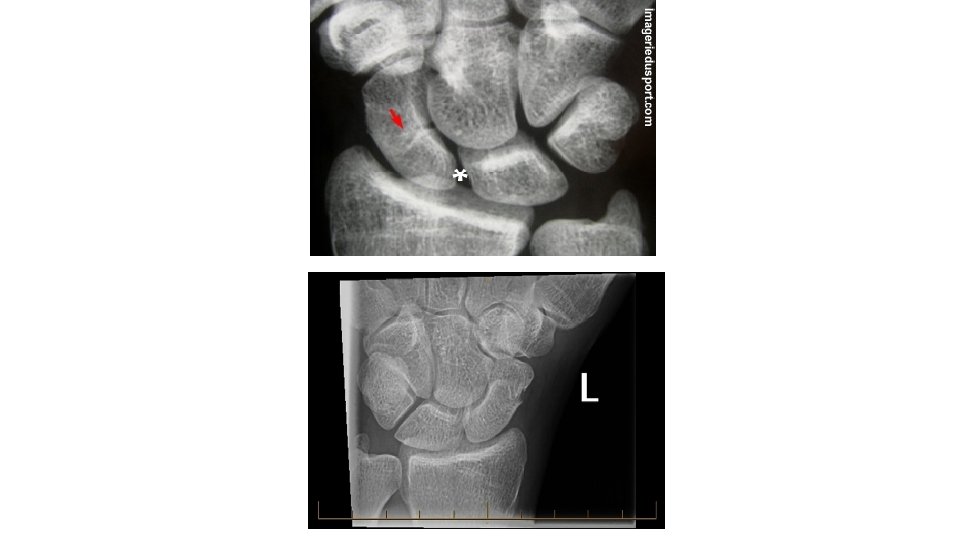
Fractures of Hand Fracture of the scaphoid often results from a fall on the palm with the hand abducted. The fracture occurs across the narrow part (“waist”) of the scaphoid. Pain occurs primarily on the lateral side of the wrist, especially during dorsiflexion and abduction of the hand. Initial radiographs of the wrist may not reveal a fracture, but radiographs taken 10 to 14 days later may reveal a fracture because bone resorption has occurred. Owing to the poor blood supply to the proximal part of the scaphoid, union of the fractured parts may take several months. Avascular necrosis of the proximal fragment of the scaphoid (pathological death of bone resulting from poor blood supply) may occur and produce degenerative joint disease of the wrist.
Thank You