
Hand infections Traumatic anurea By Dr Ahmed Rashidy
Hand infections & Traumatic anurea By Dr. Ahmed Rashidy Lecturer of General & Pediatric Surgery
Classification of Hand infections 1 - Cutaneous & subcutaneous infections: • • Paronychia. Subcuticular and Subcutaneous whitlow. Pulp space infection. Web space infection. 2 - Facial space infection: • Midpalmar space infection. • Thenar space infection. • Hypothenar space infection. • Space of parona infection. 3 - Synovial sheath infection: . Acute digital tenosynovitis. . Ulnar bursitis. • Radial bursitis. 4 - Bone & joint infections.
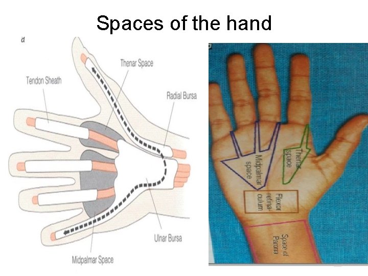
Spaces of the hand
Etiology • Causative organism: - Staph. aureus (80%), Strept, E. coli, G -ve bacilli. • Predisposing factors: 1 - More in manual workers & housewives. 2 - Bad general conditions and patients on immunosuppresive drugs 3 - Bad hygiene • Route of Infection: - Usually direct spread of infection. - Blood born infection is rare.
Clinical picture • Symptoms: Ø General : Fever, headache, anorexia, malaise Ø Local : • Pain : dull aching then become throbbing when pus is formed & Decreased by elevation of the hand. • Swelling : According to the site affected, e. g. , pulp space >ــــ distal phalanx edema at the dorsum of the hand is common, irrespective of the site of infection. • Disterbance of function: Limited movements • Signs: Ø General : fever, tachycardia Ø Local : hotness, redness, diffuse edema maximum on the dorsum of hand , Enlarged tender axillary lymph nodes
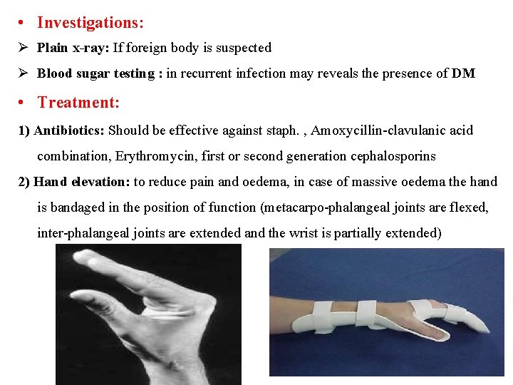
• Investigations: Ø Plain x-ray: If foreign body is suspected Ø Blood sugar testing : in recurrent infection may reveals the presence of DM • Treatment: 1) Antibiotics: Should be effective against staph. , Amoxycillin-clavulanic acid combination, Erythromycin, first or second generation cephalosporins 2) Hand elevation: to reduce pain and oedema, in case of massive oedema the hand is bandaged in the position of function (metacarpo-phalangeal joints are flexed, inter-phalangeal joints are extended and the wrist is partially extended)

3) Surgical drainage: indicated in pus formation A- Acute paronychia and distal pulp space infection are drained by local ring anaesthesia around the root of the finger, otherwise general anaesthesia is preferred. B- Pneumatic tourniquet is applied around the arm, the arm is elevated for few minutes and then the tourniquet is inflated to obtains bloodless field. C- Appropriate skin incision is used (according to the site ), pus is drained by sinus forceps. D- Pus is sampled for culture and sensetivity. 4) Post-operative: hand elevation, physiotherapy and wound dressing

Acute paronychia Definition: lnfection of the tissue surrounding the nail bed. Commonest hand infection (30% of cases). C/P: Nail bed indurated. Later on become cystic, yellowish. At first at one side latter on around the nail fold. Complications: Spread of infection under nail (subungual abscess). Incision: Oblique incision at the angle of the nail fold. U Shaped incision , if - pus present all around the nail. Excision of outer quarter of nail if pus under the nail (subungual abscess).
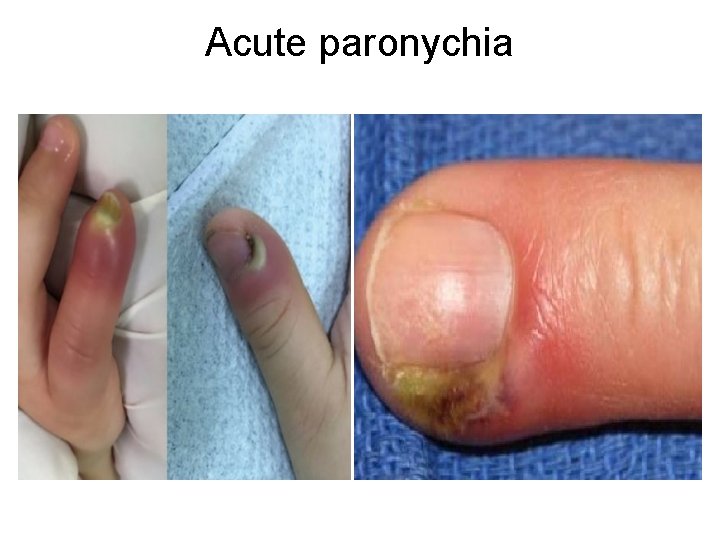
Acute paronychia
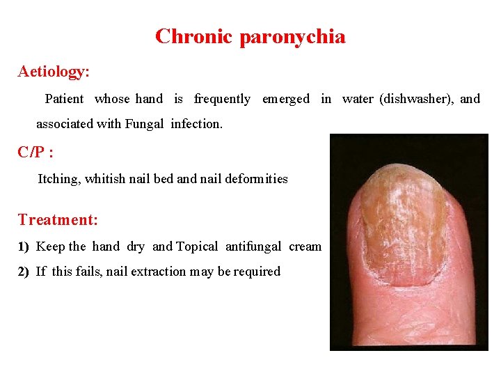
Chronic paronychia Aetiology: Patient whose hand is frequently emerged in water (dishwasher), and associated with Fungal infection. C/P : Itching, whitish nail bed and nail deformities Treatment: 1) Keep the hand dry and Topical antifungal cream 2) If this fails, nail extraction may be required

Subcuticular Whitlow - lt is pus under the cuticle within the epidermis. lt may be communicated with deeper abscess (collar stud abscess). Treatment: - The raised epidermis is incised - Gentle probing is done for a track extending to a deeper abscess if present.
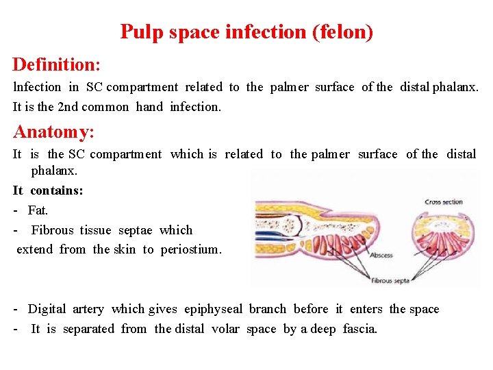
Pulp space infection (felon) Definition: lnfection in SC compartment related to the palmer surface of the distal phalanx. It is the 2 nd common hand infection. Anatomy: It is the SC compartment which is related to the palmer surface of the distal phalanx. It contains: - Fat. - Fibrous tissue septae which extend from the skin to periostium. - Digital artery which gives epiphyseal branch before it enters the space - It is separated from the distal volar space by a deep fascia.

Pulp space infection (felon) C/P: Tender swelling of the distal phalanges. The distal phalanx is first indurated, Later on become cystic, yellowish. Complications: 1) Thrombophlibitis of digital vessels results in necrosis and infection Osteomyelitis of the distal phalanx except epiphysis (Parrot peak deformity). 2) Septic arthritis.

Pulp space infection (felon) Treatment: Early surgical drainage is indicated (don’t wait for fluctuation). The incision is done in the 1) lateral side of the distal 2/3 of the distal phalanx or 2) over the most tender point • Counter incision is done in severe case • The incision should be deepened to divide all septa
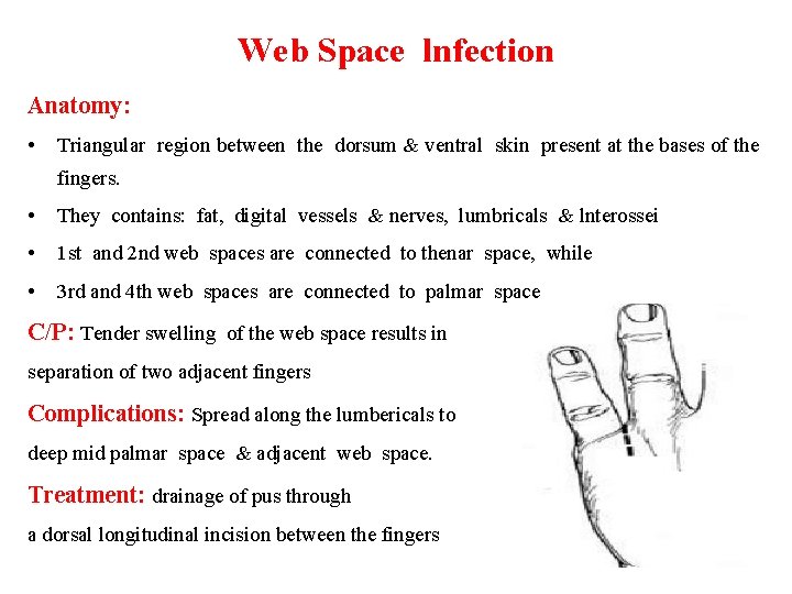
Web Space lnfection Anatomy: • Triangular region between the dorsum & ventral skin present at the bases of the fingers. • They contains: fat, digital vessels & nerves, lumbricals & lnterossei • 1 st and 2 nd web spaces are connected to thenar space, while • 3 rd and 4 th web spaces are connected to palmar space C/P: Tender swelling of the web space results in separation of two adjacent fingers Complications: Spread along the lumbericals to deep mid palmar space & adjacent web space. Treatment: drainage of pus through a dorsal longitudinal incision between the fingers

Mid-palmar space lnfection Definition: Infection occurs deep to the tough palmar aponeurosis Aetiology : either from 1) punctures 2) Spread of either web space or tendon sheath infection C/P : characterized by marked dorsal oedema Treatment : Pus is evacuated through an incision in one of the transverse palmar creases. To avoid injury of deep important structures, the incision is made through the skin only and then a sinus forceps is thrust inwards and is gently opened to let out pus (Hilton’s method)

Thenar and hypothenar space lnfection Anatomy : The thenar space is pounded anteriorly by the palmar fascia and flexor tendons of the index and thumb, posteriorly by the adductor pollicis and medially by the fibrous septum of the palm C/P : presents by ballooning of thenar eminence and oedema of the dorsum of the hand Treatment : Drained through an incision at the site of maximum tenderness or where pus points at the skin An incision that is done along the medial site of thenar eminence should stop 2 cm distal to the distal wrist crease to avoid injury of the motor branch of the median nerve that supplies thenar muscles

Thenar and hypothenar space incision
Space of parona infection Anatomy : This space lies in the distal part of the forearm between the pronator quadratus and the flexor muscles. Treatment: Drainage should be along the ulnar side of the forearm
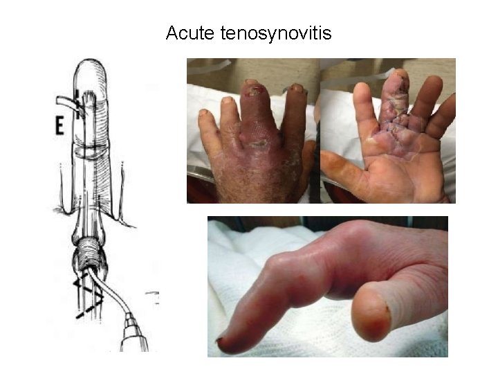
Acute tenosynovitis This is the most serious of hand infections Anatomy: Each finger of these has a separate synovial sheath. Ø Proximal end: At the level of metacarpophalyngealjoint. Ø Distal end: At the base of distal phalanges. Ø They enclose the flexor tendons. Aetiology: It is usually the sequel of deep pin pricks
Acute tenosynovitis C/p : Involvement of a digital flexor sheath produces Ø pain and swelling of the finger which the patient keeps in the semiflexed position (hook sign). Ø Any atempt at active or passive movement stretches the inflamed synovium and induce severe pain. Ø Tenderness is maximum just proximal to the crease over the metacarpo-phalangeal joints which is the proximal extent of the synovial sac
Acute tenosynovitis Ø Little finger sheath affection is likely to spread to the ulnar bursa producing marked hand swelling, semi-flexion and limitation of movement of the medial four fingers, and diffuse palm tenderness that may extend to the distal part of the forearm. The maximum point of tenderness may be on the ulnar side of the palm between the 2 palmar creases (Kanavel’s sign). Ø Radial bursitis produces similar picture in the thumb
Acute tenosynovitis Complications: 1) Thrombo-phlebitis of the tendon vessels that run along the vinculae leads to its sloughing (resulting in stiff useless fingers). 2) Infection spread to the mid-palmar space and the space of parona. 3) Osteomyelitis and arthritis
Acute tenosynovitis Treatment : Lack of response to conservative treatment is an indication for surgery. Ø The inflamed synovial sheath is drained through 2 incisions, (one at its proximal and another at its distal end). Ø A fine catheter is inserted through each incision to allow continuous drainage and irrigation of the sheath with an antibiotic solution.
Acute tenosynovitis
Ulnar bursitis Anatomy: Ø Larger than radial bursa. Ø Distally connected with synovial sheath of little finger. Ø Proximally run between the flexor retinaculum and pronator quadratus Ø Envelop the flexor tendons of the medial 4 fingers. C/P: Ø Swelling of little finger, palm, distal part of forearm. Ø Slight semiflexion of the little finger (hook sign). Ø Limitation of movements of the medial 4 fingers. Ø Edema of the dorsum of the hand Ø Tenderness over an infected ulnar bursa between transverse palmar crease and hypothenar muscles (Kanavel’s sign)
Ulnar bursitis Complications: 1) Sloughing of the tendon due to interfering with nutrition of the tendon. 2) Adhesions inside the synovial sheath (limitation of movement). 3) Oesteomyelitis and Arthritis. Treatment: Drainage by incision along radial border of hypothenar eminence In severe cases, counter incision at the lower part of forearm
Radial bursitis Anatomy: Small in size. Ø Distally connected with synovial sheath of the thumb. Ø Proximally run between the flexor retinaculum and pronator quadratus Ø Envelops the tendon of flexor pollicis longus. C/P: Tender swelling of thumb, thenar eminence & distal part of forearm.
Radial bursitis Complications: 1) Sloughing of the tendon due to interfering with nutrition of the tendon. 2) Adhesions inside the synovial sheath (limitation of movement). 3) Oesteomyelitis and Arthritis. Treatment: Drainage by incision on the ulnar side of thenar eminence stopping proximally 1. 5 inches distal to the distal crease of the wrist to avoid injury of the motor branch of median nerve In severe cases, counter incision at the lower part of forearm
Traumatic anurea (Crush syndrome) By Dr. Ahmed Rashidy Lecturer of General & Pediatric Surgery
Historical background Ø The association between crush injury, rhabdomyolysis and acute renal failure was first reported during the Second World War in victims trapped during the ‘London Blitz’. Ø Initially described by Bywaters and Beall in 1941 in a patient who initially appeared to be unharmed but subsequently died of renal failure.
Definition Ø Severe systemic manifestation of trauma and ischaemia involving soft tissues, principally skeletal muscle, due to prolonged severe crushing. Ø It leads to increased permeability of the cell membrane and to the release of potassium, enzymes and myoglobin from within cells. Ischaemic renal dysfunction secondary to hypotension and diminished renal perfusion results in acute tubular necrosis and uraemia. Ø It is also known as Bywaters' syndrome
Aetiology 1) Major trauma causing crushing of the muscles as occurs after air raids, earthquake and mining accident 2) prolonged tourniquet 3) After excessive exercise 4) When limbs have been forced into abnormal postures for prolonged periods, such as during general anaesthesia or coma induced by alcohol or drugs.
Pathophysiology Ø Prolonged crushing of a muscle mass causes death of the muscle cells, with release of myoglobin and vasoactive mediators into the circulation. Ø Injured muscle also sequesters many litres of fluid, reducing the effective intravascular volume, which results in renal vasoconstriction and ischaemia. Ø Myoglobin is concentrated in the tubules, and precipitates, leading to tubular obstruction. Ø The crushed muscle swells considerably & tension develops, impeding circulation & increasing extent ischemic damage. The limb feels tense & pain is severe.
Clinical picture • Crushing injury to a large mass of skeletal muscle. • Sensory and motor disturbances in the compressed limbs, which subsequently become tense and swollen. The limb/body part may be pulseless. • Myoglobinuria and/or haemoglobinuria, which may make the urine tea-coloured quite early on. • There may be oliguria with profound hypovolaemic shock. • Nausea, vomiting, confusion and agitation may occur as consequences of disturbed body chemistry; urea, creatinine, uric acid, potassium, phosphate and creatine kinase are elevated. There may also be hypocalcaemia.
Treatment (A) Early cases: • First aid treatment: 5 Rs. 1 - Reassurance 2 - Rapid history taking about type, time and nature of trauma. 3 - Resuscitation Air way patency, Breathing normally, Circulation adequate by arrest of bleeding (see haemorrhage), and correct shock (antishock), Disability by splinting fractures and Exposure of the patient and seek for hidden injuries. 4 - Research for other possible injuries for associated visceral, arterial, nerve or tendon injuries. 5 - Remember 3 As (antitetanic, anti-gas gangrene and antibiotics).
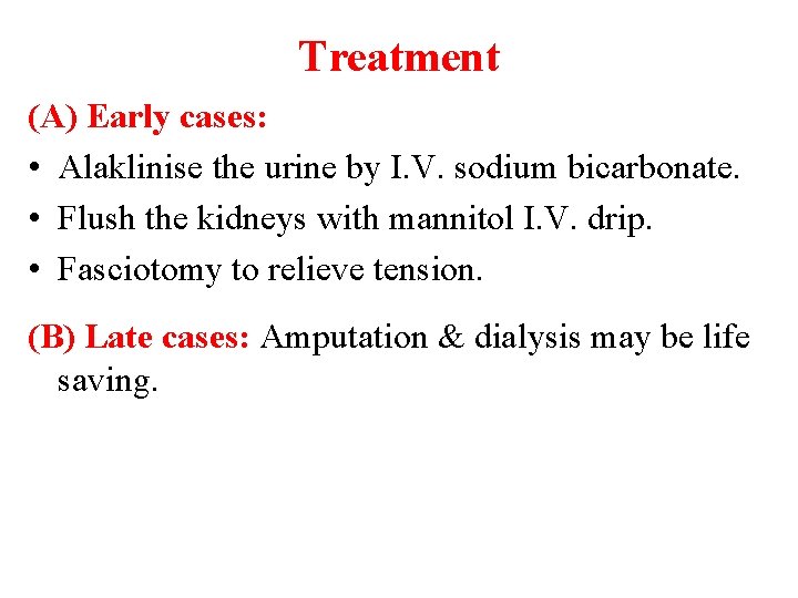
Treatment (A) Early cases: • Alaklinise the urine by I. V. sodium bicarbonate. • Flush the kidneys with mannitol I. V. drip. • Fasciotomy to relieve tension. (B) Late cases: Amputation & dialysis may be life saving.

Thank you
- Slides: 38