Hand in your homework please Detach the first
 and")


- Slides: 20
Hand in your homework please! • Detach the first section (including exam questions) and put your name on it! • Homework for MONDAY: Complete the section on ‘Methods of studying cells’ INCLUDING exam questions to hand in on MONDAY.
The Cell Cycle and Mitosis
Cell Division • Mitosis is a type of cell division that produces genetically identical copies of cells. • Not all cells can divide. • What types of cells DO divide? • Cells that can divide go through a CELL CYCLE.
The Cell Cycle Mitosis is only a small part of the ‘life’ of a cell. The process from one cell division to the next is known as the cell cycle. There are 4 parts to the cell cycle: Gap 1 These stages are all parts of Synthesis interphase. Gap 2 Mitosis
The cell cycle 5 of 32 © Boardworks Ltd 2008
Can you think of 3 reasons why mitosis is important?
A bit of background… • A chromosome is a strand of DNA. • After S-phase (DNA replication), identical copies of chromosomes remain attached – SISTER CHROMATIDS joined at the CENTROMERE. DNA replication Chromosome Centromere Sister chromatids
Reminder: • The chromosomes can’t be seen most of the time – they are tangled strands of DNA. • During cell division, the chromosomes condense – they coil up, aided by a number of proteins.
Mitosis 9 of 32 © Boardworks Ltd 2008
Interphase • The cell is engaged in metabolic activity in preparation for mitosis • Chromosomes are not visible under the light microscope
Prophase • Chromosomes condense and become visible under the light microscope • The nucleolus disappears • Centrioles begin moving to opposite ends of the cell and spindle fibres extend from the centrioles
Metaphase • The nuclear membrane dissolves, marking the beginning of metaphase. • Spindle fibres attach to the centromere of each chromosome.
Metaphase • Spindle fibres align the chromosomes along the equator of the cell • This helps ensure that during the next phase, when the chromosomes are separated, each new nucleus will receive one copy of each chromosome
Anaphase • The sister chromatids separate at the centromeres and move to opposite sides (poles) of the cell
Telophase • Chromatids arrive at opposite poles of the cell, and nuclear membranes form around them • The chromosomes are no longer visible under the light microscope
Another way to remember the stages… Invisible Apart Present Middle Two
CYTOKINESIS • The cytoplasm divides so two cells are formed. • The two daughter cells are genetically identical to each other, and to the parent cell.
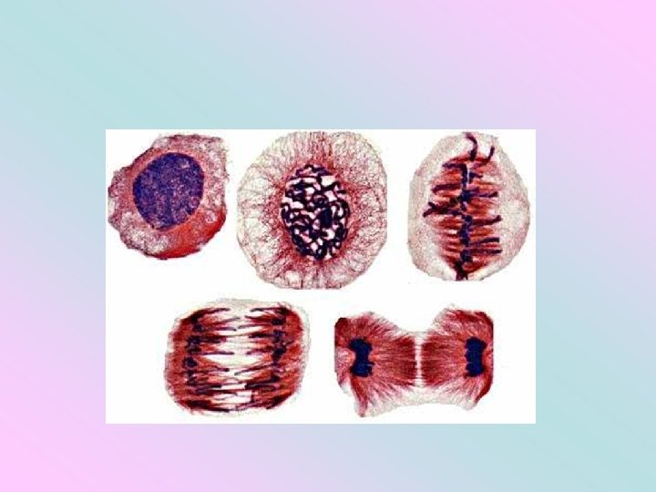
Try to identify the stages of mitosis in the two photomicrographs on the back of your sheet. Write a sentence for each one to explain how you reached your answer.
Stages of the cell cycle 20 of 32 © Boardworks Ltd 2008