GUMBORO DISEASE SYNONYMS INFECTIOUS BURSAL DISEASE INFECTIOUS BURSITIS

GUMBORO DISEASE SYNONYMS: INFECTIOUS BURSAL DISEASE, INFECTIOUS BURSITIS AND AVIAN NEPHROSIS Dr. SANJIV KUMAR ASSTT. PROFESSOR, DEPTT. OF PATHOLOGY, BVC, BASU, PATNA
 is seen in young domestic chickens worldwide and")
INTRODUCTION • Infectious bursal disease (IBD) is seen in young domestic chickens worldwide and is caused by infectious bursal disease virus (IBDV). • Symptoms of the clinical disease can include depression, watery diarrhea, ruffled feathers, and dehydration. Acute highly contagious infection of chickens • First report in Gumboro (Delware District of USA) • Economically significant, because heavy mortality in 3 – 6 wks old chickens and severe prolonged immunosuppression of chickens infected at an early age.

Etiology • Infectious bursal disease is caused by a birnavirus (infectious bursal disease virus; Birna virus ( ds RNA) - Birna = two • Two serotypes of IBDV have been identified. The serotype 1 viruses cause disease in chickens and, within them, antigenic variation can exist between strains. Antigenic drift is largely responsible for this antigenic variation, but antigenic differences can also occur through genome homologous recombination. • Serotype 2 strains of the virus infect chickens and turkeys but have not caused clinical disease or immunosuppression in these hosts.

Transmission • Mainly by contaminated feed and water. • Affected birds excrete the virus in faeces for 10 -14 days • Role of mechanical vectors (Human, wild birds, insects) • No vertical transmission and carriers

PATHOGENESIS • Clinical disease is associated to bird age with the greatest bursal mass, which occurs between 3 and 6 weeks of age. • Ig. M-bearing B-lymphocytes (lymphoblasts), the main target of infection. • Young birds at around two to eight weeks of age are more susceptible to disease. • Birds over eight weeks are resistant to challenge and will not show clinical signs unless infected by highly virulent strains. • Subclinical disease occurs in chickens infected before three weeks of age. • The B-cell destruction is usually most severe in subclinically infected young, as virus will destroy a smaller population and most cells in one place (the bursa).
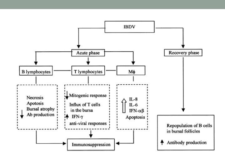

• After ingestion, the virus destroys the lymphoid follicles in the bursa of Fabricius as well as the circulating B-cells in the secondary lymphoid tissues such as GALT (gutassociated lymphoid tissue), CALT (conjunctiva), BALT (Bronchial) caecal tonsils, Harderian gland, etc. • Acute disease and death is due to the necrotizing effect of these viruses on the host tissues. • Kidney failure is a common cause of mortality. • If the bird survives and recovers from this phase of the disease, it remains immunocompromised which means it is more susceptible to other diseases.

CLINICAL SIGNS Morbidity typically reaches 100%. In the acute form birds are prostrated, debilitated and dehydrated. They produce a watery diarrhea and may have swollen feces-stained vent. Mortality rates vary with virulence of the strain involved, the challenge dose, previous immunity, presence of concurrent disease, as well as the flock's ability to mount an effective immune response. Immunosuppression of very young chickens, less than three weeks of age, is possibly the most important outcome and may not be clinically detectable (subclinical).
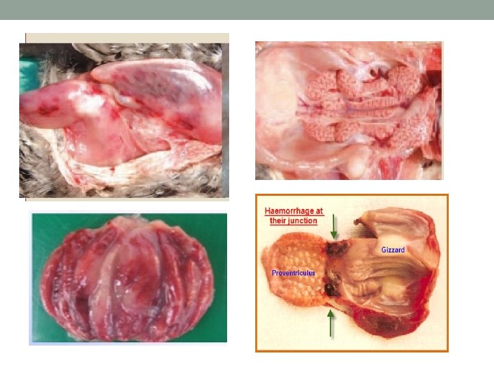
, some times at the junction of")
GROSS PATHOLOGY • Muscular haemorrhage (thigh and pectoral), some times at the junction of proventriculus and gizzard. • Haemorrhages of pectoral leg muscles are typical of IBD • Liver- Hepatomegaly and peripheral infarcts • Spleen- Splenomegaly • Kidneys- Swelling and white appearance, dilatation of tubules with urates ( cell debris, occasionally). Bursa- Enlarged, inflamed, edematous and cream coloured (early) and arophy (after 3 – 8 days). Haemorrhage on the internal and serosal surfaces. Caseous core within the lumen from sloughed epithelium

Lesions • Bursa- inflammation and destruction of lymphocytes, later regenerates. • Spleen - Moderate lymphoid cell necrosis • Thymus and caecal tonsil - Lymphoid cellular reaction (early stage), but less extensive damage • Kidneys - Non – specific, degenerative changes • Liver - Mild perivascular infiltration of monocytes.
")
DIAGNOSIS • Based on history, clinical signs and gross lesions AGPT (using macerated bursa) ELISA Immunoperoxidase Immunofluorescence Virus isolation (rarely) - Time consuming process Inoculation of suspected bursa into 10 – 11 days old embryonated eggs Some strains grow on Chick embryo fibroblast, vero cells or certain lymphoblastoid cell cultures
- Slides: 12