
Guinea Pig Diseases Antibiotic toxicity Antibiotics may suppress

Guinea Pig Diseases
Antibiotic toxicity Antibiotics may suppress clinical signs but usually do not eliminate the organism and fatal antibioticinduced enterocolitis is always a concern in treating guinea pigs. ¡ Antibiotics implicated include penicillin, aminoglycoside, erythromycin, clindamycin, lincomycin, chlonetracycline, estreptomycin, bacitracin, and dihydrostreptomycin. ¡
BACTERIAL AND FUNGAL DISEASES Bacterial and fungal agents are responsible for some of the most significant diseases of guinea pigs ¡
Bacterial diseases ¡ ¡ Several primary respiratory diseases of the guinea pig are of bacterial origin, with Bordetella bronchiseptica and Streptococcus pneumoniae being the most important. Other infectious agents act as opportunistic invaders in traumatized tissue (Streptococcus zooepidemicus), and contribute to enteric disease (Clostridium piliforme and Salmonella spp) and dermatitis (Trichophyton mentagrophytes).
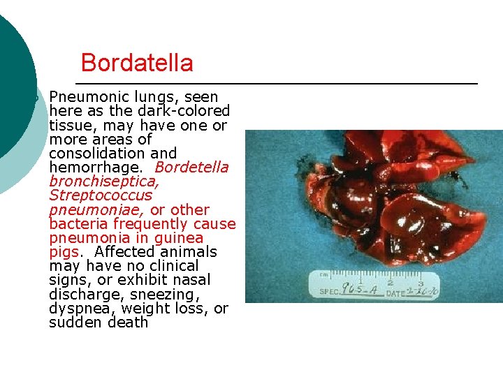
Bordatella ¡ Pneumonic lungs, seen here as the dark-colored tissue, may have one or more areas of consolidation and hemorrhage. Bordetella bronchiseptica, Streptococcus pneumoniae, or other bacteria frequently cause pneumonia in guinea pigs. Affected animals may have no clinical signs, or exhibit nasal discharge, sneezing, dyspnea, weight loss, or sudden death
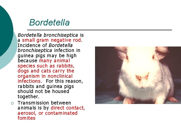
Bordetella ¡ ¡ Bordetella bronchiseptica is a small gram negative rod. Incidence of Bordetella bronchiseptica infection in guinea pigs may be high because many animal species such as rabbits, dogs and cats carry the organism in nonclinical infections. For this reason, rabbits and guinea pigs should not be housed together. Transmission between animals is by direct contact, aerosol, or contaminated fomites
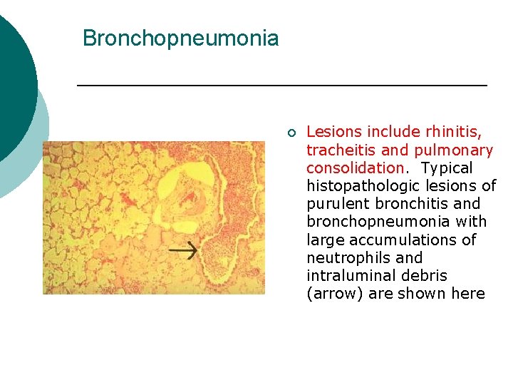
Bronchopneumonia ¡ Lesions include rhinitis, tracheitis and pulmonary consolidation. Typical histopathologic lesions of purulent bronchitis and bronchopneumonia with large accumulations of neutrophils and intraluminal debris (arrow) are shown here
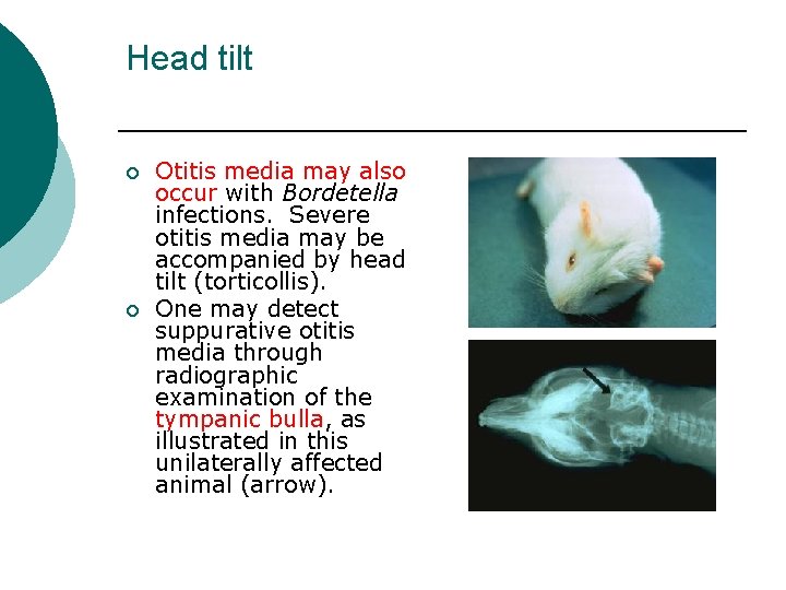
Head tilt ¡ ¡ Otitis media may also occur with Bordetella infections. Severe otitis media may be accompanied by head tilt (torticollis). One may detect suppurative otitis media through radiographic examination of the tympanic bulla, as illustrated in this unilaterally affected animal (arrow).
Bordatella Treatment of Bordetella bronchiseptica infection is often unrewarding. Antibiotics of choice include chloramphenicol, sulfamethazine and trimethoprimsulfa. ¡ Effective prevention includes good husbandry and maintenance of a closed Bordetella free colony ¡
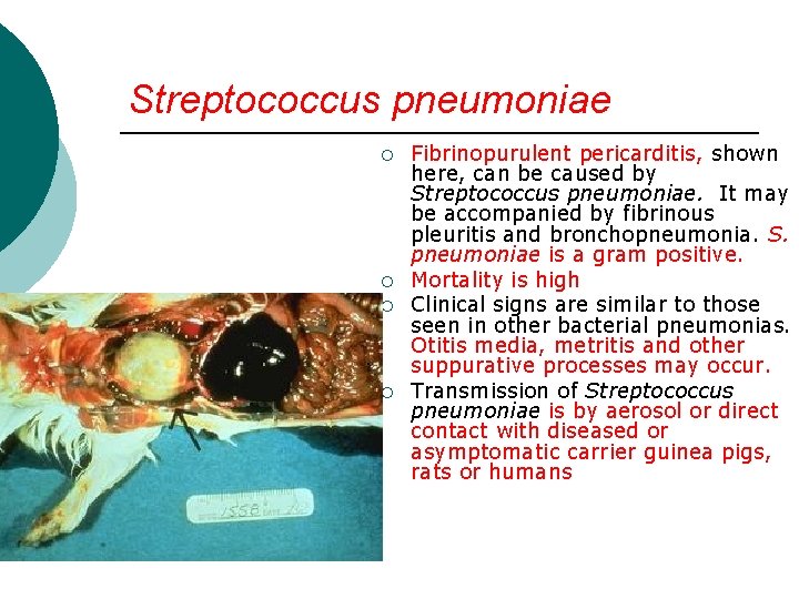
Streptococcus pneumoniae ¡ ¡ Fibrinopurulent pericarditis, shown here, can be caused by Streptococcus pneumoniae. It may be accompanied by fibrinous pleuritis and bronchopneumonia. S. pneumoniae is a gram positive. Mortality is high Clinical signs are similar to those seen in other bacterial pneumonias. Otitis media, metritis and other suppurative processes may occur. Transmission of Streptococcus pneumoniae is by aerosol or direct contact with diseased or asymptomatic carrier guinea pigs, rats or humans
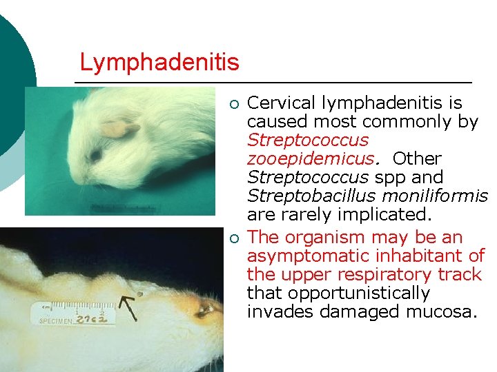
Lymphadenitis ¡ ¡ Cervical lymphadenitis is caused most commonly by Streptococcus zooepidemicus. Other Streptococcus spp and Streptobacillus moniliformis are rarely implicated. The organism may be an asymptomatic inhabitant of the upper respiratory track that opportunistically invades damaged mucosa.
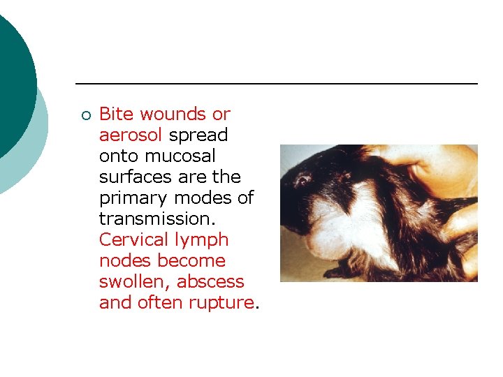
¡ Bite wounds or aerosol spread onto mucosal surfaces are the primary modes of transmission. Cervical lymph nodes become swollen, abscess and often rupture.
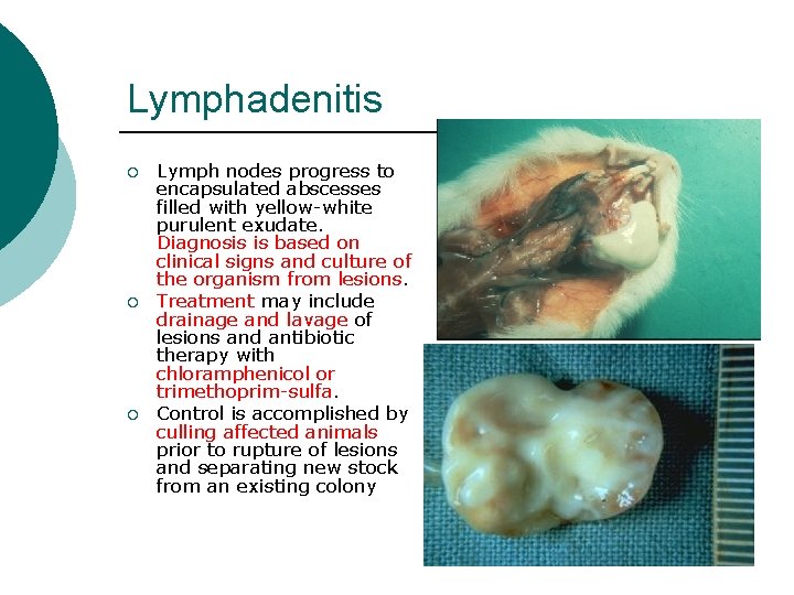
Lymphadenitis ¡ ¡ ¡ Lymph nodes progress to encapsulated abscesses filled with yellow-white purulent exudate. Diagnosis is based on clinical signs and culture of the organism from lesions. Treatment may include drainage and lavage of lesions and antibiotic therapy with chloramphenicol or trimethoprim-sulfa. Control is accomplished by culling affected animals prior to rupture of lesions and separating new stock from an existing colony
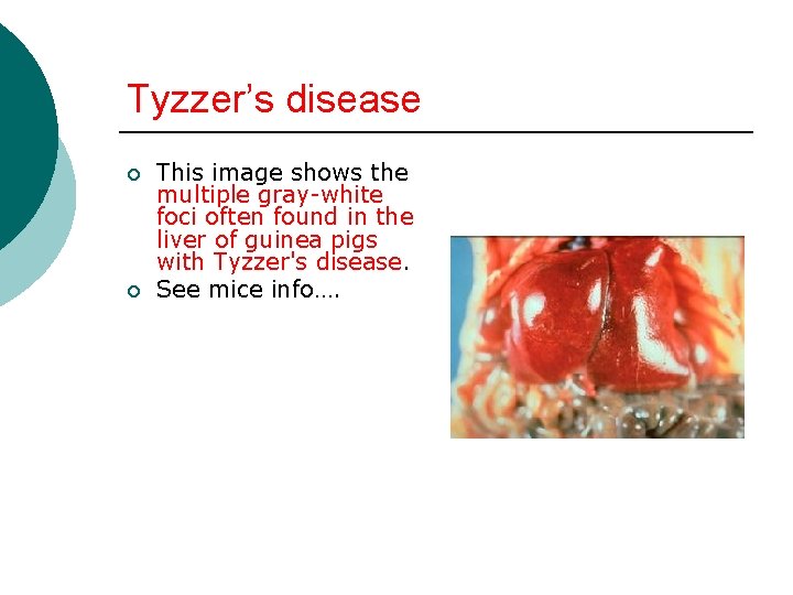
Tyzzer’s disease ¡ ¡ This image shows the multiple gray-white foci often found in the liver of guinea pigs with Tyzzer's disease. See mice info….
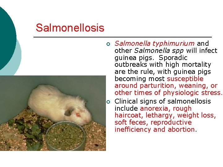
Salmonellosis ¡ ¡ Salmonella typhimurium and other Salmonella spp will infect guinea pigs. Sporadic outbreaks with high mortality are the rule, with guinea pigs becoming most susceptible around parturition, weaning, or other times of physiologic stress. Clinical signs of salmonellosis include anorexia, rough haircoat, lethargy, weight loss, soft feces, reproductive inefficiency and abortion.
Transmission ¡ Many animal species harbor Salmonella organisms which are shed intermittently in the feces. Transmission is fecal-oral; organisms often are introduced in contaminated foods such as fresh fruits and vegetables, or through the water supply.
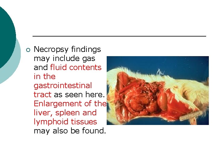
¡ Necropsy findings may include gas and fluid contents in the gastrointestinal tract as seen here. Enlargement of the liver, spleen and lymphoid tissues may also be found.
Salmonella ¡ Management Culture-positive animals should be removed from the colony. The room and equipment should be disinfected and restocked with clean animals.
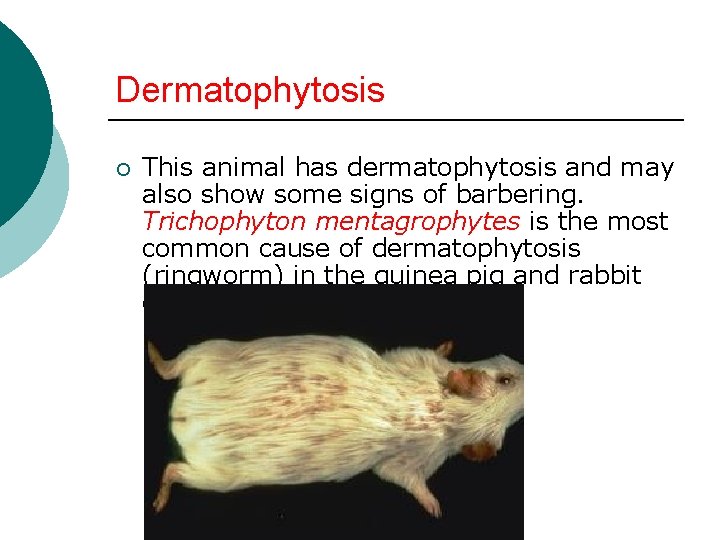
Dermatophytosis ¡ This animal has dermatophytosis and may also show some signs of barbering. Trichophyton mentagrophytes is the most common cause of dermatophytosis (ringworm) in the guinea pig and rabbit causing patchy alopecia
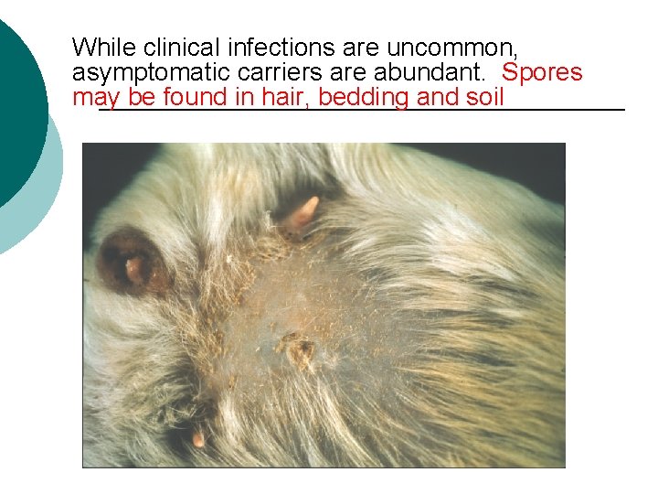
While clinical infections are uncommon, asymptomatic carriers are abundant. Spores may be found in hair, bedding and soil
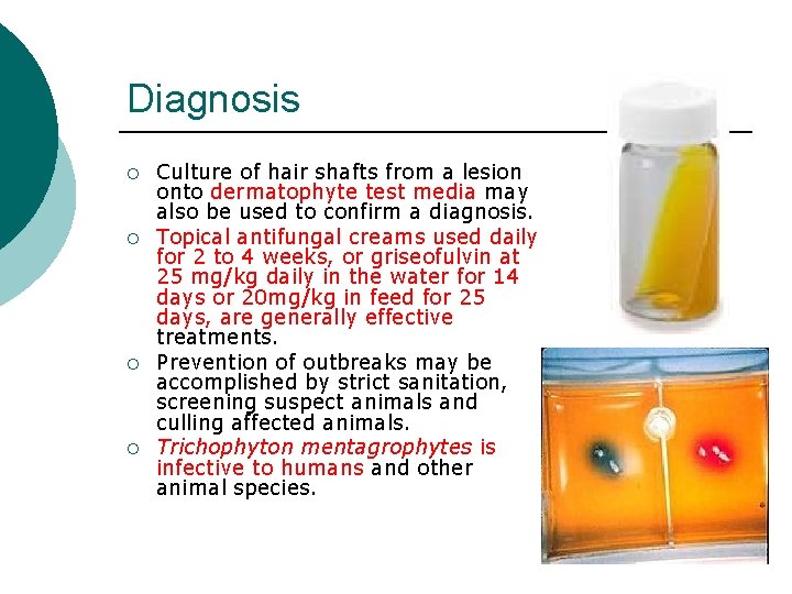
Diagnosis ¡ ¡ Culture of hair shafts from a lesion onto dermatophyte test media may also be used to confirm a diagnosis. Topical antifungal creams used daily for 2 to 4 weeks, or griseofulvin at 25 mg/kg daily in the water for 14 days or 20 mg/kg in feed for 25 days, are generally effective treatments. Prevention of outbreaks may be accomplished by strict sanitation, screening suspect animals and culling affected animals. Trichophyton mentagrophytes is infective to humans and other animal species.
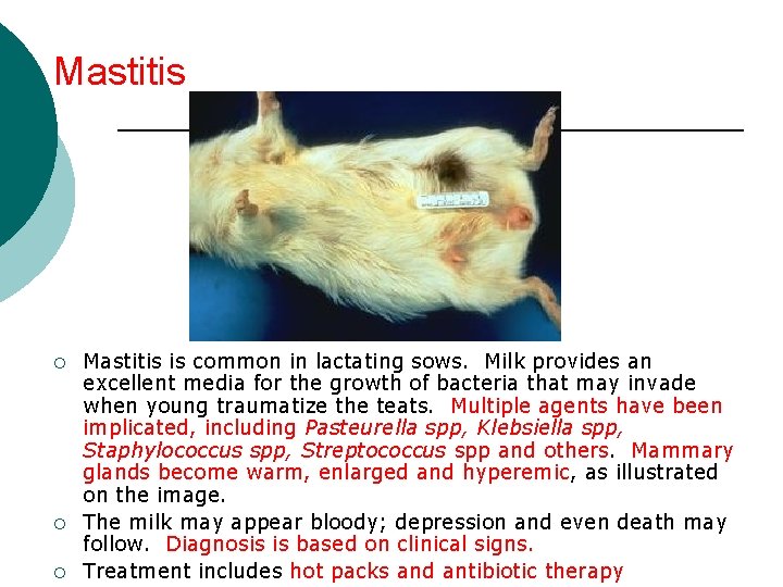
Mastitis ¡ ¡ ¡ Mastitis is common in lactating sows. Milk provides an excellent media for the growth of bacteria that may invade when young traumatize the teats. Multiple agents have been implicated, including Pasteurella spp, Klebsiella spp, Staphylococcus spp, Streptococcus spp and others. Mammary glands become warm, enlarged and hyperemic, as illustrated on the image. The milk may appear bloody; depression and even death may follow. Diagnosis is based on clinical signs. Treatment includes hot packs and antibiotic therapy
VIRAL AND CHLAMYDIAL DISEASES ¡ Some viral and chlamydial disease agents are documented, but they are of limited importance
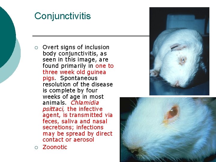
Conjunctivitis ¡ ¡ Overt signs of inclusion body conjunctivitis, as seen in this image, are found primarily in one to three week old guinea pigs. Spontaneous resolution of the disease is complete by four weeks of age in most animals. Chlamidia psittaci, the infective agent, is transmitted via feces, saliva and nasal secretions; infections may be spread by direct contact or aerosol Zoonotic
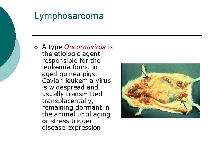
Lymphosarcoma ¡ A type Oncornavirus is the etiologic agent responsible for the leukemia found in aged guinea pigs. Cavian leukemia virus is widespread and usually transmitted transplacentally, remaining dormant in the animal until aging or stress trigger disease expression.
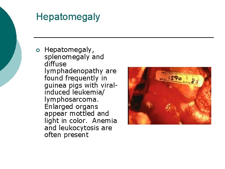
Hepatomegaly ¡ Hepatomegaly, splenomegaly and diffuse lymphadenopathy are found frequently in guinea pigs with viralinduced leukemia/ lymphosarcoma. Enlarged organs appear mottled and light in color. Anemia and leukocytosis are often present
PARASITES ¡ Many parasites are known to infect guinea pigs, but as a rule, parasites are more a problem in hobby and breeding colonies than in the research setting.
Ectoparasites ¡ Common ectoparasites of the guinea pig include the mites Trixicaris caviae and Chirodiscoides caviae, as sell as the lice, Gliricola porcelli and Gyropus ovalis.
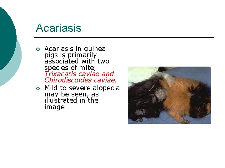
Acariasis ¡ ¡ Acariasis in guinea pigs is primarily associated with two species of mite, Trixacaris caviae and Chirodiscoides caviae. Mild to severe alopecia may be seen, as illustrated in the image
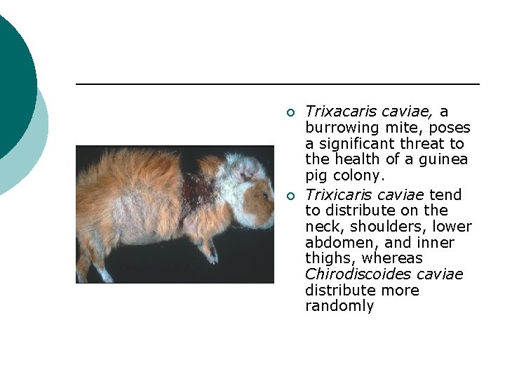
¡ ¡ Trixacaris caviae, a burrowing mite, poses a significant threat to the health of a guinea pig colony. Trixicaris caviae tend to distribute on the neck, shoulders, lower abdomen, and inner thighs, whereas Chirodiscoides caviae distribute more randomly
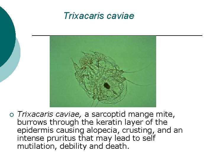
Trixacaris caviae ¡ Trixacaris caviae, a sarcoptid mange mite, burrows through the keratin layer of the epidermis causing alopecia, crusting, and an intense pruritus that may lead to self mutilation, debility and death.
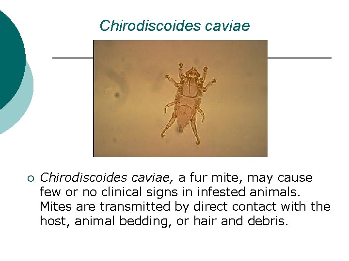
Chirodiscoides caviae ¡ Chirodiscoides caviae, a fur mite, may cause few or no clinical signs in infested animals. Mites are transmitted by direct contact with the host, animal bedding, or hair and debris.
Treatments ¡ Reported treatments for acariasis include pyrethroid flea powder dusts, lime sulfur dips and injections with ivermectin.
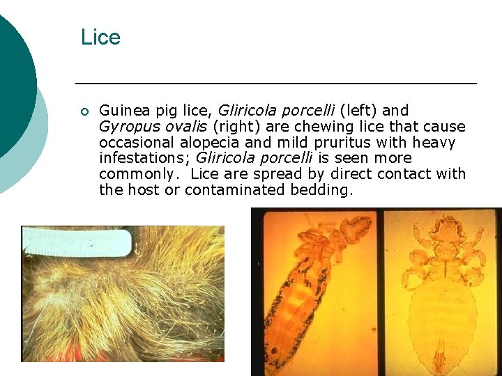
Lice ¡ Guinea pig lice, Gliricola porcelli (left) and Gyropus ovalis (right) are chewing lice that cause occasional alopecia and mild pruritus with heavy infestations; Gliricola porcelli is seen more commonly. Lice are spread by direct contact with the host or contaminated bedding.
Noninfectious Disease
NONINFECTIOUS DISEASES OF GUINEA PIGS The noninfectious diseases of guinea pigs may be placed in four categories: ¡ Nutritional and metabolic diseases ¡ Management-related diseases ¡ Other noninfectious diseases ¡ Neoplasia
NUTRITIONAL AND METABOLIC DISEASES ¡ Nutritional and metabolic diseases, our first category, represent a major cause of illness in guinea pigs. These diseases present as clinical or, usually, subclinical entities. The diseases discussed here are hypovitaminosis C (or scurvy), pregnancy toxemia and metastatic mineralization.
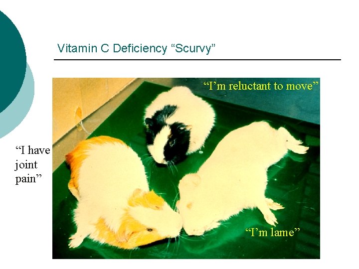
Vitamin C Deficiency “Scurvy” “I’m reluctant to move” “I have joint pain” “I’m lame”
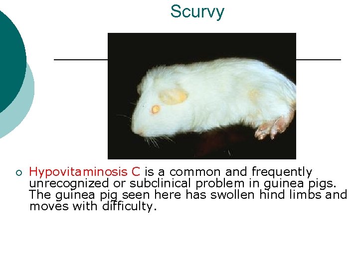
Scurvy ¡ Hypovitaminosis C is a common and frequently unrecognized or subclinical problem in guinea pigs. The guinea pig seen here has swollen hind limbs and moves with difficulty.
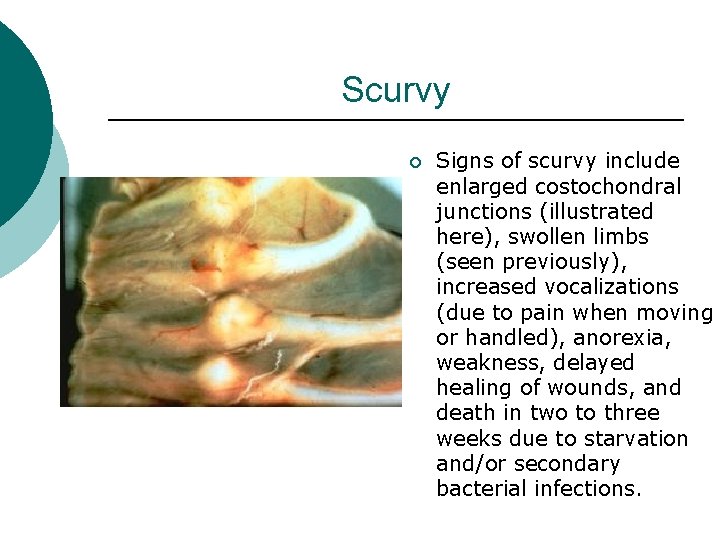
Scurvy ¡ Signs of scurvy include enlarged costochondral junctions (illustrated here), swollen limbs (seen previously), increased vocalizations (due to pain when moving or handled), anorexia, weakness, delayed healing of wounds, and death in two to three weeks due to starvation and/or secondary bacterial infections.
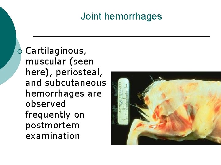
Joint hemorrhages ¡ Cartilaginous, muscular (seen here), periosteal, and subcutaneous hemorrhages are observed frequently on postmortem examination
Treatment/ prevention ¡ ¡ Clinically ill guinea pigs should receive vitamin C daily until improvement is seen. The amount of vitamin C added to the diet will vary, but is approximately 50 mg/kg. A daily source of ascorbic acid should be provided to every guinea pig at a dosage rate of 10 mg/kg per day for normal guinea pigs and 30 mg/kg per day for pregnant sows.
Sources of vitamin C ¡ A daily source of vitamin C is provided by use of high quality, commercially available guinea pig diets that have been stored in a cool, dry place. The diet should be used within 90 days of the milling date. Good sources of vitamin C include a handful of kale or cabbage, 1/4 fresh orange or green pepper, or the addition of ascorbic acid to the drinking water (200 -400 mg per liter of water, prepared daily).
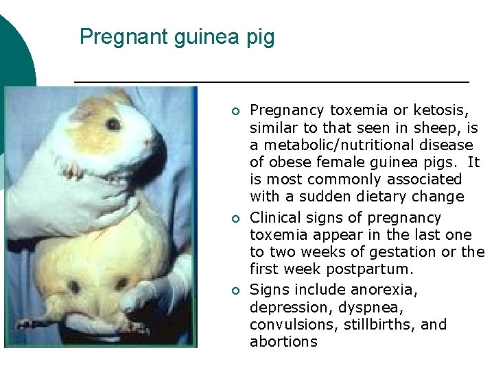
Pregnant guinea pig ¡ ¡ ¡ Pregnancy toxemia or ketosis, similar to that seen in sheep, is a metabolic/nutritional disease of obese female guinea pigs. It is most commonly associated with a sudden dietary change Clinical signs of pregnancy toxemia appear in the last one to two weeks of gestation or the first week postpartum. Signs include anorexia, depression, dyspnea, convulsions, stillbirths, and abortions
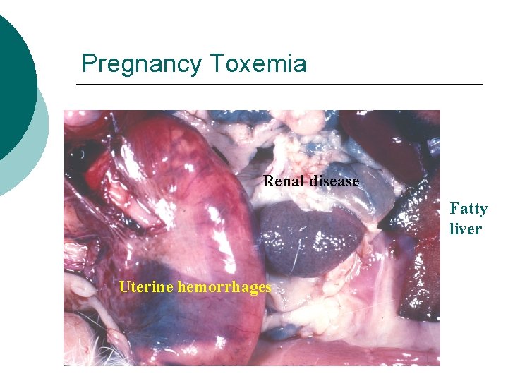
Pregnancy Toxemia Renal disease Fatty liver Uterine hemorrhages
MANAGEMENT-RELATED DISEASES ¡ A number of other noninfectious diseases occur in guinea pigs, many of which are related to the management of the animals. Those to be discussed include antibioticinduced enterotoxemia, heat stress, pododermatitis, cage trauma, vaginitis, and preputial plugs
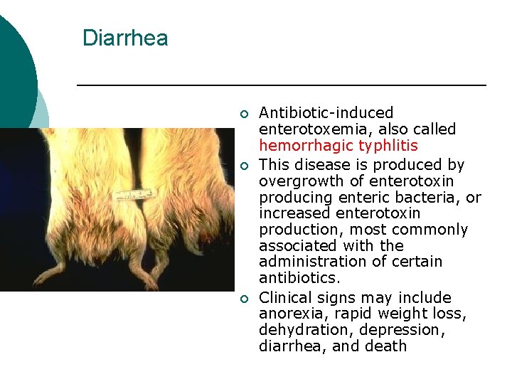
Diarrhea ¡ ¡ ¡ Antibiotic-induced enterotoxemia, also called hemorrhagic typhlitis This disease is produced by overgrowth of enterotoxin producing enteric bacteria, or increased enterotoxin production, most commonly associated with the administration of certain antibiotics. Clinical signs may include anorexia, rapid weight loss, dehydration, depression, diarrhea, and death
Antibiotics to avoid ¡ Antibiotics implicated include penicillin, erythromycin, lincomycin, chlortetracycline, oxytetracycline, bacitracin, and dihydrostreptomycin
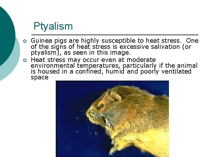
Ptyalism ¡ ¡ Guinea pigs are highly susceptible to heat stress. One of the signs of heat stress is excessive salivation (or ptyalism), as seen in this image. Heat stress may occur even at moderate environmental temperatures, particularly if the animal is housed in a confined, humid and poorly ventilated space
Treatment ¡ Treatment is usually unrewarding but would include rapid cooling with cool running water or ice water, steroids and administration of supportive care.
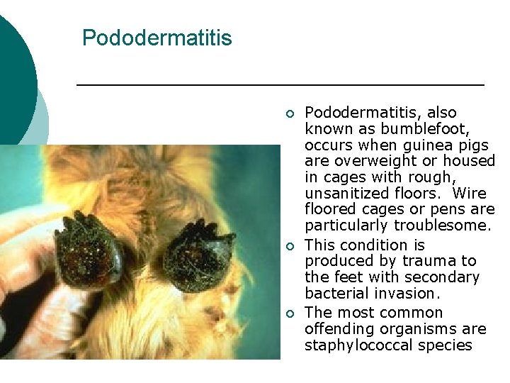
Pododermatitis ¡ ¡ ¡ Pododermatitis, also known as bumblefoot, occurs when guinea pigs are overweight or housed in cages with rough, unsanitized floors. Wire floored cages or pens are particularly troublesome. This condition is produced by trauma to the feet with secondary bacterial invasion. The most common offending organisms are staphylococcal species
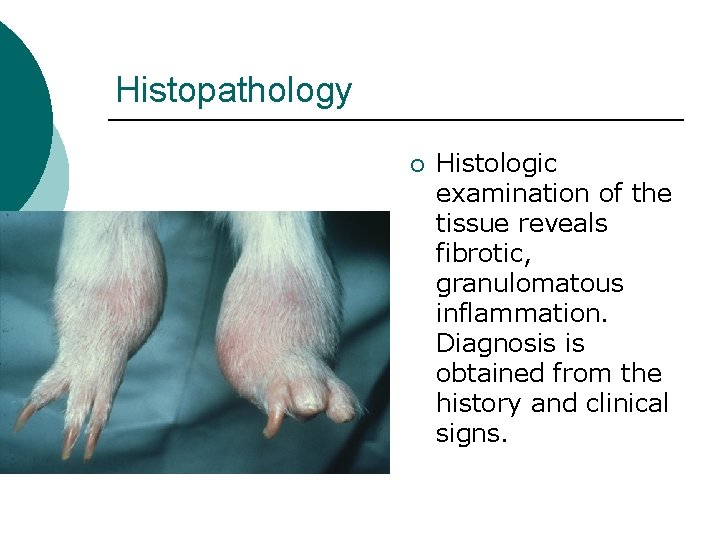
Histopathology ¡ Histologic examination of the tissue reveals fibrotic, granulomatous inflammation. Diagnosis is obtained from the history and clinical signs.
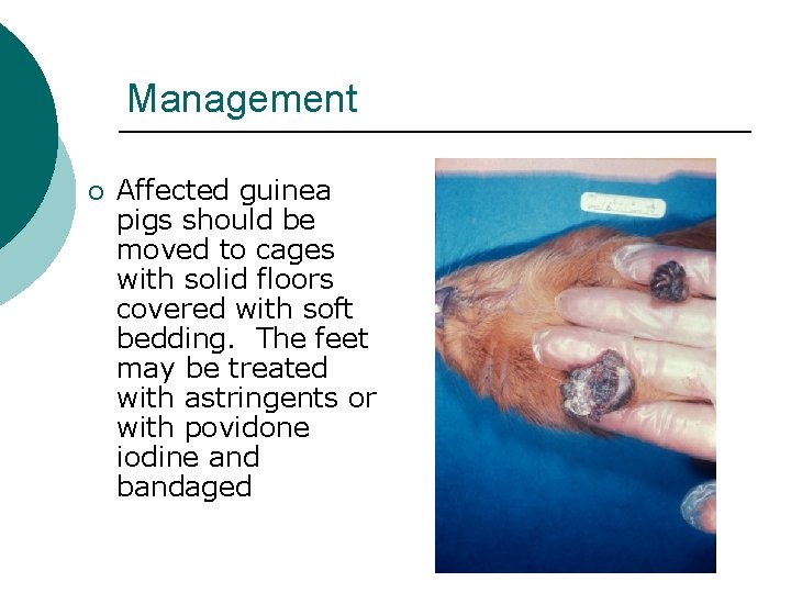
Management ¡ Affected guinea pigs should be moved to cages with solid floors covered with soft bedding. The feet may be treated with astringents or with povidone iodine and bandaged
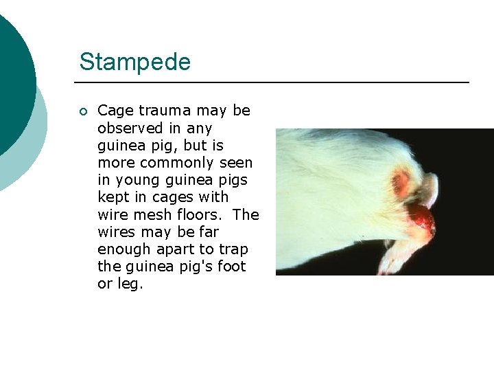
Stampede ¡ Cage trauma may be observed in any guinea pig, but is more commonly seen in young guinea pigs kept in cages with wire mesh floors. The wires may be far enough apart to trap the guinea pig's foot or leg.
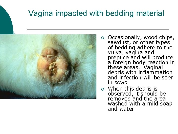
Vagina impacted with bedding material ¡ ¡ Occasionally, wood chips, sawdust, or other types of bedding adhere to the vulva, vagina and prepuce and will produce a foreign body reaction in these areas. Vaginal debris with inflammation and infection will be seen in sows. When this debris is observed, it should be removed and the area washed with a mild soap and water
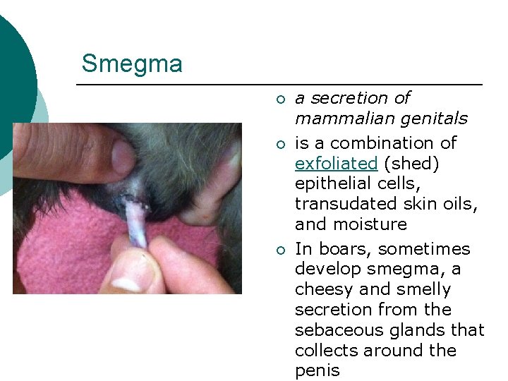
Smegma ¡ ¡ ¡ a secretion of mammalian genitals is a combination of exfoliated (shed) epithelial cells, transudated skin oils, and moisture In boars, sometimes develop smegma, a cheesy and smelly secretion from the sebaceous glands that collects around the penis

Barbered guinea pig ¡ Barbering or hair pulling among cage mates and transient hair loss of weanlings will affect small areas, regions, or the entire body. A diagnosis of a specific type of alopecia is based on history and clinical signs. Treatment involves separating offending guinea pigs
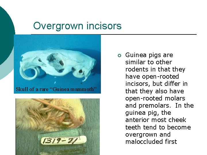
Overgrown incisors ¡ Skull of a rare “Guinea mammoth” Guinea pigs are similar to other rodents in that they have open-rooted incisors, but differ in that they also have open-rooted molars and premolars. In the guinea pig, the anterior most cheek teeth tend to become overgrown and maloccluded first
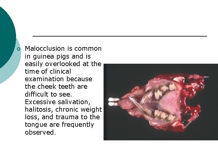
¡ Malocclusion is common in guinea pigs and is easily overlooked at the time of clinical examination because the cheek teeth are difficult to see. Excessive salivation, halitosis, chronic weight loss, and trauma to the tongue are frequently observed.
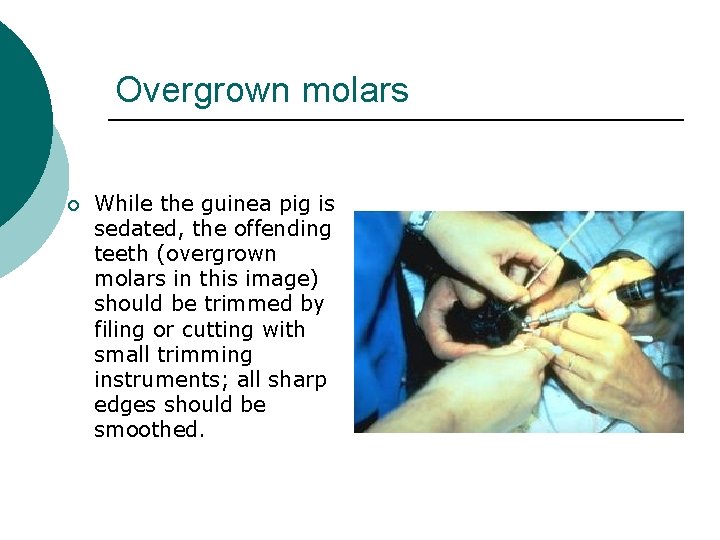
Overgrown molars ¡ While the guinea pig is sedated, the offending teeth (overgrown molars in this image) should be trimmed by filing or cutting with small trimming instruments; all sharp edges should be smoothed.
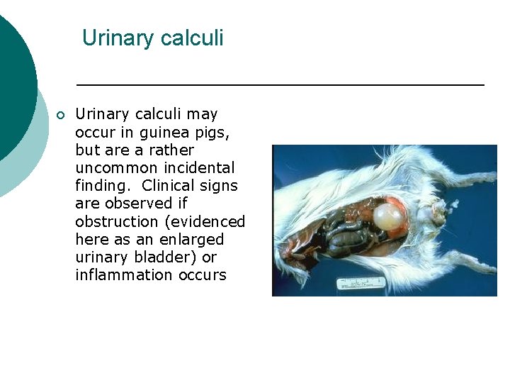
Urinary calculi ¡ Urinary calculi may occur in guinea pigs, but are a rather uncommon incidental finding. Clinical signs are observed if obstruction (evidenced here as an enlarged urinary bladder) or inflammation occurs
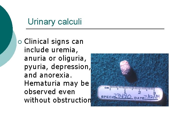
Urinary calculi ¡ Clinical signs can include uremia, anuria or oliguria, pyuria, depression, and anorexia. Hematuria may be observed even without obstruction.
Resources ¡ Center for Laboratory Animal Medicine and Care University of Texas Houston Health Sciences Center
- Slides: 63