Great vessels of the thorax abdomen and pelvis

Great vessels of the thorax, abdomen and pelvis Dr Amal Albtoosh Al-Rawashdeh

Lecture plan v Types of blood vessels v The differences between arteries and veins v Some terms related to arteries nomination v Great blood vessels of the thorax v Great blood vessels of the abdomin v. Great blood vessels of the pelvis v list the MAIN Aorta branches in the human body

Blood vessels • Blood vessels are found throughout the body. • There are five main types of blood vessels: üArteries, üArterioles, üCapillaries, ü Venules üAnd veins.
 • Carry blood AWAY from")
ARTERIES VS. VEINS Arteries ( red ? ? ) • Carry blood AWAY from the heart • Are usually positioned deeper within the body • More muscular than veins • Generally remain open if blood flow stopped, due to their thick muscular layer. Vein ( blue ? ? ) • Carry blood BACK to the heart • Are usually positioned closer beneath the surface of the skin/ still we have deep veins • less muscular than arteries. Contain valves to help keep blood flowing in the right direction • Would collapse if blood flow stops.

Arteries terms • COMMON, TRUNK , RECURRENT, COLLATERAL ARTERIES • Other terms: circumflex, perforating, nutrients

Arteries terms cont. Common: shared by, coming from, or done by two or more people, groups, or things. Anatomy: will branch off at least to two main braches Example: The common iliac arteries are two large arteries that originate from the aortic bifurcation at the level of the fourth lumbar vertebra. They end in front of the sacroiliac joint, one on either side, and each bifurcates into the external and internal iliac arteries.
The main woody stem of a tree as")
Arteries terms cont v Trunk: (dic. )The main woody stem of a tree as distinct from its branches and roots. v Trunk of the human body: a person's or animal's body apart from the limbs and head. (synonym: torso) v So in arteries it will be the stem that branches off the main artery and then gives 3 or more branches
 turning back so")
Arteries terms cont. • Recurrent: (of a nerve or blood vessel) turning back so as to reverse direction. Example: radial recurrent artery. An artery with its origin in the radial artery that ascends around the lateral side of the elbow joint

Arteries terms • Collateral: additional but subordinate; secondary. Example: Radial collateral artery

Recurrent vs collateral • A collateral artery is generally one that participates in an anastomosis. A recurrent artery is one that branches off and then turns so that the direction of flow is opposite that of its parent artery.

Great blood vessels of the thorax • Pulmonary trunk • Aorta • Superior vena cava • Inferior vena cava • Pulmonary veins

The pulmonary trunk v. Is the first and largest vessel within the pulmonary arterial tree. v. It passes deoxygenated blood from the heart to the lungs. v It originates from the right ventricle and passes superiorly and posteriorly for about 4 -5 cm. v. During this course, it is ensheathed by the pericardial sac and initially is anterior to the ascending aorta.

Aorta v. The aorta is the first and the largest artery in the human body. v. Diameter : 3. 5 cm at its origin (ascending aorta), 2. 5 cm in the descending portion (thoracic aorta), and 1. 82 cm in the abdomen (abdominal aorta). v. Blood flow: 4900 milliliters per minute

Aorta v. The aorta can be divided into four sections: Øthe ascending aorta, Øthe aortic arch, Ø the thoracic (descending) aorta Ø the abdominal aorta. v. It terminates at the level of L 4 by bifurcating into the left and right common iliac arteries.
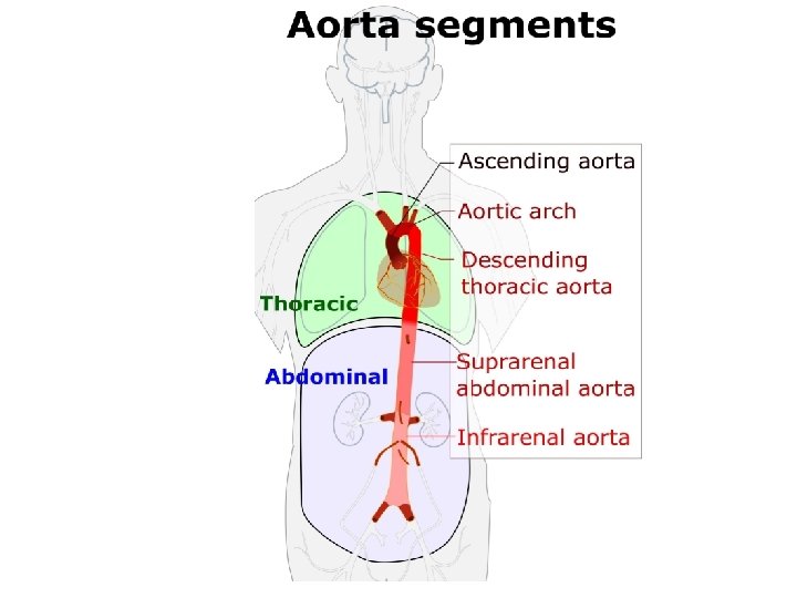

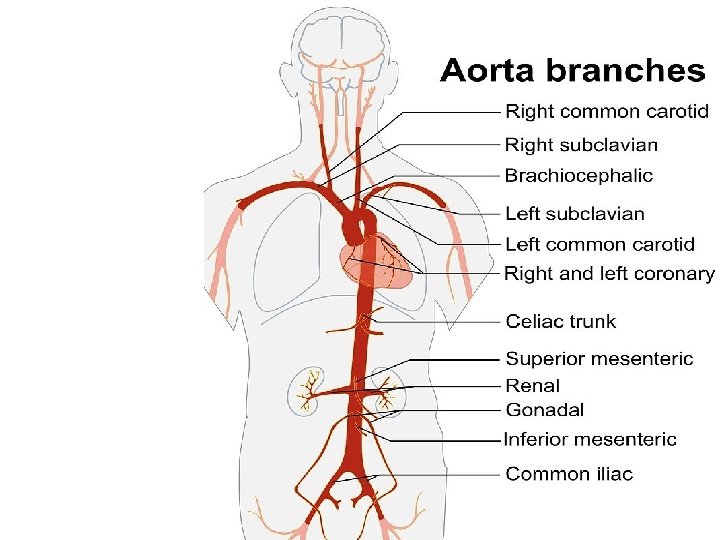
Ascending Aorta v arises from the aortic orifice from the left ventricle v ascends to become the aortic arch. v travels (with the pulmonary trunk) in the pericardial sheath. Branches Ø the left coronary Øthe right coronary arteries Both supply the myocardium. q The dilated, sinus portion, or aortic root segment of the ascending aorta continues superiorly with the tubular portion of the ascending aorta. The area of transition between these two components is marked by a sharp crease known as the sinotubular junction (STJ).

Aortic Arch v is a continuation of the ascending aorta v BEGINS at the level of the second sternocostal joint. v ENDS at the level of the T 4 vertebra v The arch is still connected to the pulmonary trunk by the ligamentum arteriosum (remnant of the foetal ductus arteriosus). Branches (three major branches) • Brachiocephalic trunk: The first and largest branch that ascends laterally to split into the right common carotid and right subclavian arteries. These arteries supply the right side of the head and neck, and the right upper limb. • Left common carotid artery: Supplies the left side of the head and neck. • Left subclavian artery: Supplies the left upper limb.

Descending Thoracic Aorta v. STARTS at the lower border of T 4 as a continuation of the aortic arch. v It passes through aortic opening of the diaphragm at T 12 v Relations with the esophagus A) Upper part: On the right side B) Middle part: anterior C) Lower part: on the left side
")
Branches of the descending (thoracic aorta)

The superior vena cava vis a large vein, v diameter of up to 2 cm v length of approximately 7 cm. v. It arises from the union of the left and right brachiocephalic veins, posterior to the first right costal cartilage. v. It descends vertically through the superior mediastinum, behind the intercostal spaces and to the right of the aorta and trachea. v. At the level of the second costal cartilage, the SVC enters the middle mediastinum and becomes surrounded by the fibrous pericardium. v It terminates by emptying into the SUPERIOR aspect of the right atrium at the level of the third costal cartilage.

Tributaries v. The superior vena cava contains venous blood from the head, neck, both upper limbs and from structures within the thorax v At the level of T 4, the superior vena cava receives the azygous vein, which drains the upper lumbar region and thoracic wall. The SVC receives tributaries from several minor vein groups: ØMediastinal veins ØOesophageal veins ØPericardial veins

Pulmonary Veins vreceive oxygenated blood from the lungs, delivering it to the left side of the heart to be pumped back around the body. v. There are four pulmonary veins, with one superior and one inferior for each of the lungs. v. They enter the pericardium to drain into the superior left atrium, on the posterior surface. v. The inferior left pulmonary vein is found at the hilum of the lung, vwhile the right inferior pulmonary vein runs posteriorly to the superior vena cava and the right atrium.

Inferior Vena Cava v. The inferior vena cava receives deoxygenated blood from the lower body (all structures inferior to the diaphragm), delivering it back to the heart. v. It is initially formed in the pelvis by the common iliac veins joining together. v It travels through the abdomen, collecting blood from the hepatic, lumbar, gonadal, renal and phrenic veins. v. The inferior vena cava then passes through the diaphragm, entering the pericardium at the level of T 8. v It drains into the inferior portion of the right atrium.

Great blood vessels of the abdomen and pelvis v. The abdomen and pelvis contain the majority of internal organs. v these regions supplied by an extensive network of arteries and veins. v. All arterial blood delivered to this region comes via branches of the ABDOMINAL AORTA, v. All venous blood eventually finds its way back to inferior vena cava (IVC).
 v. Common iliac veins at the level of L 5")
Inferior vena cava (IVC) v. Common iliac veins at the level of L 5 unite to form IVC to right atrium v Tributaries of IVC: 1 - Inferior phrenic veins (right and left). 2 - Right suprarenal vein (the left ends in the left renal vein). 3 - Renal veins (right and left). 4 - Right gonadal (testicular or ovarian) vein (the left ends in the left renal vein). 5 - The Lumbar veins (right and left). 6 - Common iliac veins (right and left). 7 - Hepatic veins (right and left).
 v. There")
Abdominal aorta v. Enters abdomen through aortic aperture of diaphragm (T 12) v. There are 10 major branches of the abdominal aorta. v Bifurcates at L 4 into common iliac to supply lower limb and pelvis

Paired branches Single branches Coeliac trunk T 12 Inferior phrenic artery Superior mesenteric artery L 1 Middle suprarenal artery ﻣﻦ ﻣﺤﺎﺿﺮﺓ ﺩ ﻳﻮﺳﻒ ﺣﺴﻴﻦ L 2 Renal artery Inferior mesenteric artery L 3 Gonadal artery Median sacral artery Abdominal aorta L 4 Lumbar arteries Common iliac artery

Blood supply to the pelvis v. Aorta ends by splitting into right, left common iliac arteries. v. Each common iliac splits into internal and external iliac arteries. v. External iliac passes under inguinal ligament to lower limb v. Internal iliac enters pelvis v. It is divided into anterior and posterior trunk v. The anterior trunk supplies muscles, visceral organ v. The posterior trunk supplies pelvic parietal structure.

- Slides: 30