
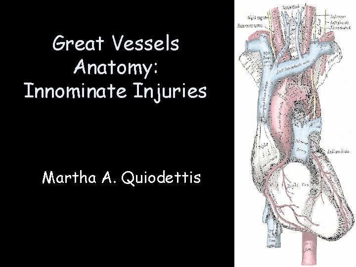
Great Vessels Anatomy Innominate Injuries Martha A Quiodettis
- Slides: 48
Great Vessels Anatomy: Innominate Injuries Martha A. Quiodettis
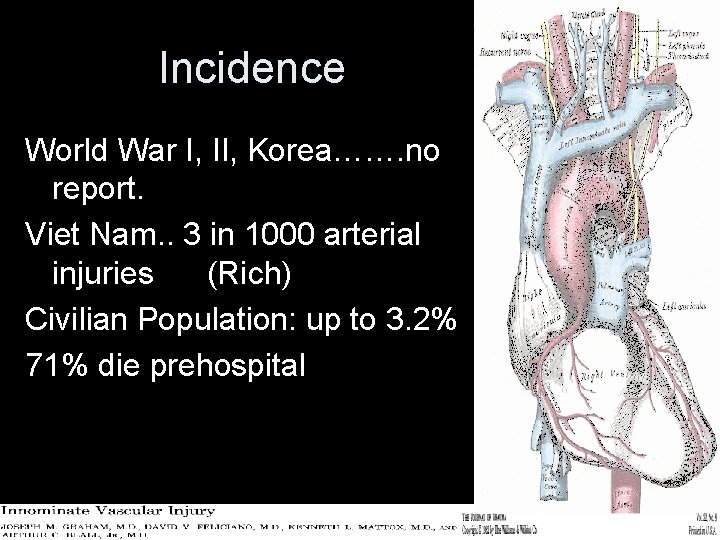
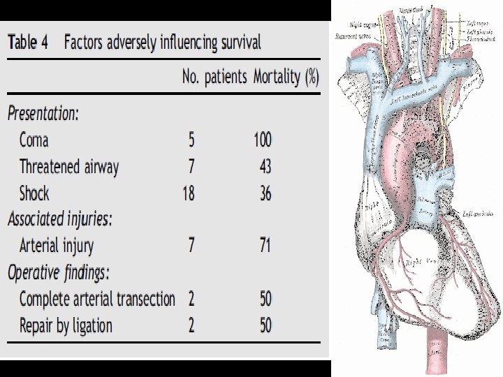
Incidence World War I, II, Korea……. no report. Viet Nam. . 3 in 1000 arterial injuries (Rich) Civilian Population: up to 3. 2% 71% die prehospital
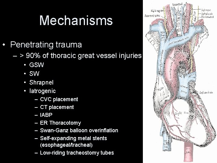
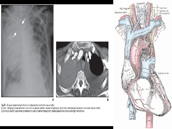
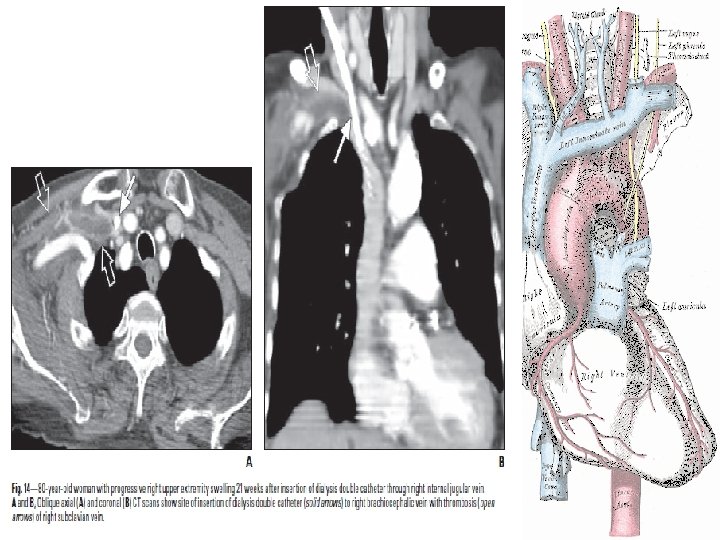
Mechanisms • Penetrating trauma – > 90% of thoracic great vessel injuries • • GSW SW Shrapnel Iatrogenic – – – CVC placement CT placement IABP ER Thoracotomy Swan-Ganz balloon overinflation Self-expanding metal stents (esophageal/tracheal) – Low-riding tracheostomy tubes
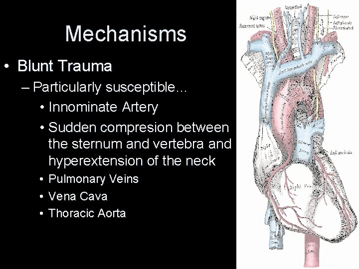
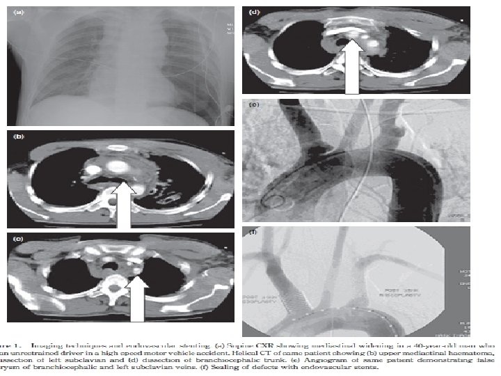
Mechanisms • Blunt Trauma – Particularly susceptible… • Innominate Artery • Sudden compresion between the sternum and vertebra and hyperextension of the neck • • • Pulmonary Veins Vena Cava Thoracic Aorta
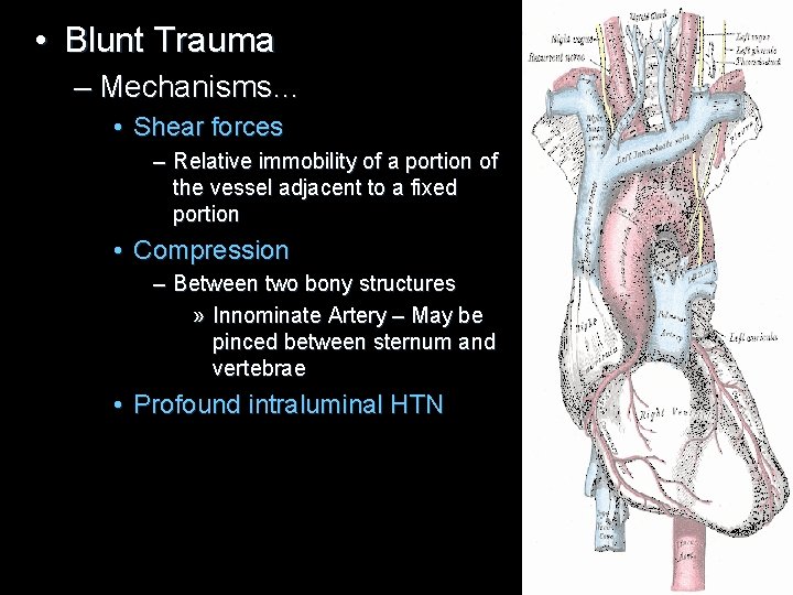
• Blunt Trauma – Mechanisms… • Shear forces – Relative immobility of a portion of the vessel adjacent to a fixed portion • Compression – Between two bony structures » Innominate Artery – May be pinced between sternum and vertebrae • Profound intraluminal HTN
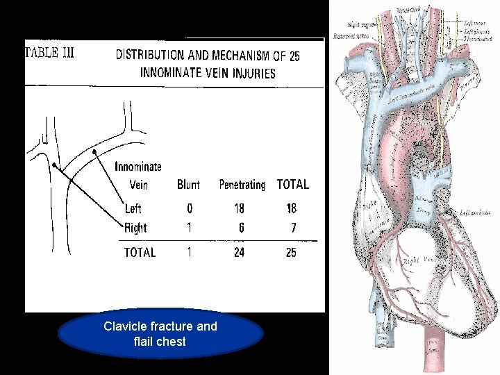
Clavicle fracture and flail chest
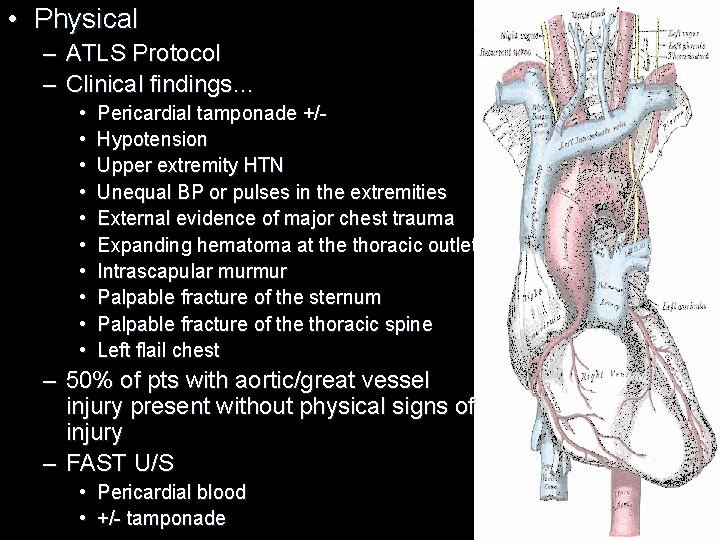
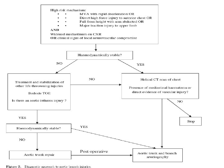
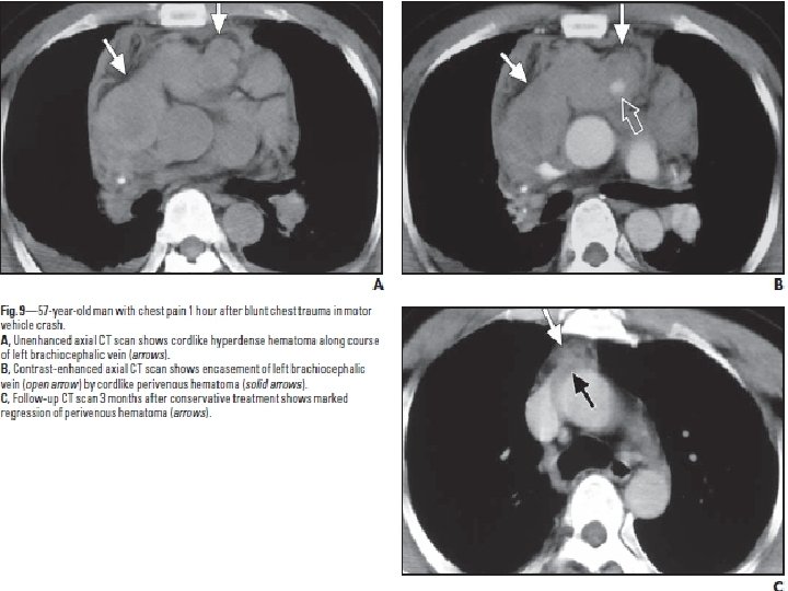
• Physical – ATLS Protocol – Clinical findings… • • • Pericardial tamponade +/Hypotension Upper extremity HTN Unequal BP or pulses in the extremities External evidence of major chest trauma Expanding hematoma at the thoracic outlet Intrascapular murmur Palpable fracture of the sternum Palpable fracture of the thoracic spine Left flail chest – 50% of pts with aortic/great vessel injury present without physical signs of injury – FAST U/S • Pericardial blood • +/- tamponade
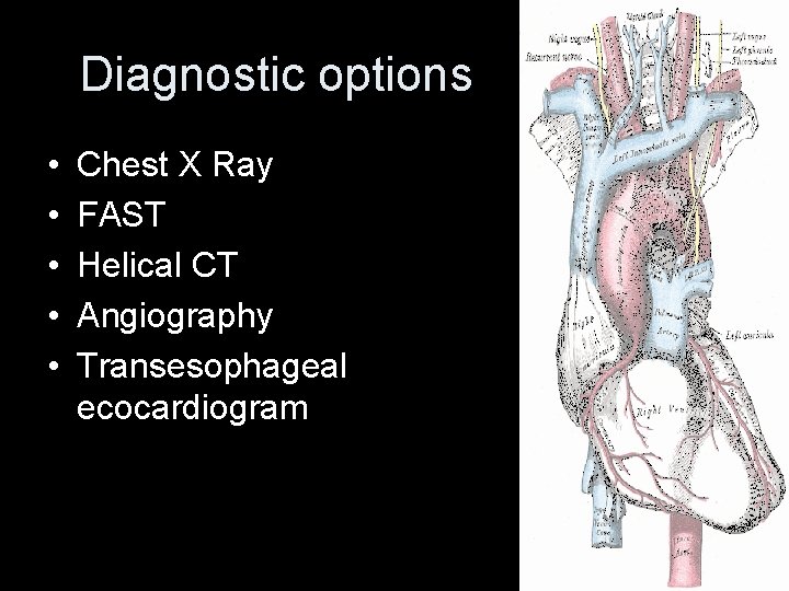
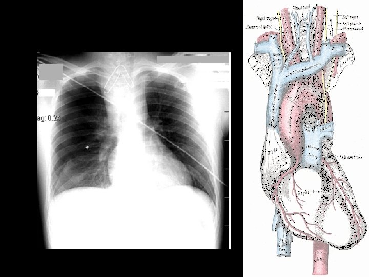
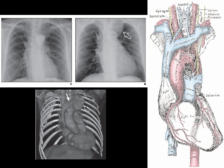
Diagnostic options • • • Chest X Ray FAST Helical CT Angiography Transesophageal ecocardiogram
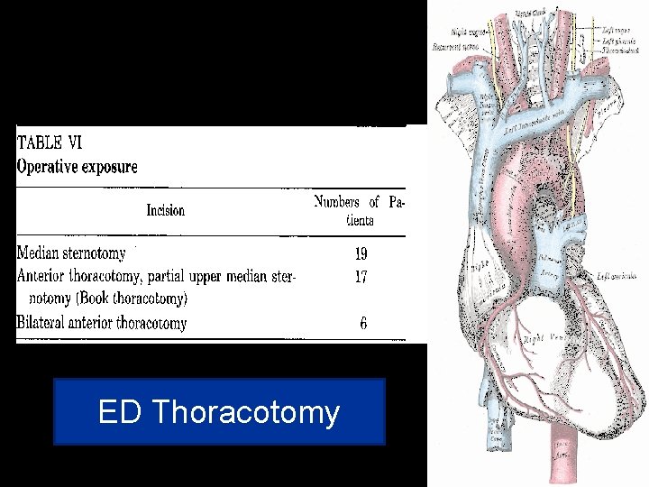
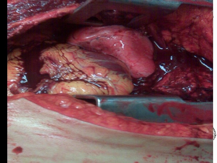
ED Thoracotomy
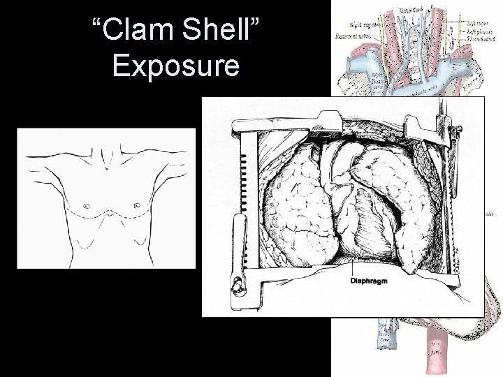
“Clam Shell” Exposure
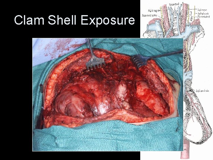
Clam Shell Exposure
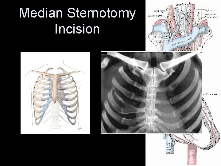
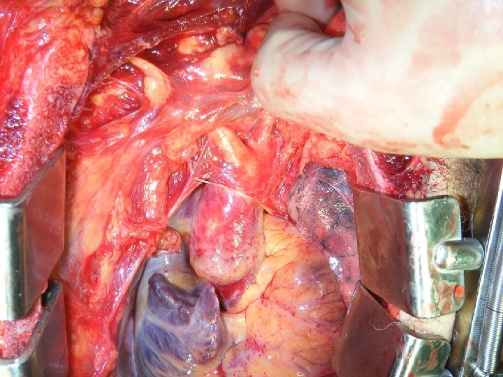
Median Sternotomy Incision
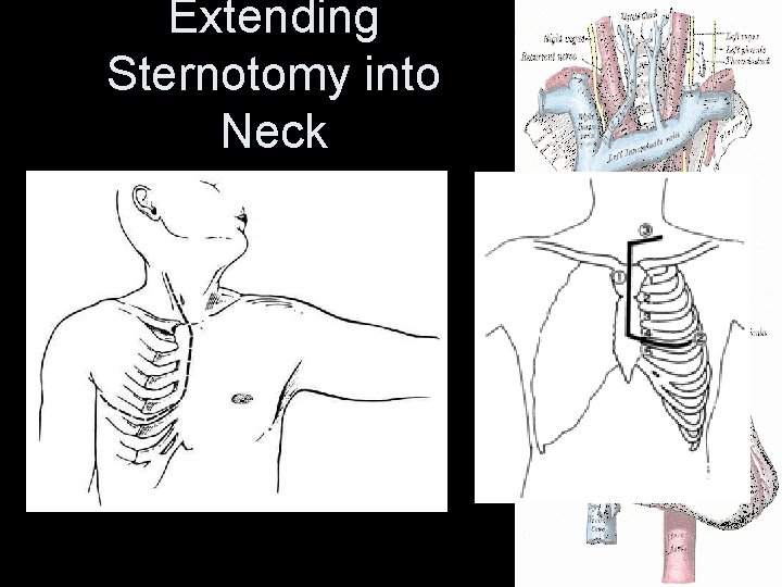
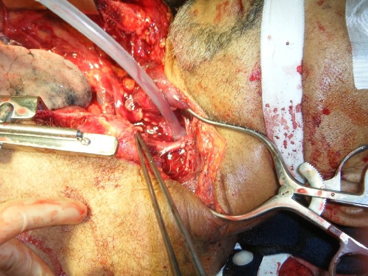
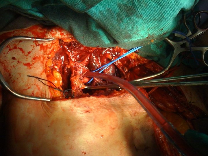
Extending Sternotomy into Neck
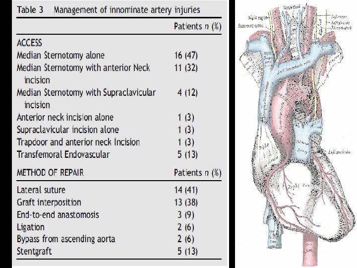
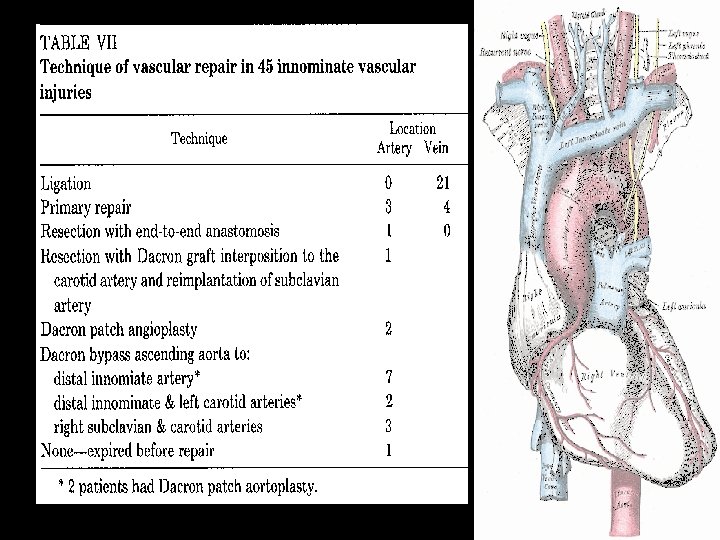
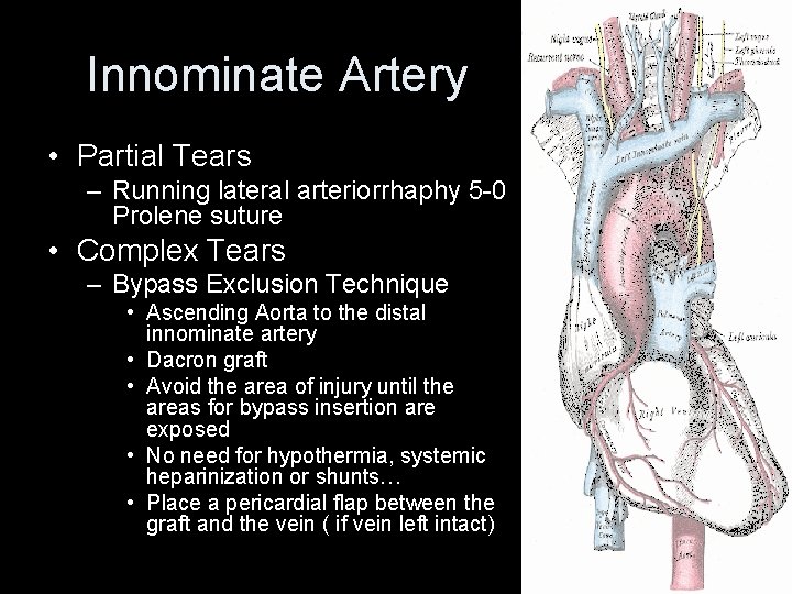
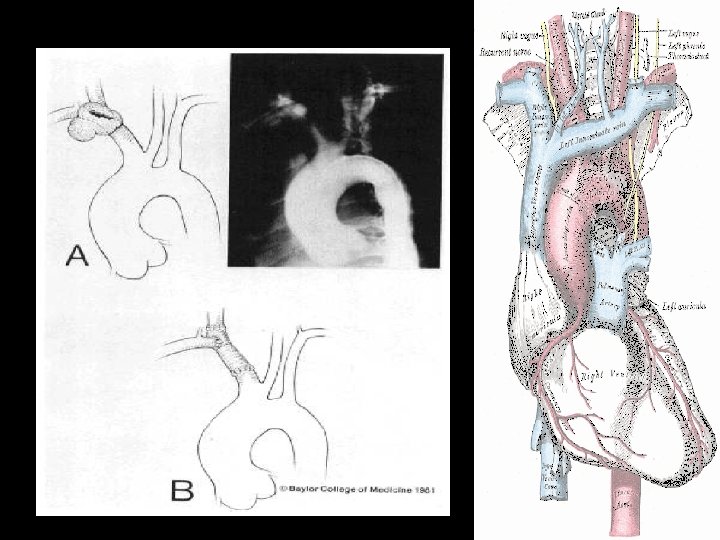
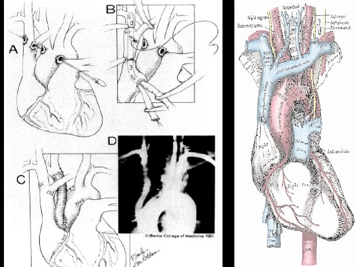
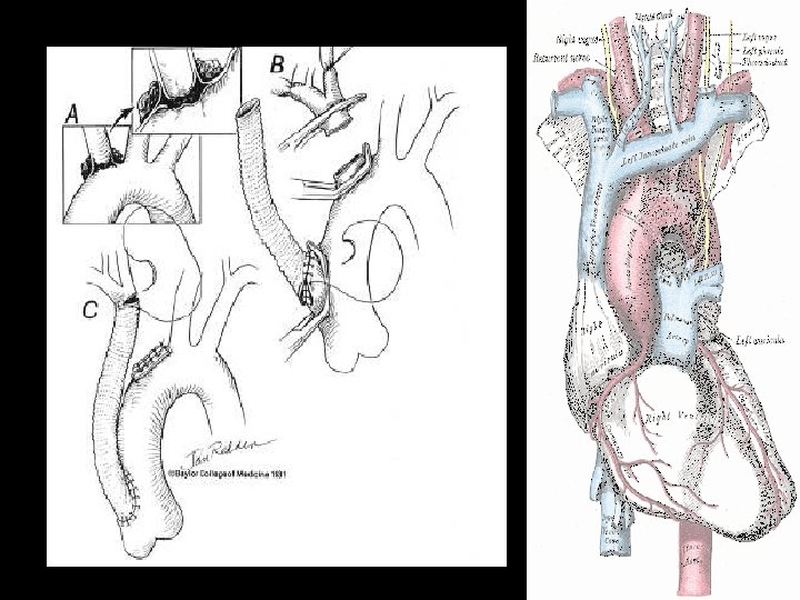
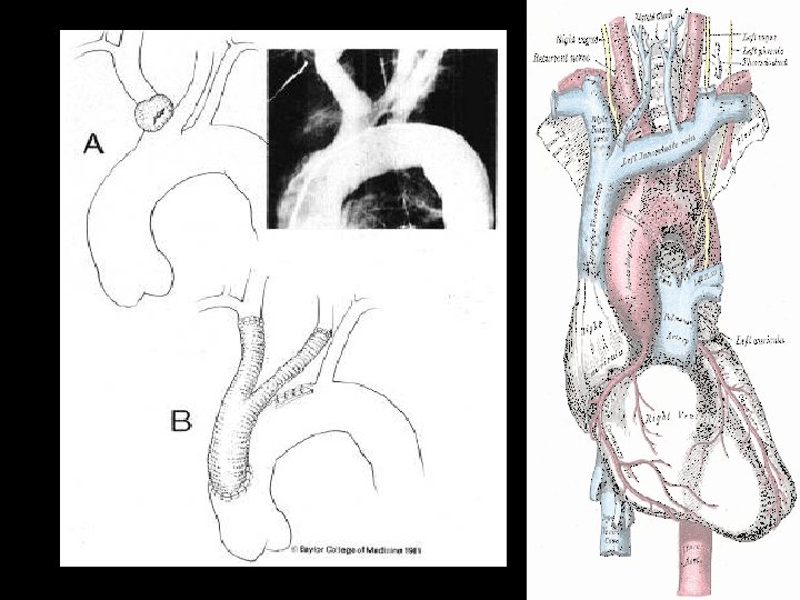
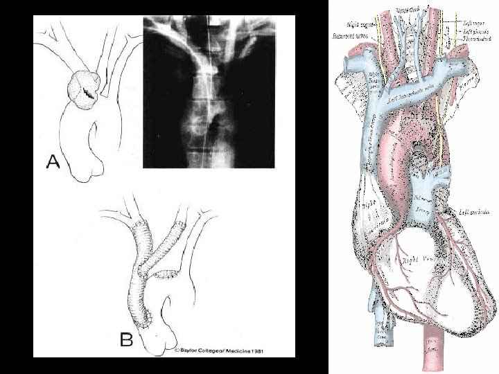
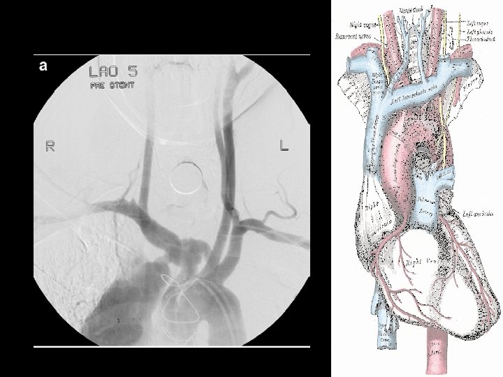
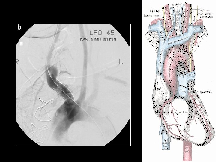
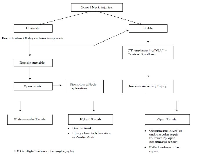
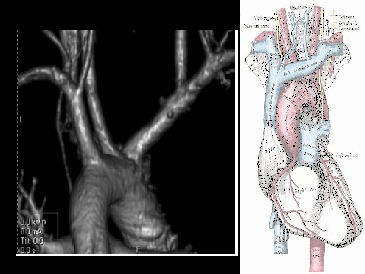
Innominate Artery • Partial Tears – Running lateral arteriorrhaphy 5 -0 Prolene suture • Complex Tears – Bypass Exclusion Technique • Ascending Aorta to the distal innominate artery • Dacron graft • Avoid the area of injury until the areas for bypass insertion are exposed • No need for hypothermia, systemic heparinization or shunts… • Place a pericardial flap between the graft and the vein ( if vein left intact)
Tips from the Masters • Proximal and distal control before entering the hematoma • Keep MAP at least 60 • Fragile structure. 5 -0 or less. • 8 -12 mm Dacron graft • Routine use of shunts or CP bypass are not mandatory • Be aware of anatomic variants (Up to 30%)
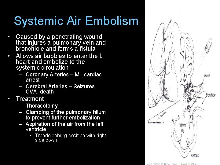
Systemic Air Embolism • Caused by a penetrating wound that injures a pulmonary vein and bronchiole and forms a fistula • Allows air bubbles to enter the L heart and embolize to the systemic circulation – Coronary Arteries – MI, cardiac arrest – Cerebral Arteries – Seizures, CVA, death • Treatment – Thoracotomy – Clamping of the pulmonary hilum to prevent further embolization – Aspiration of the air from the left ventricle • Trendelenburg position with right side down
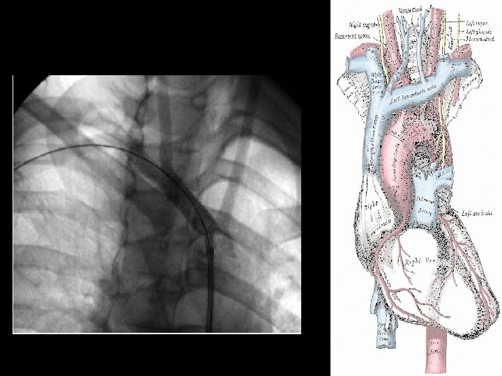
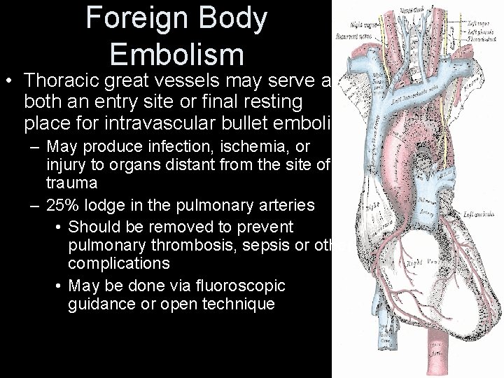
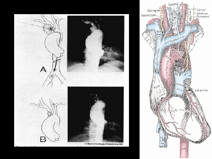
Foreign Body Embolism • Thoracic great vessels may serve as both an entry site or final resting place for intravascular bullet emboli – May produce infection, ischemia, or injury to organs distant from the site of trauma – 25% lodge in the pulmonary arteries • Should be removed to prevent pulmonary thrombosis, sepsis or other complications • May be done via fluoroscopic guidance or open technique
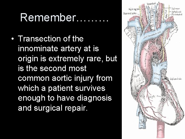
Remember……… • Transection of the innominate artery at is origin is extremely rare, but is the second most common aortic injury from which a patient survives enough to have diagnosis and surgical repair.
QUESTIONS? ? ?