Gram Positive cocci Staphylococci Introduction The staphylococci are


 strains isolates contained the vancomycin resistance gene")


 enterotoxins. 50% of S aureus strains")


 plasma diluted 1: 5 is mixed with an")


 that are")
-there are three antigenically distinct streptococcal pyrogenic exotoxins: A,")


 • Extensive and very rapidly spreading necrosis of the skin")


 • They are anaerobic")


- Slides: 86
Gram Positive cocci Staphylococci
Introduction • • The staphylococci are: gram-positive , spherical cells, arranged in grape-like irregular clusters.
• They grow readily on many types of media. ferment carbohydrates • produce pigments that vary from white to deep yellow. • Some are members of the normal flora of the skin and mucous membranes of humans. • Those found in the environment are called Micrococci.
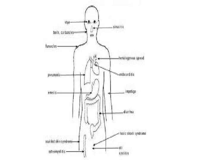
Diseases wounds , abscess formation, pimple, " hair follicle infection. bacteremia, endocarditis, osteomyelitis, meningitis, or pulmonary infection. Toxic shock syndrome, fever, vomiting, diarrhea, myalgias, a scarlatiniform rash, and hypotension, cardiac and renal failure, occurs in children or in men with staphylococcal wound infections, in wounds infections, or in the throat, a variety of pyogenic infections, even septicemia, Food poisoning.
Some Skin infections
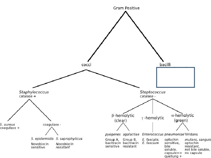
Pathogenic forms • The three main species of clinical importance are • Staphylococcus aureus, = coagulase-positive • Staphylococcus epidermidis, and Staphylococcus saprophyticus= which together are called Staph albus.
Morphology & Identification • Typical cell of the Organism • are spherical cells about 1 µm in diameter arranged in irregular clusters … • Single cocci, pairs, tetrads, and chains forms • in liquid cultures.
• Young cocci stain strongly gram-positive; on aging, many cells become gram-negative. • Staphylococci are non-motile • do not form spores.
Gram stain +ve cocci in clusters
Gram positive cocci in clusters
Culture • Staphylococci grow readily on most bacteriologic media under aerobic or microaerophilic conditions. • at 37 °C but form pigment best at room temperature (20– 25 °C). • Colonies on solid media are round, smooth, raised, and glistening. • S aureus usually forms gray to deep golden yellow colonies.
Staph colonies on BA
• S epidermidis colonies usually are gray to white on primary isolation; many colonies develop pigment only upon prolonged incubation. • No pigment is produced in anaerobic cultures or in broth.
• Various degrees of hemolysis are produced by S aureus and occasionally by other species. • Peptostreptococcus species, are anaerobic cocci, which often resemble staphylococci in morphology
Characteristics • The staphylococci produce catalase, which differentiates them from the streptococci which are catalase negative. • ferment many carbohydrates, producing lactic acid but not gas. • Proteolytic enzymes activity varies greatly among. • Resistant to drying, heat (they withstand 50 °C for 30 minutes), and 9% sodium chloride but are readily inhibited by certain chemicals, eg, 3% hexachlorophene.
• Staphylococci are variably sensitive to many antimicrobial drugs. Resistance falls into several classes: • 1. β-Lactamase production is common, is under plasmid control, so resistant to many penicillins (penicillin G, ampicillin, ticarcillin, piperacillin, and similar drugs) by transduction & conjugation. • 2. Resistance to nafcillin (and to methicillin and oxacillin) is due to the mec. A gene on the chromosome which encodes a low-affinity penicillin binding protein (PBP 2 or PBP 2 a).
S aureus is considered to be susceptible to vancomycin but resistant strains are developing. These are often known as vancomycin-intermediate S aureus, or "VISA. The mechanism of resistance is associated with increased cell wall synthesis and alterations in the cell wall Vancomycin resistant S aureus strains are nafcillin-resistant but generally are susceptible to oxazolidinones and to quinupristin/dalfopristin.
Several isolates of vancomycin-resistant S aureus (VRSA) strains isolates contained the vancomycin resistance gene van. A from enterococci and the nafcillin resistance gene mec. A. Vancomycin resistance in S aureus is of major concern worldwide.
Tolerance • Staphylococci are inhibited by a drug but not killed by it. • Note— there is great difference between minimal inhibitory and minimal lethal concentrations of an antimicrobial drug.
Variation • A culture of staphylococci may contain some bacteria that differ from the bulk of the population in expression of colony characteristics (such colony size, pigment, hemolysis), in enzyme elaboration, in drug resistance, and in pathogenicity.
Antigenic Structure • Staphylococci contain antigenic • polysaccharides and • proteins as well as other substances important in cell wall structure. • Peptidoglycan, a polysaccharide polymer containing linked subunits, provides the rigid exoskeleton of the cell wall. • Peptidoglycan is destroyed by strong acid or exposure to lysozyme. It is important in the pathogenesis of infection: It elicits production of interleukin-1 (endogenous pyrogen) and opsonic antibodies by monocytes, and it can be a chemoattractant for polymorphonuclear leukocytes, have endotoxin-like activity, and activate complement.
• Teichoic acids, in the peptidoglycan and can be antigenic. • Protein A is a cell wall component of many S aureus strains that binds to Ig. G molecules (except Ig. G 3). • Protein A -: Protein A with attached Ig. G molecules directed against a specific bacterial antigen agglutinates bacteria that have the antigen ("coagglutination").
Staph aureus • S aureus have coagulase, or clumping factor on the cell wall surface. • coagulase binds to fibrinogen in plasma (not serum) forming aggregation of the bacteria.
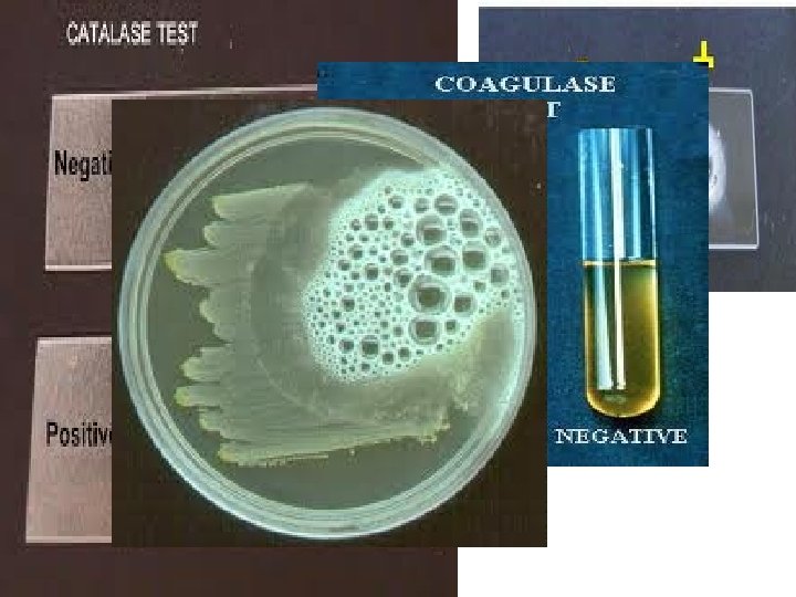
Enzymes & Toxins • Catalase—this test differentiate Staph from Streptococci. • Coagulase —test identifies Staph aureus (is +ve). • Clumping factor is responsible for adherence of the organisms to fibrinogen and fibrin. When mixed with plasma, S aureus forms clumps. Clumping factor is distinct from coagulase
Other Enzymes • Other enzymes produced by staphylococci include • hyaluronidase, or spreading factor; • staphylokinase resulting in fibrinolysis • proteinases; • lipases; and β-lactamases
Exotoxins • The α-toxin is a potent hemolysin many kinds of cells, including human red blood cells. • These protein toxins are capable of efficiently lysing white blood cells. • Leukocidin • This toxin of S aureus has two components-kill white blood cells of humans and rabbits.
Exfoliative Toxins • These epidermolytic toxins of S aureus. • Epidermolytic toxin A is a chromosomal gene product and is heat-stable. • Epidermolytic toxin B is plasmid-mediated and heat-labile. • Together they cause generalized desquamation of the skin called staphylococcal scalded skin syndrome. • . . they are superantigens.
Toxic Shock Syndrome Toxin • Most S aureus strains isolated from patients with toxic shock syndrome produce a toxin called toxic shock syndrome toxin-1 (TSST-1), which is the same as enterotoxin F. • TSST-1 is a typical superantigen. • The toxin is associated with fever, shock, and multisystem involvement, including a desquamative skin rash. The gene for TSST-1 is found in about 20% of S aureus isolates.
Enterotoxins • There are multiple (A–E, G–I, K–M) enterotoxins. 50% of S aureus strains can produce & causes food poisoning. • Like TSST-1, the enterotoxins are superantigens. The enterotoxins are heat-stable. • Enterotoxins are produced when S aureus grows in carbohydrate and protein foods. • Ingestion of 25 g of enterotoxin B results in vomiting and diarrhea. The emetic (vomiting) effect of enterotoxin is probably the result of central nervous system. • The exfoliative toxins, TSST-1, and the enterotoxin genes are on a chromosomal element called a pathogenicity island.
Diagnostic Laboratory Tests • Specimens: • • Surface swab pus, blood, tracheal aspirate, or spinal fluid for culture,
Smears • Prepare smear and do Gram stain. • Typical staphylococci appear as gram positive cocci in clusters in Gram-stained smears of pus or sputum
Culture • Grow on blood agar plates in 18 hours at 37 °C, hemolysis and pigment production may not occur until several days later and are optimal at room temperature. • S aureus but not other staphylococci ferment mannitol. Specimens contaminated with a mixed flora can be cultured on media containing 7. 5% Na. Cl. Mannitol salt agar is selective.
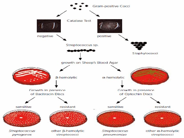
Catalase Test • A drop of 3% hydrogen peroxide solution is placed on a slide, and a small amount of the bacterial growth is placed in the solution. The formation of bubbles (the release of oxygen) indicates a positive test.
Coagulase Test Citrated rabbit (or human) plasma diluted 1: 5 is mixed with an equal volume of broth culture or growth from colonies on agar and incubated at 37 °C. A tube of plasma mixed with sterile broth is included as a control. If clots form in 1– 4 hours, the test is positive. Slide test? Coagulase-positive staphylococci are considered pathogenic for humans. Infections of prosthetic devices can be caused by organisms of the coagulase-negative S epidermidis group.
Susceptibility Testing • Staph are resistant to many antibiotics. • Test many drugs including penicillins and cephalosporins.
Prevention
Gram positive cocci STREPTOCOCCI
• • The streptococci are gram-positive spherical bacteria form pairs, chains during growth.
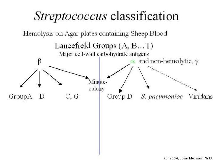
Classification of Streptococci • The classification has been based on observations over many years: • 1. colony morphology and hemolytic reactions on blood agar. • 2. serologic specificity of the cell wall antigens and capsular antigens • 3. biochemical reactions and resistance to physical and chemical factors. • 4. ecologic features.
Haemolysis • Complete disruption of erythrocytes with clearing of the blood around the bacterial growth is called βhaemolysis. • Incomplete lysis of erythrocytes with reduction of hemoglobin and the formation of green pigment is called α-haemolysis. • Some other streptococci are non-haemolytic (sometimes called gamma or γ-haemolysis).
Lancefield Classification • The cell wall has many amino sugars or groupspecific carbohydrate. These are used for grouping into Lancefield groups A–H and K–U. Group A streptococci- rhamnose-N-acetylglucosamine Group B - rhamnose-glucosamine polysaccharide Group C -rhamnose-N-acetylgalactosamine; Group D -glycerol teichoic acid containing D-alanine and glucose Group F- glucopyranosyl-N-acetylgalactosamine
Approach to Lancefield Classification • • • Many methods were used. Culture was treated with : hot hydrochloric acid, nitrous acid, formamide; enzymatic lysis(eg, with pepsin or trypsin). These extracts contain the carbohydrate group–specific antigens/ substance that yield precipitin reactions specific antisera. This permits arrangement of many streptococci into groups A–H and K–U. Typing is generally done only for groups A, B, C, F, and G.
Capsular Polysaccharides • The antigenic specificity of the capsular polysaccharides is used to classify Strept pneumoniae into over 90 types • to type the group B streptococci also called Streptococcus agalactiae.
Toxins & Enzymes • More than 20 extracellular products (Toxins & Enzymes) that are antigenic are produced by S pyogenes. • Streptokinase (Fibrinolysin)- digests fibrin • Streptodornase or Streptococcal deoxyribonuclease)- for ezymatic debridement of purulent exudates. • Hyaluronidase— dissioves tissue so spreading factor
• Pyrogenic Exotoxins (Erythrogenic Toxin)-there are three antigenically distinct streptococcal pyrogenic exotoxins: A, B, and C. • ‘ A’ causes streptococcal toxic shock syndrome and scarlet fever. • Diphosphopyridine Nucleotidase-ability to kill leukocytes • Haemolysins also called streptolysins-Streptolysin O • -causes haemolysis on BA. Antistreptolysin O. . titre of 160 -200 units is diagnostic of current or recent strep infection.
Pathogenesis & Clinical Findings • The diseases are many
Diseases Attributable to Invasion by S pyogenes, β-Hemolytic Group A Streptococci • Erysipelas
Symptoms of erysipelas • • • Symptoms in the infected area Skin sore with a raised edge Pain Swelling Redness Hardened appearance Bodily symptoms High fever Prolonged muscle stiffness Confusion or mental status changes, particularly in elderly patients
Cellulitis • Streptococcal cellulitis is an acute, rapidly spreading infection of the skin and subcutaneous tissues. It follows infection associated with mild trauma, burns, wounds, or surgical incisions. • Pain, tenderness, swelling, and erythema occur. • Cellulitis is differentiated from erysipelas is In cellulitis, the lesion is not raised.
Necrotizing Fasciitis (Streptococcal Gangrene) • Extensive and very rapidly spreading necrosis of the skin and subcutaneous tissues. • Group A streptococci that causes the disease is called "flesh-eating bacteria“.
Puerperal Fever • If the streptococci enter the uterus after delivery, puerperal fever develops, which is essentially a septicemia originating in the infected wound (endometritis).
Bacteremia/Sepsis • Infection of traumatic or surgical wounds with streptococci results in bacteremia, which rapidly can be fatal. • S pyogenes bacteremia can also follow skin infections, such as cellulitis and rarely pharyngitis
Local Infection with S pyogenes and Their By-Products • Streptococcal Sore Throat • nasopharyngitis, • tonsillitis
Streptococcal Pyoderma • Local infection of superficial layers of skin, especially in children, is called impetigo
Invasive Group A Streptococcal Infections, Streptococcal Toxic Shock Syndrome, and Scarlet Fever • Fulminant, invasive S pyogenes infections with streptococcal toxic shock syndrome are characterized by shock, bacteremia, respiratory failure.
Poststreptococcal Diseases • Rheumatic Fever-- In acute nephritis, there is blood and protein in the urine, edema, high blood pressure, and urea nitrogen retention; serum complement levels are also low. •
Post Strept • Glomerulonephritis--In acute nephritis, there is blood and protein in the urine, edema, high blood pressure, and urea nitrogen retention; serum complement levels are also low. A few patients die; some develop chronic glomerulonephritis with ultimate kidney failure; and the majority recover completely.
Rheumatic Fever • This is the most serious sequela of S pyogenes because it results in damage to heart muscle and valves. • Sequela is condition following as a consequence of disease
Diagnostic Laboratory Tests • Specimens. • A throat swab, pus, or blood is obtained for culture. Serum is obtained for antibody determinations.
Smears • Smears from pus often show single cocci or pairs rather than definite chains. • gram-positive. • Smears of throat swabs masked by viridans streptococci.
Culture • Cultured on blood agar plates. If anaerobes are suspected, suitable anaerobic media must also be inoculated. Incubation in 10% CO 2 often speeds haemolysis. • Chocolate agar –look for haemolysis
• Streptococci belonging to group A may be identified by inhibition of growth by bacitracin.
Other Streptococci • Streptococcus agalactiae • These are the group B streptococci. • Are -haemolytic and produce zones of haemolysis.
Groups C and G • These are in the nasopharynx and may cause pharyngitis, sinusitis, bacteremia, or endocarditis. • They often look like group A S pyogenes on blood agar medium and are -hemolytic.
Streptococcus bovis • These are among the nonenterococcal group D streptococci • Are part of the enteric flora, occasionally cause endocarditis, and sometimes cause bacteremia in patients with colon carcinoma. They are nonhemolytic and PYR-negative
Streptococcus anginosus Group • Other species names in the S anginosus group are S constellatus and S intermedius. They are sometimes referred to as the S milleri group. These streptococci are part of the normal flora. They may be β-, α-, or nonhemolytic. S anginosus group includes -hemolytic streptococci
Group N Streptococci • They are rarely found in human disease states but produce normal coagulation ("souring") of milk. • Groups E, F, G, H, and K–U Streptococci • These streptococci occur primarily in animals. One of the multiple species of group G streptococci, S canis, can cause skin infections of dogs but uncommonly infects humans; other species of group G streptococci infect humans.
Viridans Streptococci • • • The viridans streptococci include S mitis, S mutans, S salivarius, S sanguis. Typically they are α-hemolytic
Peptostreptococcus (Many Species) • They are anaerobic
Streptococcus pneumoniae • • • Morphology & Identification Typical Organisms are gram-positive, lancet-shaped diplococci Lysis of pneumococci occurs in a few minutes when ox bile (10%) or sodium deoxycholate (2%) is added to its broth culture
Pneumococci –Gram stain
Quellung Reaction • When pneumococci of a certain type are mixed with specific antipolysaccharide serum of the same type—or with polyvalent antiserum—on a microscope slide, the capsule swells markedly, and the organisms agglutinate by cross-linking of the antibodies. This reaction is useful for rapid identification and for typing of the organisms, either in sputum or in cultures
Diseases • Production of Disease • no toxins of significance • capsule has the virulence power
Pathology • Pneumococcal infection causes an outpouring of fibrinous edema fluid into the alveoli, followed by red cells and leukocytes, • in consolidation of portions of the lung. bloodstream via the lymphatic drainage of the lungs. mononuclear cells actively phagocytose the debris, The pneumococci are taken up by phagocytes and digested intracellularly.
Diagnostic Laboratory Tests • Blood is drawn for culture; CSF and sputum are collected for demonstration of pneumococci by smear and culture. • Serum antibody tests are impractical.
Stained Smears • A Gram-stained film of rusty-red sputum shows typical organisms, many polymorphonuclear neutrophils, and many red cells.
Capsule Swelling Tests • Fresh emulsified sputum mixed with antiserum causes capsule swelling (the quellung reaction) for identification of pneumococci.
Culture • The culture is created by sputum cultured on blood agar and incubated in CO 2 or a candle jar.
Treatment • Since pneumococci are sensitive to many antimicrobial drugs
Enterococci • The enterococci have the group D groupspecific substance and were previously classified as group D streptococci.