
Grafts Flaps and Tissue Transplantation Yamur AYDIN M
- Slides: 68
Grafts, Flaps and Tissue Transplantation Yağmur AYDIN, M. D. University of Istanbul, Cerrahpasa Medical Faculty Department of Plastic, Reconstructive and Aesthetic Surgery
Causes of Tissue Deficiency Trauma Tumoral resection Congenital anamolies RECONSTRUCTION Tissue transplantation
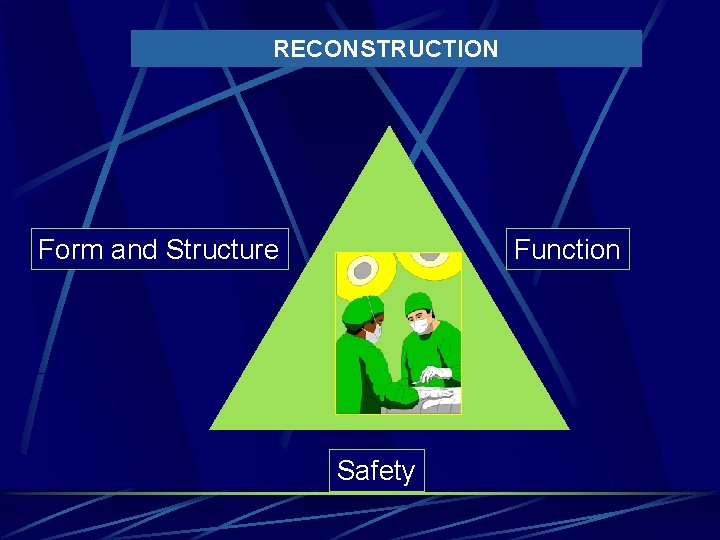
RECONSTRUCTION Form and Structure Function Safety
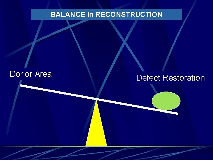
BALANCE in RECONSTRUCTION Donor Area Defect Restoration
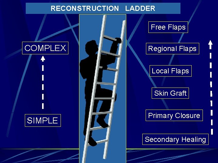
RECONSTRUCTION LADDER Free Flaps COMPLEX Regional Flaps Local Flaps Skin Graft SIMPLE Primary Closure Secondary Healing
Nomenclature Graft: tissue separated from its donor bed and relies on ingrowth of new vessels from the recipient tissues Vascularized graft (or flap): remains attached to donor blood supply or becomes revascularized via microvascular anastomoses to recipient vessels Autograft : tissue transplanted from one location to another within the same individual. İzograft : tissue transplanted from a genetically identical donor to the recipient (syngeneic mice or human monozygotic twins) Allogreft (homograft) : tissue transplanted between unrelated individuals of the same species Xenogreft (hetereograft) : tissue transplanted between different species
Tissue Transplantation Autologous l l l Skin Dermis, fat, fascia Cartilage Bone Muscle Nerve Allogeneic Xenogeneic Alloplastic materials l l l Metallic Seramic Polimeric
Tissue Transplantation Basis of modern Plastic Surgery Limited donör area for autologous tissue transplantation Nonautologous tissues (Allogeneic, Xenogeneic) may be used for tissue deficiency They are rejected because of foreign body antigens Long term immunosupression need to survive longer
Advantages of Autologous Tissue Transplantation Easy integration No rejection response No fibrous capsule formation around the transplant
Disadvantages of autologous tissue transplantation Donor area morbidity Limited supply More complex and longer operation Resorption and deformation
Immunologic Response to Allogeneic and Xenogeneic Tisuues Cellular response (T cells) Humoral immunologic response(B lymphocytes) Matching of HL-A, HL-B ve HL-DR antigens are important factor in long term survival Hyperacute rejection occurs within the first few minutes to hours after transplantation Rejection response is less to tissues which have few cells and lesser vascularity (cornea, cartilage)
Biomaterials 1. Metals: used in plating systems for craniomaxillofacial internal fixation (Stainless steel, cobalt-chromium, pure titanium, titanium alloys, and gold ) 2. Calcium ceramics: used as bone graft substitutes (Hydroxyapatite, Tricalcium phosphate, hydroxyapatite cement) 3. Polymers: used in both bone and soft tissue reconstruction and augmentation (silicone, polyurethane, polyesters, nylon, polyethylene, polypropylene, cyanoacrylates) 4. Biologic materials: used in the treatment of depressed scars and facial wrinkles (collagen, fibrel, hyaluronic acid)
Advantages of Biomaterials No donor site morbidity Less operative time Easy availability and unlimited supply Fabricated according to patient needs No resorption or deformation
Ideal Implant Biocompatible Nontoxic Nonallergenic Noncarcinogenic Easy to shape, remove, and sterilize Resistant to strain Able to be fabricated into specifically required forms Productive of no foreign-body inflamatuary response Mechanically reliable Resistant to resorption and deformation Nonsupportive of growth of microorganism Radiolucent ( not interfere with CT and MR imaging)
Disadvantages of Biomaterials Rejection Infection Implant malposition or extrusion Implant defects (broken, punctured) Fibrosis around the implant because of foreign body response
Tissue Transplantation in Plastic Surgery Skin l Autograft, allograft, xenograft Bone l Autograft, allograft Cartilage l Autograft, allograft, xenograft Nerve l Autograft, allograft Tendon l Only Autograft Fascia l Autograft, allograft
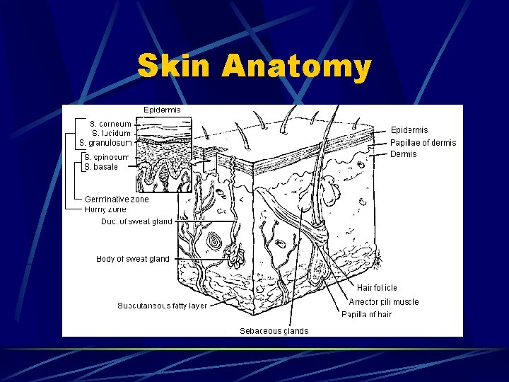
Skin Anatomy
Skin Embryology Derived form both ectoderm and mesoderm Ectodermal skin appendages develop with formation of epidermis at 11 weeks of gestation and complete at 5 months Suface of Ectoderm : Epidermis, Pilosebaceous glands, Apocrine and eccrine sweat glands, Hair follicles, Nail units l Neuroectoderm: melamocytes, nerves, and specialized sensory units Mesoderm : Sructural components of dermis (macrophages, mast cells, Langerhan’s cells, Merkel cells, fibroblasts, blood and lymphatic vessels)
Skin Functions The skin is the largest organ of the body Protect underlying structures from enviromental trauma by entry of pathogens and potentially toxic substances Must allow considerable compressions and extentions Passive regulation of intracellular fluid balance and active regulation of body temperature
Skin Grafts
Skin Autograft full thickness or partial thickness require a recipient bed that is well vascularized and free of devitalized tissue and no bacterial contamination (<105 microorganisms per gram of tissue) Close contact between the skin graft and its recipient bed is essential hematoma beneath the graft and insufficent immobilization are common causes of graft failure To optimize take of a skin graft, the recipient site must be prepared before grafting
Skin Graft Survival The transplanted skin derives its initial nutrition via serum from the recipient site in a process called “plasmatic imbibition” last for 24 to 48 hours The graft then gains blood supply from the recipient bed by ingrowths of blood vessels. This process of “inosculation“ begins within 48 hours
Skin Autograft full thickness partial thickness
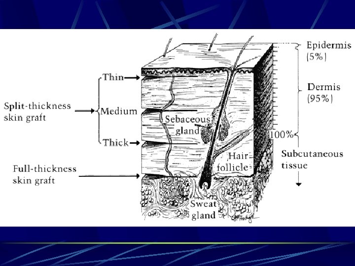
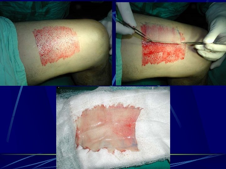
Partial Thickness Skin Grafts Consist of entire epidermis and a portion of dermis A thin split-thickness harvest site will generally heal within 7 days SSG’s can be taken from anywhere on the body The abdominal wall, buttocks, and thigh are common donor sites for SSG’s
Skin Graft Donor Site Healing The donor site epidermis regenerates from the immigration of epidermal cells originating in the hair follicle shafts and adnexal structures left in the dermis A thin split-thickness harvest site (less than 10/1, 000 of an inch) will generally heal within 7 days Full-thickness skin graft harvest sites heal by primary intention
Most Common Causes of Autolous Skin Graft Failure Hematoma, Seroma Infection (> 105 organism/1 gr tissue) Shear force ( inadequate immobilization) Poor vascularized bed (fibrozis, radiotherapy; exposed bone, cartilage, or tendon devoid of its periosteum, perichondrium, or paratenon)
Full Thickness Skin Graft contains the entire dermis (adnexal structures such as sweat glands, sebaceous glands, hair follicles, and capillaries) Usually harvest from skin is thin(upper eyelid, postauricular area, or supraclavicular area). Other harvest sites are hairless groin, antecubital fossa, distal forearm, prepuce FSG harvest sites can be closed primarily or applied a SSSG from another body part
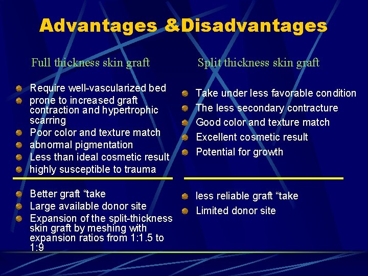
Advantages &Disadvantages Full thickness skin graft Split thickness skin graft Require well-vascularized bed prone to increased graft contraction and hypertrophic scarring Poor color and texture match abnormal pigmentation Less than ideal cosmetic result highly susceptible to trauma Take under less favorable condition The less secondary contracture Good color and texture match Excellent cosmetic result Potential for growth Better graft “take Large available donor site Expansion of the split-thickness skin graft by meshing with expansion ratios from 1: 1. 5 to 1: 9 less reliable graft “take Limited donor site
Sensory return Graft sensation is regained as nerves grow into the graft Sensory recovery begins at around 4 -5 weeks and is completed by 12 -24 months Pain, light touch, and temperature return in that order
Skin Allografts Skin allograft was the first “organ” transplant achieved and constituted the foundation of modern transplant immunology strongly antigenic and is subject to rejection ( 10 days in burns) Obtained from relatives or human corpse (frozen and stored) beneficial in large burns (> % 50) as a biologic dressing Frozen and stored or may be used immediately with cyclosporine immunusupression
Skin Xenografts Pig skin grafts can be used as temporary biologic dressings in large burns Hyperacute rejection occurs within the first few minutes to hours after transplantation Advantages l Cheap, easy availablility, easy storage and sterility
Skin Flaps
Skin Flaps Unlike a graft, a flap has its own blood supply Consist of skin and subcutaneous tissue that are transferred from one part of the body to another with a vascular pedicle or attachment to the body being maintained for nourishment
When skin flaps are used? Covering recipient beds that have poor vascularity Reconstructing the full thickness of the eyelids, lips, ears, nose, and cheeks; and padding body prominencies (i. e. , for bulk and contour) It is necessary to operate through the wound at a later date to repair underlying structures Muscle flaps may provide a functional motor unit or a means of controlling infection in the recipient area
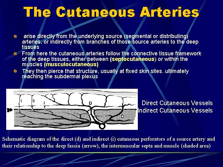
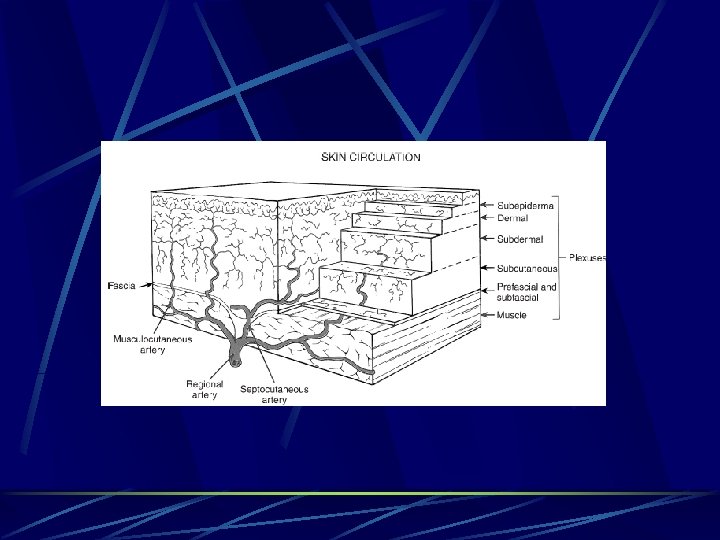
The Cutaneous Arteries arise directly from the underlying source (segmental or distributing) arteries, or indirectly from branches of those source arteries to the deep tissues From here the cutaneous arteries follow the connective tissue framework of the deep tissues, either between (septocutaneous) or within the muscles (musculocutaneous) They then pierce that structure, usually at fixed skin sites. ultimately reaching the subdermal plexus Direct Cutaneous Vessels Indirect Cutaneous Vessels Schematic diagram of the direct (d) and indirect (i) cutaneous perforators of a source artery and their relationship to the deep fascia (arrow), the intermuscular septa and muscle (shaded area)
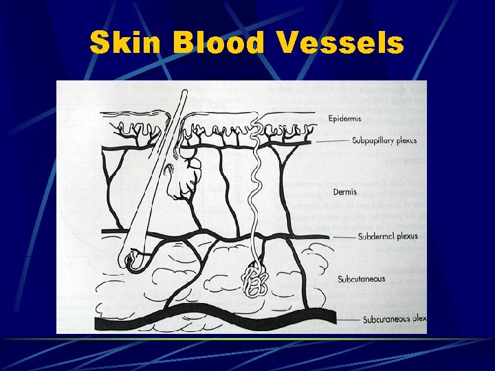
Skin Blood Vessels
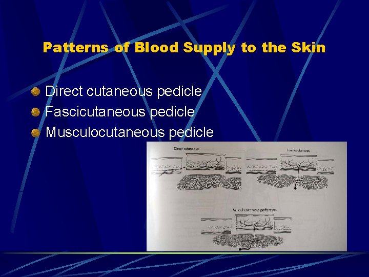
Patterns of Blood Supply to the Skin Direct cutaneous pedicle Fascicutaneous pedicle Musculocutaneous pedicle
Skin Flap Classification Proximity to defect : Local Distant
Skin Flap Classification Composition: Cutaneous Fasciocutaneous Musculocutaneous Osteocutaneous
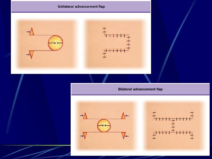
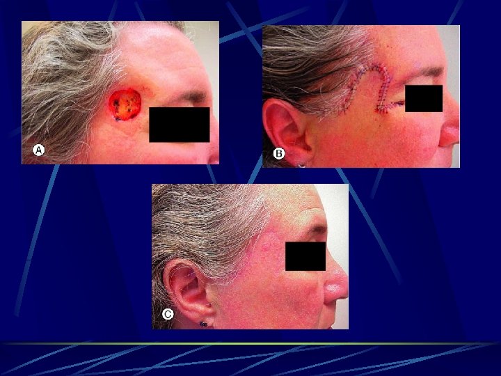
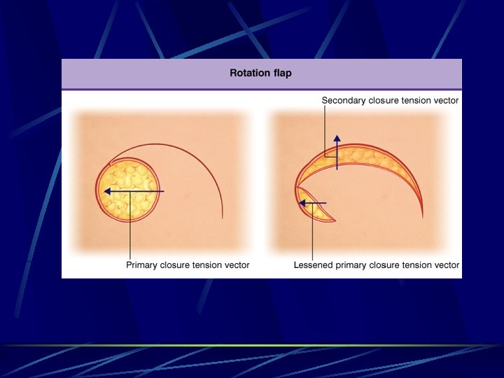
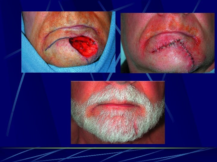
Skin Flap Classification Method of movement: Advancement Rotation Transposition Interpolation Free
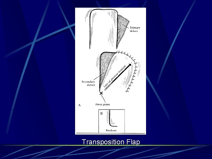
Transposition Flap
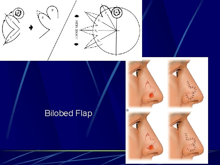
Bilobed Flap
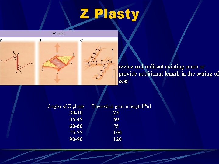
Z Plasty revise and redirect existing scars or provide additional length in the setting of scar Angles of Z-plasty 30 -30 45 -45 60 -60 75 -75 90 -90 Theoretical gain in length(%) 25 50 75 100 120
Skin Flap Classification Specialized : Sensory Tendon Hair bearing
Skin Flap Classification Blood supply : Random Axial pattern Fasciocutaneus Musculocutaneous
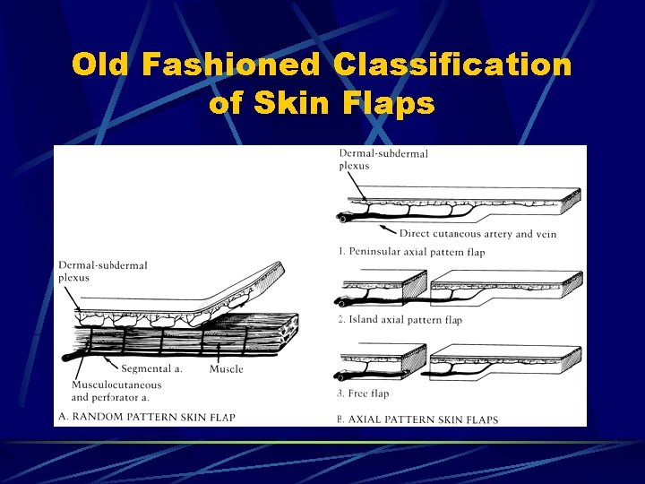
Old Fashioned Classification of Skin Flaps
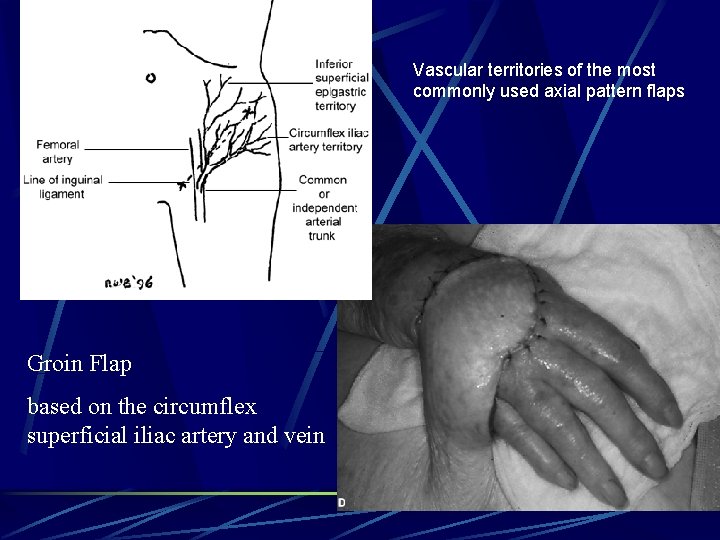
Vascular territories of the most commonly used axial pattern flaps Groin Flap based on the circumflex superficial iliac artery and vein
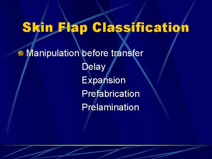
Skin Flap Classification Manipulation before transfer Delay Expansion Prefabrication Prelamination
Other Flaps Muscular Bone Visceral organ (jejenum, sigmoid colon) Omentum
Free Composite Grafts Contain two or more tissue (dermis-cartilage, dermofat, skin-muscle, pulpa) Need well-vascularized bed Poor vascularization and graft taking Stasis and necrosis in the graft because of insufficent venous and lymphatic return Results is not optimal l Limited size l Contraction l Contur problem because of bowing
Enhancing Survival of Composite Grafts Well vascularized bed, no fibrosis Atrumatic technique Postoperative cooling > 5 mm distant from the nearest vascular bed is at risk for necrosis Center of graft is never more than 5 -8 mm away from a blood supply
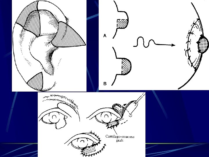
Composite Grafts in Plastic Surgery Nose (from ear or nasal septum) l l l Nasal ala Columella Lateral nasal wall Nasa roof and lining reconstruction Short nose Septal perforation Ear l l l Helical rim Chonca Tragus Eyebrow (scalp) Nipple (opposite nipple or ear lobule) Eyelid (septal chondromucosal graft)
Bone Transplantation Both bone autograft and allografts are used for bone defect reconstruction Bone xenografts are not used nowadays because of sequester of all viable osteocyte Cortical or cancellous bone graft Revascularization of cortical grafts may take a few months Revascularization of cancellous bone grafts are more rapid Healing of vascularized bone grafts are better. Particularly suitable in a field after trauma, cronic scarring, or prior radiation. Biomecanically are superior to nonvascularized grafts
Bone Graft Donor Areas Cranium (cortical) Thorax (split rib grafts) İliac ( good quality cortical and cancellous bone source) Tibia (cancellous ) Others l l l Distal radİUs, pro. Ximal ulna (hand surgery) Fibula (esp. vascularized flap) Metatars
Tendon Grafts Only if primary or delayed primary repair is not feasible Contrindicated if there is stiff joints, adherent extensor tendons, and inadequate skin cover Only autograft Unacceptable amount of host reaction and adhesion after allografts and xenografts
Donor Areas for Tendon Graft Palmaris longus (usually) Plantaris Middle 3 toes extensor tendons
Cartilage Grafts Cartilage has no intrinsic blood supply The use of cartilage autografts is widespread and includes nasal, auricular, craniofacial skeleton, and joint reconstruction Cartilage is immunologically privileged due to the shielding of chondrocytes by its matrix, which is only weakly antigenic Both chondrocytes and matrix are subject to xenogeneic mechanisms of rejection with a generally poorer outcome in comparison. There is only small number of usage
Donor Areas for Cartilage Graft Choose according to aim l Costal cartilage(7, 8 ve 9. ribs) l l l Ear cartilage: l l l Ear reconstruction Nasal dorsal and alar area reconstruction Lower eyelid support Nipple-aerola reconstruction Orbita floor reconstruction Temporomandibular joint repair Nasal septal cartilage l Aestetic Rhinoplasty and Nasal reconstruction
Nerve Grafts The nerve graft acts as a biologic conduit for the regenerating axons Vascularized nerve grafts are theoretically advantageous particularly in scarred beds Other “conduits” used as nerve grafts have included autologous vein, silicone tube seeded with Schwann cells, and freeze fractured autologous muscle
Donor Areas for Nerve Graft Sural sinir (most often) N. Safeneous Lateral femoral cutaneous nerve Medial antebrachial cutaneous nerve Lateral antebrachial cutaneous nerve Dorsal antebrachial cutaneous nerve Superficial radialal nerve Servikal plexus cutaneous nerves Interkostal nerve