GRAFTS AND FLAPS BY Dr Mahmoud Abdalla Hifny

GRAFTS AND FLAPS BY Dr. Mahmoud Abdalla Hifny Lecturer of Plastic Surgery

Flap

Flaps are elevated from a donor site and transferred to the recipient site with an intact vascular supply. It survives by carrying its own blood supply until new blood vessels from the recipient site are generated in which the native blood supply (pedicle) can be divided. Flaps can be used when the wound bed is unable to support a skin graft or when a more complex reconstruction is needed.

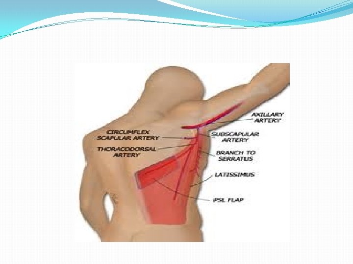
Flap Classification A. By composition — Flaps can be classified by the type of tissue transferred 1. Single component a. Skin flap — i. e. Parascapular flap b. Muscle flap — i. e. Rectus muscle flap or latissimus dorsi muscle flap c. Bone flap — i. e. Fibula flap d. Fascia flap — i. e. Serratus fascia flap

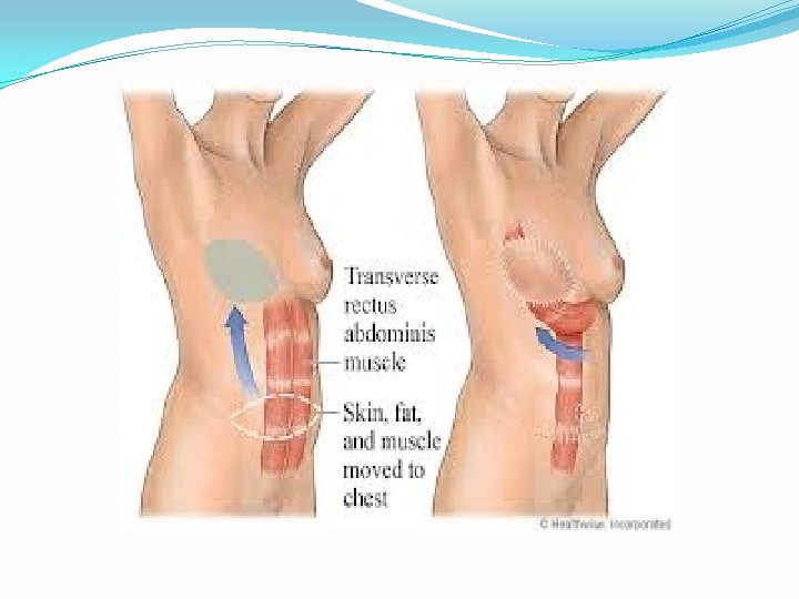
2. Multiple components a. Fasciocutaneous — anterolateral thigh flap Radial forearm flap or b. Myocutaneous — Transverse rectus abdominis myocutaneous flap c. Osseoseptocutaneous — Fibula with a skin paddle

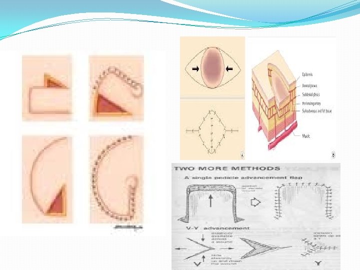
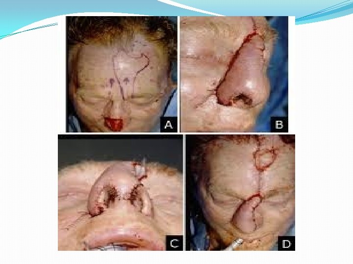
B. By location — Flaps can be described by the proximity to the primary defect that needs to be reconstructed. The harvest leaves a secondary defect that needs to be closed 1. Local flaps — Local flaps are raised from the tissue adjacent to the primary defect. Its movement into the defect can be described as advancement, rotation, or transposition. Specific examples of local skin flaps are the V-Y, rhomboid, and bilobed flaps

2. Regional — Regional flaps are raised from tissue in the vicinity but not directly adjacent to the primary defect. The movement is described as transposition or interpolation 3. Distant — Distant flaps are raised from tissue at a distance from the primary defect. This usually requires re-anastamosis of the blood vessels to recipient blood vessels in the primary defect. These are called free flaps

C. By vascular pattern: 1. Random vs. Axial: a. Random pattern flaps do not have a specific or named blood vessel incorporated in the base of the flap. Because of the random nature of the vascular pattern, it is limited in dimensions, specifically in the length: breadth ratio

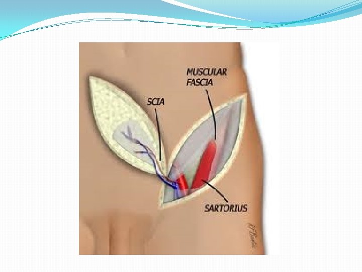
b. Axial pattern flaps are designed with a specific named vascular system that enters the base and runs along its axis. This allows the flap to be designed as long and as wide as the territory the axial artery supplies


2. Pedicled vs. Free a. Pedicled flaps remain attached to the body at the harvest site. The pedicle is the base that remains attached and includes the blood supply. It is transferred to the defect with its vascular pedicle acting as a leash. b. Free flaps are detached at the vascular pedicle and transferred from the donor site to the recipient site. They require re-anastamosis of the artery and vein to recipient vessels at the recipient site.

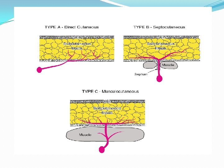
CLASSIFICATION OF FASCIAL AND FASCIOCUTANEOUS FLAPS: MATHES AND NAHAI Type A Direct cutaneous pedicle Type B Septocutaneous pedicle Type C Musculocutaneous pedicle

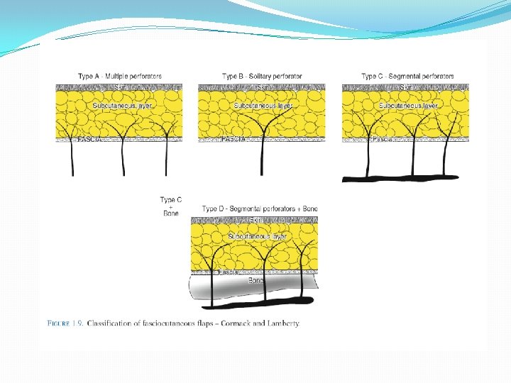
CLASSIFICATION OF FASCIOCUTANEOUS FLAPS: CORMACK AND LAMBERTY The flap from fasciocutaneous vessels running from deep arteries of the body. Most flaps raised in a limb have a fasciocutaneous pattern of blood supply. Type A Multiple, unnamed fasciocutaneous vessels entering the base of the flap E. g. , Ponten lower leg flaps Type B Single fasciocutaneous vessel running along the axis of the flap E. g. , Scapular, parascapular flaps Type C Multiple perforating vessels from a deep artery in the septum between muscles E. g. , Radial forearm flap

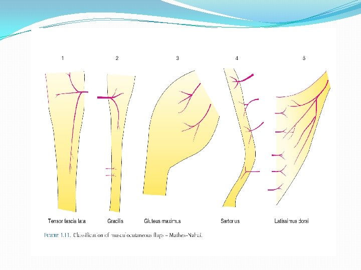
CLASSIFICATION OF MUSCULOCUTANEOUS FLAPS: MATHES AND NAHAI Type I One vascular pedicle nourishes the whole flap (Gastrocnemius, Tensor fascia lata, Abductor digiti minimi) Type II Dominant vascular pedicle with additional minor vascular pedicles (Trapezius, Gracilis) Type III Two dominant vessels (Rectus abdominus, Gluteus Maximus, Serratus, Temporalis) Type IV Segmental supply (Sartorius, Tibialis anterior, Flexor hallucis longus) Type V Dominant pedicle but alternative minor pedicles which can support the flap (Latissimus Dorsi, Pectoralis Major)

THANK YOU
- Slides: 23