Government arts and science college women formerly Bharathidasan
 (formerly Bharathidasan university constituent college for women) orathanadu")
Government arts and science college (women) (formerly Bharathidasan university constituent college for women) orathanadu 614 625 Department of biochemistry Presented by Dr. R. PRIYA Guest lecturer Department of Biochemistry

I B. Sc. , Biochemistry Subject : Core course II Human physiology subject code: semester 16 SCCBC 2 : II

Human heart anatomy and physiology

Synopsis Introduction Structure of heart Blood flow through the heart Circulation of heart Composition of blood 1. Plasma protein 2. Formed element 3. Hematopoiesis 4. Erythropoiesis 5. leukocytes 6. Platelets Cardiac cycle Electrocardiogram Function of heart Function of blood

INTRODUCTION TO THE CIRCULATORY SYSTEM BLOOD Is a fluid connective tissues a specialized body fluid Blood is a mixture of about 55 percent plasma and 45 percent blood cells About 7 to 8 percent of total body weight is blood It has four main components 1. plasma 2. Red blood cells 3. White blood cells 4. Platelets FUNCTION Transport oxygen an nutrients to the lungs and tissues Form blood clots to prevent excess blood loss Carry cells and antibodies that fight infection Bring waste products to the kidneys and liver to filter blood Regulate body temperature

Plasma The liquid component of blood Mixture of water sugar fat protein and salts Trasnports blood cells throughout the body Besides nutrients waste products antibodies clotting proteins Chemical messengers such as hormones and proteins That help maintain body fluid balance RED BLOOD CELLS(Erythrocytes) Bright red coloured cells The most abundant cell in the blood The shape of red blood cell is a biconcave disc

Small size-Helps red blood cells pass through narrow capillaries Flattened disc shape-provides a large surface area, allowing rapid diffusion of oxygen Contains special protein haemoglobin-it absorbs oxygen in the lungs and release oxygen in the rest of the body Does not contain a nucleus-increases amount of space inside the cell for haemoglobin Production of red blood cells is controlled by erythropoietin (a hormone produced primarily by the kidneys) White Blood Cells (Leukocytes) White blood cells protect the body from infection Much fewer in number than red blood cells (about 1 percent of blood) The most common type of white blood cell is the neutrophil Account for 55 to 70 percent of the total WBC count Each neutrophil lives less than a day So bone marrow constantly makes new neutrophils

Components of blood Unlike red and white blood cells Platelets are not actually cells Rather small fragments of cells Platelets help the blood clotting(coagulation) Assemble at the site of an injury Stick to the lining of the injured blood vessel Form a platform on which blood coagulation can take place

THE HEART Is about 4. 8 inches tall and 3. 35 inches wide Weighs about. 68 Ib. In men and. 56 Ib. In women Beats about 100, 000 times per day Beats 2. 5 billion time in an average 70 yr. Lifetime Pumps about 2000 gallons of blood each day Circulates blood completely 1000 times each day Pumps blood through 62, 000 miles of vessels Suffers 7. 2 mil. CAD deaths worldwide each year

STRUCTURE OF HEART

Outer covering of the heart The heart is a large muscular organ comprised of four different layers (from the outside to the inside) Pericardium Epicardium Myocardium Endocardium PERICARDIUM A tough double layered fibro serous sac Contain the heart and the roots of the great vessels The pericardial sac has two layers 1. Serous layer 2. Fibrous layer The space between the two layers of serous pericardium is Pericardial cavity which is filled with serous fluid The pericardial fluid It protects the heart from any kind of external jerk or shock

Fibrous pericardium It is the most external layer made of dense connective tissue Function It protects the heart Anchor it to the surrounding walls Prevent it from over filling with blood Serous pericardium It has two layers 1. Parietal pericardium Combined to and inseparable from the fibrous pericardium 2. Visceral pericardium Is a part the epicardium The visceral layer extends till the opening of the great vessels it joins with the parietal layer where the aorta and pulmonary arteries leave the heart and the venacavae and pulmonary veins enter the heart Function Help to lubricate heart to avoid friction during heart activity

Epicardium A thin layer of connective tissue and fat Provide an extra layer of protection for the heart It is considered an extension of serous pericardium Myocardium Muscle tissue of the heart Composed of cardiac muscle cells(Cardiomyocytes) Receives nervous stimulation from the Sinoatrial node (SA) and Atrioventricular (AV) node via Purkinje fibers

Endocardium Innermost layer of tissue Simple squamous endothelial Is continuous with Endothelial lining of great blood vessels Function Lines the inside of the heart Offer a smooth lining for chambers of the heart Cover the valves A SECTION OF THE HEART WALL

Four chambers of the heart ATRIA Thin walled upper chambers Separated by atrial septum Right side of septum has oval depression, fossa ovalis cordis, remnant of the foramen ovale Act as receiving chamber for blood returning from the body and lungs VENTRICLES Lower chambers which make up the bulk of the muscle mass of the heart left ventricle 2/3 larger than right ventricle Right ventricle is a thin walled and oblong, like pocket attached to left ventricle Contraction of left ventricle pulls in right ventricle, aiding its contraction (termed left ventricular aid) Separated by intraventricular septum

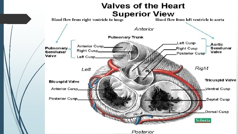
Four Valves of the heart Tricuspid valve Separates right atrium from right ventricle Pulmonic semilunar valve Separates right ventricle from pulmonary artery Bicuspid (Mitral) Valve Separates left atrium from left ventricle Aortic semilunar valve Separates left ventricle from aorta Chordae tendineae cordis Anchor free ends of A-V valves to papillary muscles Prevent A-V valves from pushing upward into atria during ventricular contraction
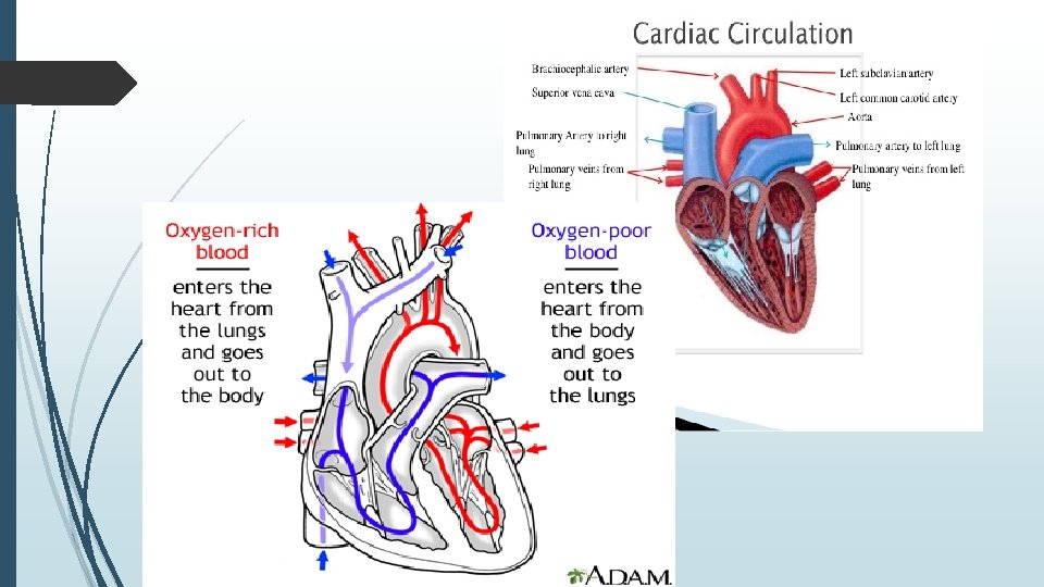

Blood flow through the heart The right and left sides of the heart work together causing blood to flow continuously to the heart lungs and body RIGHT SIDE OF THE HEART Blood enters the heart through two large veins The inferior and superior vena cava emptying oxygen poor blood(from the body) into the right atrium As the right atrium contracts blood flows into right ventricle(through the open tricuspid valve) When the ventricle is full the tricuspid valve shuts Prevents blood from flowing backward into the right atrium while the right ventricle contracts As the ventricle contracts blood leaves the heart (through the pulmonic valve) Into the pulmonary artery and to the lungs where it is oxygenated

The oxygenated blood then returns to the heart through the pulmonary veins LEFT SIDE OF THE HEART Pulmonary vein empty Oxygen-rich blood from the lungs into the left atrium As the atrium contracts Blood flows from atrium into the ventricle (through the open mitral valve) When the ventricle is full (the mitral valve shuts) Prevent blood from flowing backward into the atrium(while the ventricle contracts) As the ventricle contracts Blood leaves the heart (through the aortic valve) Into the aorta and to the body

Circulation of the blood Blood enters the heart through the inferior and superior vena cava, flowing into the right atrium The bloof passes through the tricuspid valve into the right ventricle It then passes through the pulmonic semilunar valve, entering the pulmonary artery of the pulmonary circulation It flows through the pulmonary bed of the right and left lungs to the pulmonary vein, reentering the heart al the left atrium It then flows through the bicuspid valve into the left ventricle passing through the aortic semilunar valve, the blood enters the aorta and systemic vascular system

The heart as a double pump The pulmonary and systemic circuits

Double circulation Mammals have a double circulatory system which means one circuit links the heart and lungs the other circuit links the heart with the rest of the body During a single cycle blood goes twice in the heart This means that there are two loops in our body in which blood circulates One is oxygenated blood (it has little or no oxygen but a lot of carbon dioxide) IMPORTANCE OF DOUBLE CIRCULATION It is necessary to separate oxygenated and de-oxygenated blood It makes the circulatory system more efficient Higher blood pressure so a greater flow of blood to the tissues Helps to maintain a constant body temperature

Posterior view of heart

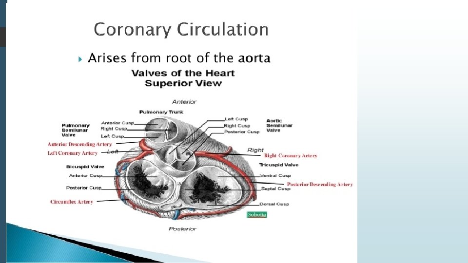
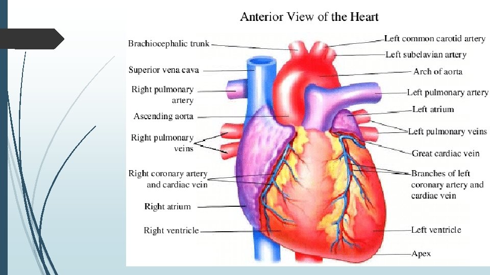
Blood vessels of human heart
 Suspended and carried in plasma (fluid")
Composition of blood Consists of formed elements (cells) Suspended and carried in plasma (fluid part) Total blood volume 60 -80 m. L/kg of body weight Plasma is straw-colored liquid consisting of 90% water and dissolved solutes Include ions, metabolites, hormones, antibodies and proteins

Plasma protein Constitute 7 -9% of plasma Three types of plasma proteins 1. Albumins 2. Globulins 3. Fibrinogen Albumin accounts for 60 -80% Creates colloid osmotic pressure that draws water from interstitial fluid into capillaries to maintain blood volume and pressure Globulins carry lipids Gamma globulins are antibodies Fibrinogen serves as clotting factor Converted to fibrin when clotting blood Serum is fluid left when blood clots
 and Leukocytes (WBC) RBC ARE FLATTENED BICONCAVE DISCS")
Formed elements Composed of Erytrhocytes (RBC) and Leukocytes (WBC) RBC ARE FLATTENED BICONCAVE DISCS Generated in the red bone marrow by the process of erythropoiesis from the hemocytoblast, a common stem cell Shape provides increased surface area for diffusion Lack nuclei and mitochondria Has semi-permeable membrane Contains hemoglobin molecule that transport oxygen Approx. 30 trillion in the body
")
Hematopoiesis Is the formation of blood cells from stem cells in marrow (mueloid tissue) and lymphoid tissue RBCs increase in number above normal with chronic hypoxia Erythropoisis is formation of RBCs Stimulated by erythropoietin (EPO) from kidney Leukopoiesis if formation of WBCs Stimulated by variety of cytokines

Erythropoiesis 2. 5 million RBCs are developed Lifespan 120 days Old RBCs removed from blood by phagocytic cells in liver, spleen and bone marrow Iron recycled back into hemoglobin production

Leukocytes Have nucleus, mitochondria and amoeboid ability Formed in the myeloid tissue Can squeeze through capillary walls (Diapedesis) Granular leukocytes help detoxify foreign substances and release heparin include eosinophils, basophils and neutrophils Eosinophils Basophil Neutrophil

Monocyte LEUKOCYTES Agranular leukocytes are phagocytic and produce antibodies Include lymphocytes and monocytes lymphocytes

MEGAKARYOCYTE Specialized type of blood cell Fragments into small irregular pieces of protoplasm called thrombocytes and platelets Have no nucleus Have a granular cytoplasm Function in clot formation Platelets (Thrombocytes) Are smallest of formed elements, lack nucleus Constitute most of mass of blood clots Release serotonin to vasoconstrict and reduce blood flow to clot area Secrete growth factors to maintain integrity of blood vessel wall Survive 5 -9 days

Cardiac cycle The cardiac cycle consists of one heartbeat or one cycle of contraction and relaxation of the cardiac muscle The cardiac cycle can be divided into two parts Atrial cycle (0. 8) Atrial systole ()0. 1 Atrial diastole (0. 7) Ventricular cycle (0. 8) Ventricular systole (0. 3) Ventricular diastole (0. 5)
 Atrial cycle Coincide with last rapid filling phase of ventricle Before")
Atrial systole(0. 1) Atrial cycle Coincide with last rapid filling phase of ventricle Before this valves are open, ventricle relaxed with already 75% blood Contraction add only remaining 25% blood Effects of Atrial systole Intraatrial pressure Right -4 -6 mm. Hg Left-7 -8 mm Hg Intravetricular pressure Narrowing of origin of great veins-Decresing venous return Atrial Diastole (0. 7) Coincide with ventricular systole and most of the ventricular diastole Atria Relax-gradual filling of atria-pressure slowly increases Ventricular systole(0. 3)-phases Phase of Iso-Volumic (Iso-metric) Contraction Phase of ventricular ejection Rapid phase Slow phase Ventricular Cycle
 Phase of Iso-Volumic (Iso-metric) contraction(0. 05) When intra-ventricular pressure")
Ventricular Cycle (CONt…. . ) Phase of Iso-Volumic (Iso-metric) contraction(0. 05) When intra-ventricular pressure rises-closes AV valves semilunar valves not yet open so contracts as closed chamber No change in volume so called Iso-Volumic contraction Sharp rise in Intraventricular pressure Phase of ventricular ejection (0. 25)-begins with opening of semilunar valves Rapid phase (0. 1)-2/3 rd of stroke volume ejected Rt ventricles velocity is less than left but duration is more Slow phase (0. 15)-1/3 rd of stroke volume ejected Ventrucular Diastole (0. 5)-phases Protodiastole Isovolumic or Isometric relaxation phase Rapid passive filling phase Reduced filling and Diastosis Last rapid filling phase iso-Volumic contraction
 Ventricular systole ends-ventricular relax-Intraventricular pressure falls-blood comes back from")
PROTODIASTOLE (0. 04 Sec. ) Ventricular systole ends-ventricular relax-Intraventricular pressure falls-blood comes back from vessels to ventricles -semilunar valves closes-2 nd heart sound Causes Diacrotic Notch in pulse Reduced filling and Diastosis As ventricles filling continues pressure differences reduces-so filling rate decrease-Diastasis Total blood transferred with rapid and slow filling is 75% of total atrial blood Isovolumic or Isometric lasts for 0. 06 Sec Begins with closure of semilunar valves A-V valves not yet open relax as closed chamber as volumic relaxation. Ends with opening of A-V Valves Rapid passive filling phase AS A-V valves open atria till now in diastole filled with venous return with increased pressure causes rapid passive filling of ventricles(3 rd heart sound) Last rapid filling phase As said earlier it coincide with atrial systole add remaining 25% of blood to ventricles With this ventricular cycle completes

Cardiac cycle
 is a representation of the electrical events of the cardiac cycle")
Electrocardiogram The electrocardiogram(ECG) is a representation of the electrical events of the cardiac cycle Each events has a distinctive waveform The study of waveform can lead to greater insight into a patient’s cardiac patho-physiology

The “PQRST” P Wave-Atrial depolarization T Wave-Ventriculat repolarization QRS-Ventricular depolarization

PR INTERVAL If PR interval is short, suggest excitation takes place through AV conduction or spread/conduction is abnormally fast from atrium to ventricle

QRS complex Duration of QRS complex suggest the time taken for impulse to excite through ventricle Normally=0. 12 or less if it is more QRS complex get widen
 to start of ventricular repolarization (T")
ST Segment End of Ventricular depolarization (QRS complex) to start of ventricular repolarization (T wave) Represents early repolarization of the ventricles Usually isoelectric, but may vary from 0. 5 mm below to 1 mm above baseline Nonspecific ST segment: Slight(<1 mm) ST segment depression or elevation

Function of heart The function of the heart in any organism is to maintain a constant flow of blood throughout the body. This replenishes oxygen and circulates nutrients among the cells and tissues. Also, because the human heart is a homologous organ, it functions no differently from any other vertebrates that possess a heart. Following are the main functions of the heart: One of the primary function of the human heart is to pump blood throughout the body. Blood delivers oxygen, hormones, glucose and other components to various parts of the body, including the human heart. The heart also ensures that adequate blood pressure is maintained in the body

Function of blood The body contains 4 to 6 liters of blood with an average p. H of 7. 35 to 7. 45 Functions include Transport oxygen, carbondioxide, nutrients, hormones and metabolic wastes Regulation of p. H, body temperature and water content of cells Production against blood loss through clotting Production against diseases through phagocytic white blood cells and antibodies

Thank you
- Slides: 48