Good morning Nasal cavity Introduction Primarily meant for


")


- Slides: 25
Good morning
• Nasal cavity
Introduction • Primarily meant for olfaction • Respiration • Acts as air conditioning chamber by adding humidity and temperature to inspired air • Filtering foreign particles by the coarse hairs of vestibule. • Elimination of secretions from paranasal sinuses and nasolacrimal ducts
Nose Divided into two regions : • The external nose • The internal nasal cavity
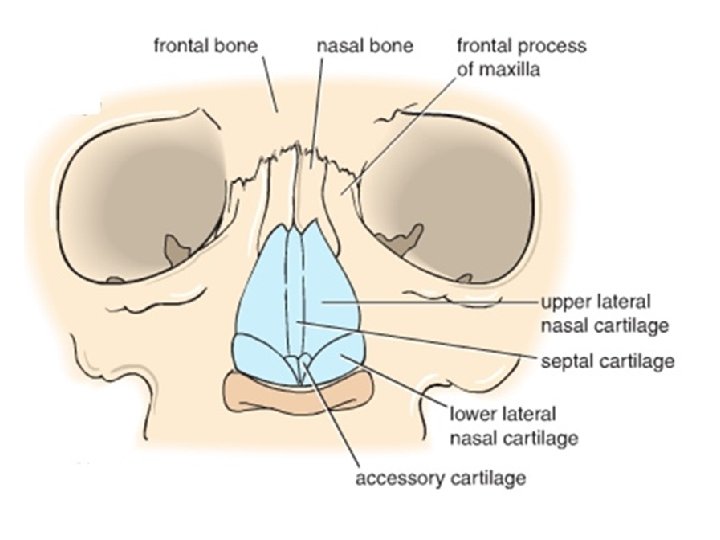
External Nose: • Pyramidal projection on the face. • Presents – • a free tip/apex. • Root at its junction with forehead. • Dorsum – from apex to the root • Skeletal framework that is partly bony and partly cartilaginous
• Upper part – bones as follows: Nasal, frontal process of maxilla and nasal notch of frontal bone. • Lower part – cartilages: • Anterior border of septal cartilage. • Upper lateral nasal cartilage –continuous with septal cartilage. • Alar cartilage • Minor alar cartilages • Fibro fatty tissue.
• Blood supply : – Dorsal nasal branch of ophthalmic artery – Alar & septal branches of facial artery – Infraorbital branch of maxillary artery • Nerve supply : • External nasal & infratrochlear branches of ophthalmic nerve. • Infraorbital branch of maxillary nerve.
Nasal cavity • Introduction: – Extends from external nares /nostrils on the face to posterior nasal aperture/choanae in the nasopharynx – Divided into two equal halves by nasal septum. – Has a – roof – floor – medial wall – Lateral wall.
Nasal cavity
• Roof: – Anterior Slope: frontal and nasal bones, nasal cartilages. – Middle Horizontal: Cribriform plate of ethmoid bone – Posterior slope: anterior and Inferior surface of body of the sphenoid bone.
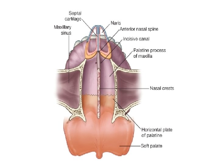
• Floor: 7. 5 cm long, 1. 5 cm wide Formed by: Upper surface of hard palate : • Palatine process of the maxilla • Horizontal plate of the palatine. • The naris opens anteriorly into the floor.
• Medial Wall/Nasal Septum: – oriented vertically in median sagittal plane and separates right and left nasal cavities. – Osseocartilagenous – Covered by mucous membrane. 3 parts: • Bony • Cartilaginous • Cuticular
• Bony part: – Vomer – Perpendicular plate of the ethmoid – Nasal spine of the frontal bone, rostrum of the sphenoid and nasal crest of the nasal bone.
• Cartilagenous Part: – Septal Cartilage – Septal process of the inferior nasal(alar) cartilage. – Occasionally Vomero-nasal cartilage • Cuticular part/ vestibule: – Fibro fatty tissue covered by skin. – Mobile – Lower margin is called columnella.
• Arterial Supply: – Anterior ethmoidal artery. – Posterior ethmoidal artery. – Superior labial branch of facial artery. – Greater palatine artery branch of maxillary artery. – Sphenopalatine artery.
Kiesselbach’s /little’s area of epistaxis. – Highly vascular area at the anteroinferior part – anastomosis of following arteries – Septal branches of 1. Anterior ethmoidal, 2. sphenopalatine, 3. greater palatine and 4. superior labial arteries – small ulcer-profuse arterial haemorrhage.
• Venous drainage: A sub mucosal plexus of veins which drains into the – facial vein, – pterygoid venous plexus and – ophthalmic vein. • Dangerous area of the face – anteroinferior part-facial vein-deep facial vein. Cavernous sinus
• Lymphatic Drainage: – Anterior Half: Submandibular nodes. – Posterior Half: Retropharyngeal and deep cervical nodes.
• Nerve Supply: – General Sensory nerves • Anterosuperior part: Internal nasal branch of the ethmoidal nerve. • Anteroinferior part: Anterior superior alveolar nerve • Posterosuperior part: posterior superiormedial nasal branches of the pterygopalatine ganglion. • Posteroinferior part: Nasopalatine branch of the pterygopalatine ganglion. – Special sensory Nerves/ Olfactory nerves to the olfactory area
Clinical anatomy • Deviated nasal septum
• Little’s area/keisselbach’s plexus
• Dangerous area of face
Thank you