Good morning Lymphatic system Introduction Components and their


")


 – Largest lymphatic organ – Located between the stomach & diaphragm. –")


- Slides: 32
Good morning
Lymphatic system
• Introduction • Components and their function -Lymph vessels -Central lymphoid organs -Peripheral lymphoid organs • Applied anatomy
Introduction • A drainage system which is accessory to venous system.
Components of lymphatic system �Lymph � Lymph vessels �Lymph capillaries �Lymph vessels proper �Terminal lymph ducts �Lymphoid tissue 1. Central • Bone marrow and Thymus • Spleen – fetal life 2. Peripheral �Lymph nodes �Spleen �Tonsil 5
Lymph �Tissue fluid that enters the lymphatic system. • It is usually a clear, colorless fluid, similar to blood plasma but low in protein. • Its composition varies from place to place; after a meal, for example, lymph draining from the small intestine, takes on a milky appearance, due to lipid content(‘’chyle’’)
Composition of lymph �Macromolecules – proteins �Particulate matters �Dust �Carbon �Bacteria , macrophages, viruses, �Cancer cells Functions • Return excess tissue fluid to the bloodstream • Transport fats from the digestive tract to the bloodstream. • Carries colloid and particulate matter. • Surveillance & defense
Circulation
Lymph vessels Lymph capillaries • Begins blindly in tissue spaces around blood capillaries. • Flattened endothelium. • Devoid of basal lamina. • Permeable to macromolecules such as colloid(protein) and particulate matter(Eg : bacteria).
Lymph vessels • Properties of lymphatic vessels – One way system toward the heart – No pump – Lymph moves toward the heart • Milking action of skeletal muscle • Rhythmic contraction of smooth muscle in vessel walls • Suction action of diaphragm • Provided with valves –beaded appearance • Interrupted by the lymph nodes on its course, hence classified into afferent and efferent lymph vessels
Terminal lymph ducts
Lymphoid tissue • Modified connective tissue • Supporting structure: Reticular fibres • Cells: Fixed cells-reticular cells Free cellslymphoblasts lymphocytes plasma cells
Distribution – 3 types • Diffuse lymphatic tissue – Found in connective tissue of almost all organs – Macrophages-connective tissue, monocytes-blood, kupffer cells-liver etc • Lymphatic nodules/follicles – Spherical collection of lymphocytes with pale germinal centre where lymphocytes are loosely arranged. – Found singly or in clusters – Respiratory and gastrointestinal tract. • Lymphatic organs – Central – bone marrow , Thymus. – Peripheral -Lymph nodes, spleen, tonsil
Lymphatics – Originate as lymph capillaries – Capillaries unite to form larger vessels • Resemble veins in structure • Connect to lymph nodes at various intervals – Lymphatics ultimately deliver lymph into 2 main channels • Right lymphatic duct – Drains right side of head & neck, right arm, right thorax – Empties into the right subclavian vein • Thoracic duct – Drains the rest of the body – Empties into the left subclavian vein
Central Fetal life – spleen. Thymus - puberty Bone marrow – throughout. Stem cells – differentiate in bone marrow – B Lymphocytes - Humoral immunity-antibodies. • Stem cells – Differentiate in Thymus – T Lymphocytes – cell mediated immunity – phagocytosis. • •
Peripheral lymphoid tissue • • Primary lymphatic follicles Lymph nodes Spleen Tonsil 16
Primary lymphatic follicles • Spherical collection of lymphocytes with pale germinal centre where lymphocytes are loosely arranged. • No capsule present. • Both B and T lymphocytes are seen. • Found singly or in clusters • Respiratory and gastrointestinal tract.
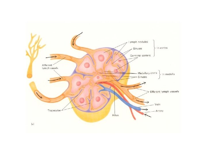
Lymph Node • kidney-shaped, Oval structures located along lymphatics. • Arranged in groups along the blood vessels. • Bean shaped and presents a hilum which transmits the efferent lymph vessel. • Lymph enters nodes through afferent lymphatics, flows through sinuses, exits through efferent lymphatics.
• Structure: • 1. capsule and the sub-capsular sinus(receives the afferent lymph vessels) • 2. cortex – • Trabeculae. • The reticular fibres and • Primary lymphatic follicle with germinal centre
• 3. medulla – the trabeculae is divides into numerous septa. • The spaces between the septa is occupied by irregular cords of lymphocytes known as the medullary cords. • Numerous blood vessels invests the trabecular septa and converts the latter into medullary sinuses
Functions of lymph nodes • Filters the lymph and remove the particulate matters such as carbon, bacteria , dust , cancer cells by phagocytic action and hence prevents its entry in the blood stream. • Produces B and T lymphocytes. • The organs, house critical immune cells such as lymphocytes which carryout our body defense against infection and disease as well as offer ACQUIRED IMMUNITY. – Macrophages – engulf and destroy foreign substances – Lymphocytes – provide immune response to antigens
Spleen(Haemal node) – Largest lymphatic organ – Located between the stomach & diaphragm. – Functions • Filters blood by removing the worn out RBC, WBC, and platelets. • Stores blood and platelets. • filters blood of bacteria and antigen-filled cells. • Produces lymphocytes.
– Histology • Capsule , trabeculae and reticular fibres are present. • Red pulp contains all the components of circulating blood. • White pulp is contains lymphatic nodules with an eccentric arteriole. • Both B and T lymphocytes are seen.
Thymus
Thymus Gland – Location – behind the sternum in the mediastinum – The capsule divides it into 2 lobes – Development • Infant – conspicuous • Puberty – maximum size • Maturity – decreases in size – Function • Differentiation and maturation of T lymphocytes. • Produces hormones
• Thymus is made up of an outer capsule • It has incomplete lobules. • Individual lobule is made up of outer cortex and inner medulla • Cortex has lymphocytes • Medulla has Hassall's corpuscles
Tonsil Multiple groups of large lymphatic nodules that surround the opening of respiratory and digestive tubes Location – mucous membrane of the oral and pharyngeal cavities Types • Pharyngeal tonsil • Tubal tonsil • Lingual tonsil • Palatine tonsil
Palatine tonsil Function -Traps bacteria and other microbes in throat.
Applied anatomy • First line of defence of the body. • Inflammation lymph vessels-lymphangitis • Lymph nodes – lymphadenitis. • Elephantiasis. • Route of spread of cancer cells.
THANK YOU
31