
GOOD MORNING Dr Tabrez Development of Female Reproductive

GOOD MORNING Dr. Tabrez

Development of Female Reproductive System
Contents …. Ø Introduction Ø Development of primitive gonads Ø Differentiation of the Gonads –Ovaries Ø Descent of ovaries & Anomalies Ø Development Of Sexual Duct Systems with its Anomalies Ø Development of External Genitalia with its Anomalies Ø References
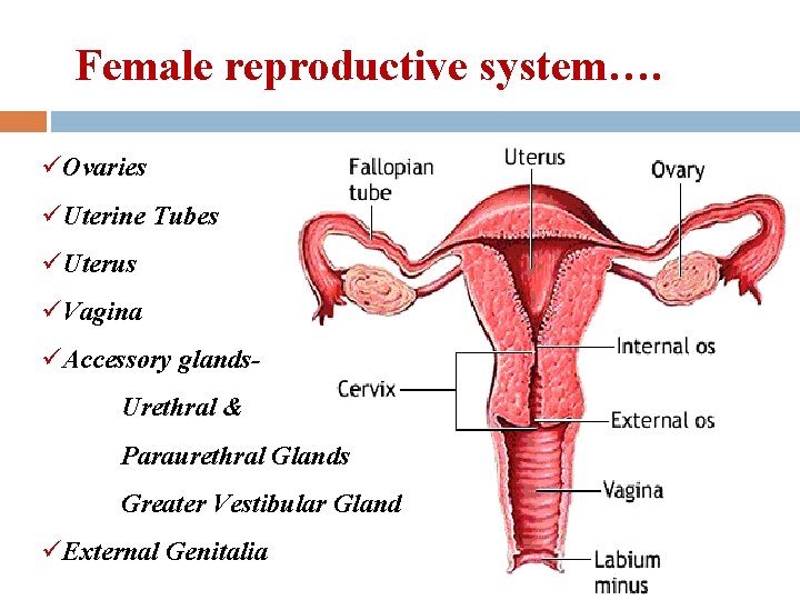
Female reproductive system…. üOvaries üUterine Tubes üUterus üVagina üAccessory glands. Urethral & Paraurethral Glands Greater Vestibular Gland üExternal Genitalia
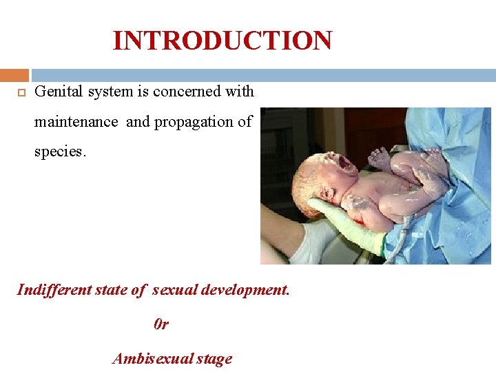
INTRODUCTION Genital system is concerned with maintenance and propagation of species. Indifferent state of sexual development. 0 r Ambisexual stage

Phenotypic differentiation of embryo…. . Gonad s Sexual ducts system Differention of external genitalia Secondary sexual character
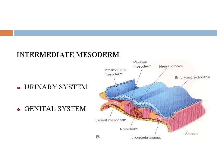
INTERMEDIATE MESODERM URINARY SYSTEM GENITAL SYSTEM
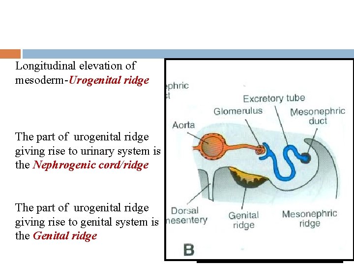
Longitudinal elevation of mesoderm-Urogenital ridge The part of urogenital ridge giving rise to urinary system is the Nephrogenic cord/ridge The part of urogenital ridge giving rise to genital system is the Genital ridge
Development of Early Gonad Proliferating Coelomic epithelium. Mesonephric Mesenchyme Primordial Germ Cells Angiogenic Mesenchyme
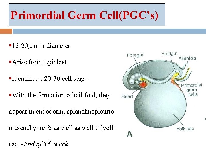
Primordial Germ Cell(PGC’s) § 12 -20µm in diameter §Arise from Epiblast. §Identified : 20 -30 cell stage §With the formation of tail fold, they appear in endoderm, splanchnopleuric mesenchyme & as well as wall of yolk sac. -End of 3 rd week.
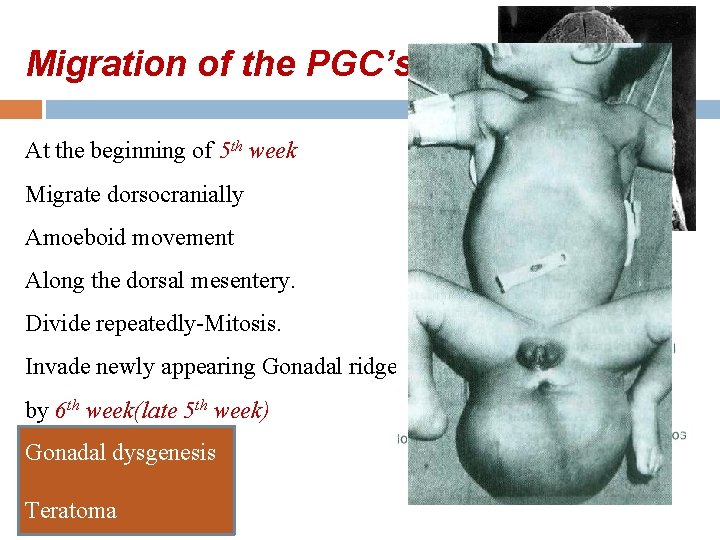
Migration of the PGC’s At the beginning of 5 th week Migrate dorsocranially Amoeboid movement Along the dorsal mesentery. Divide repeatedly-Mitosis. Invade newly appearing Gonadal ridge by 6 th week(late 5 th week) Gonadal dysgenesis Teratoma
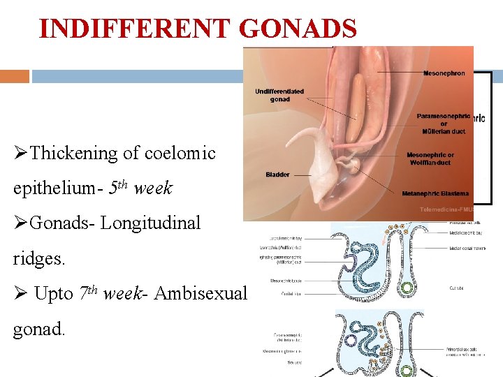
INDIFFERENT GONADS ØThickening of coelomic epithelium- 5 th week ØGonads- Longitudinal ridges. Ø Upto 7 th week- Ambisexual gonad.
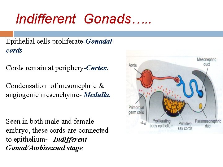
Indifferent Gonads…. . Epithelial cells proliferate-Gonadal cords Cords remain at periphery-Cortex. Condensation of mesonephric & angiogenic mesenchyme- Medulla. Seen in both male and female embryo, these cords are connected to epithelium- Indifferent Gonad/Ambisexual stage
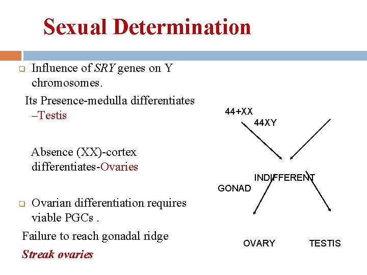
Sexual Determination Influence of SRY genes on Y chromosomes. Its Presence-medulla differentiates –Testis q 44+XX 44 XY Absence (XX)-cortex differentiates-Ovaries INDIFFERENT GONAD Ovarian differentiation requires viable PGCs. Failure to reach gonadal ridge Streak ovaries q OVARY TESTIS
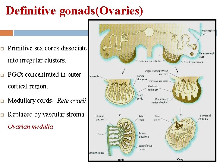
Definitive gonads(Ovaries) Primitive sex cords dissociate into irregular clusters. PGCs concentrated in outer cortical region. Medullary cords- Rete ovarii Replaced by vascular stroma. Ovarian medulla
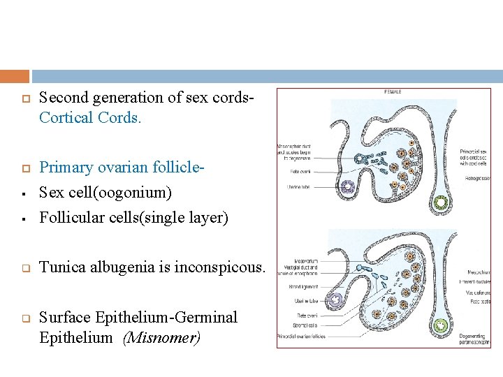
Second generation of sex cords. Cortical Cords. § Primary ovarian follicle. Sex cell(oogonium) Follicular cells(single layer) q Tunica albugenia is inconspicous. § q Surface Epithelium-Germinal Epithelium (Misnomer)
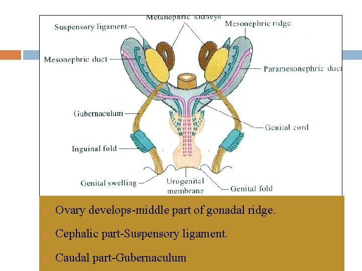
Ovary develops-middle part of gonadal ridge. Cephalic part-Suspensory ligament. Caudal part-Gubernaculum
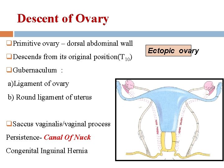
Descent of Ovary q. Primitive ovary – dorsal abdominal wall q. Descends from its original position(T 10) q. Gubernaculum : a)Ligament of ovary b) Round ligament of uterus q. Saccus vaginalis/vaginal process Persistence- Canal Of Nuck Congenital Inguinal Hernia Ectopic ovary
Summar y Genital ridge appears-5 th week. PGC’s migrate along dorsal mesentery & invade genital ridge -6 th week. Indifferent gonads differentiation- Cortex Medulla. Cortex develops-Ovaries Descent of Ovaries
Gonadal Dysgenesis: Oocytes absent Ovaries appear as streak gonads XY Female Gonadal Dysgenesis (Swyer Syndrome) Defects of sexual differentiation Total Gonadal Dysgenesis Cause: Mutation or Deletion of SRY gene Features : Phenotypically female No secondary sexual characters Treatment : Estrogen &progesterone supplement. Streak gonads are removed. Cannot generate ova, embryo transfer into Uterus.
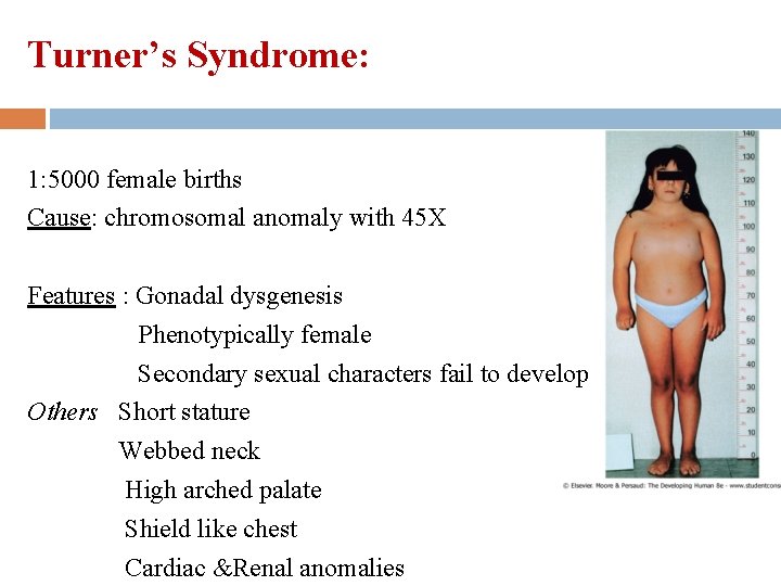
Turner’s Syndrome: 1: 5000 female births Cause: chromosomal anomaly with 45 X Features : Gonadal dysgenesis Phenotypically female Secondary sexual characters fail to develop Others Short stature Webbed neck High arched palate Shield like chest Cardiac &Renal anomalies
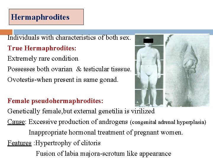
Hermaphrodites Individuals with characteristics of both sex. True Hermaphrodites: Extremely rare condition Possesses both ovarian & testicular tisssue. Ovotestis-when present in same gonad. Female pseudohermaphrodites: Genetically female, but external genetilia is virilized Cause: Excessive production of androgens (congenital adrenal hyperplasia) Inappropriate hormonal treatment of pregnant women. Features : Hypertrophy of clitoris Fusion of labia majora-scrotum like appearance
Development Of Duct System
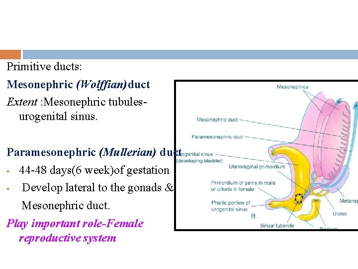
Primitive ducts: Mesonephric (Wolffian)duct Extent : Mesonephric tubulesurogenital sinus. Paramesonephric (Mullerian) duct § 44 -48 days(6 week)of gestation § Develop lateral to the gonads & Mesonephric duct. Play important role-Female reproductive system
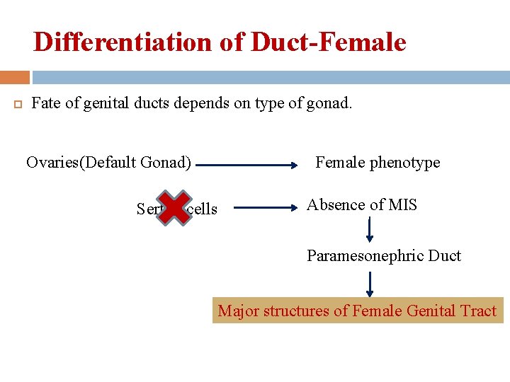
Differentiation of Duct-Female Fate of genital ducts depends on type of gonad. Ovaries(Default Gonad) Sertoli cells Female phenotype Absence of MIS Paramesonephric Duct Major structures of Female Genital Tract
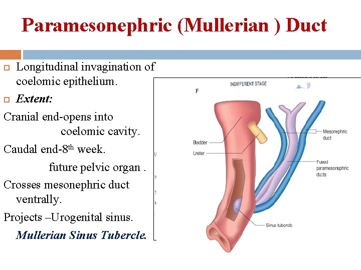
Paramesonephric (Mullerian ) Duct Longitudinal invagination of coelomic epithelium. Extent: Cranial end-opens into coelomic cavity. Caudal end-8 th week. future pelvic organ. Crosses mesonephric duct ventrally. Projects –Urogenital sinus. Mullerian Sinus Tubercle.
Derivatives Of The Duct Derivatives: Uterine tube Appendix of testis Uterus Prostatic Utricle Vagina(? )
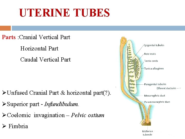
UTERINE TUBES Parts : Cranial Vertical Part Horizontal Part Caudal Vertical Part ØUnfused Cranial Part & horizontal part(? ). ØSuperior part - Infundibulum. ØCoelomic invagination – Pelvic ostium Ø Fimbria
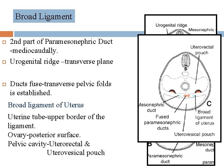
Broad Ligament 2 nd part of Paramesonephric Duct -mediocaudally. Urogenital ridge –transverse plane Ducts fuse-transverse pelvic folds is established. Broad ligament of Uterus Uterine tube-upper border of the ligament. Ovary-posterior surface. Pelvic cavity-Uterorectal & Uterovesical pouch
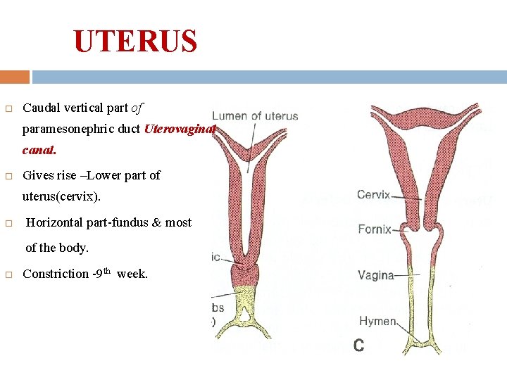
UTERUS Caudal vertical part of paramesonephric duct Uterovaginal canal. Gives rise –Lower part of uterus(cervix). Horizontal part-fundus & most of the body. Constriction -9 th week.
Paramesonephric ducts-Epithelium of endometrium & cervicalcanal. Genital septum -Myometrium and endometrial stroma. Uterus is entirely mesenchymal in origin.
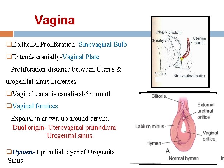
Vagina q. Epithelial Proliferation- Sinovaginal Bulb q. Extends cranially-Vaginal Plate Proliferation-distance between Uterus & urogenital sinus increases. q. Vaginal canal is canalised-5 th month q. Vaginal fornices Expansion grown up around cervix. Dual origin- Uterovaginal primodium Urogenital sinus. q. Hymen- Epithelial layer of Urogenital Sinus.
Summary Ø Uterine tube -Cephalic vertical part&(Most of Horizontal part of Paramesonephric Duct? ) Ø Uterus -Cephalic part of Uterovaginal canal (Fusion of vertical part + incorporation of segment of horizontal part) Ø Vagina-Upper part-uterovaginal primodium Lower part-Endoderm of urogenital sinus External vaginal orifice-ectoderm of genital folds, after the rupture of urogenital membrane.
References: Gray’s Anatomy; 39 th edition; Susan Standring Medical embryology; 11 th edition ; Langman The developing Human; 7 th edition; Keith L. Moore Essentials of Human Embryology ; 6 th edition , A. K. Datta Human embryology and developmental biology; 2 nd edition ; Bruce M. Carlson Human embryology; 3 th edition; William J. Larsen

THANK YOU
- Slides: 35