
Goniometry Michelle Reinink PT DPT OCS CIMT PT




- Slides: 39
Goniometry Michelle Reinink PT, DPT, OCS, CIMT PT 6280
Goniometry ■ Definition - the measurement of angles created at joints by the BONEY structures of the body. ■ Historically ■ ■ Visual approximation Accurate and standardized measurements
Purpose ■ Component of examination ■ Determinant of joint position ■ Identifies available motion at a joint ■ A/AA/PROM ■ Joint information (an inference) ■ Normal vs abnormal
Types of ROM ■ PROM ■ Non-voluntary ■ produced by an external force ■ AAROM ■ Voluntary ■ External force ■ AROM ■ Voluntary Kisner & Colby
AROM ■ Assessed first ■ Screening tool ■ Voluntary motion ■ Active contraction of muscle ■ Unassisted ■ Information regarding willingness to move ■ Pain status, guarding
PROM ■ Non-voluntary ■ Produced by an external force ■ Performed after AROM assessment ■ When abnormalities are found in AROM assessment ■ Enables assessment of passive structures ■ Tissue limited ROM
AAROM ■ Combination ■ Voluntary: prime mover muscles contract ■ External force: assistance provided to complete full arc of motion
PROM ■ Indications ■ Post-operative precautions/contradindications ■ Inflammatory conditions where AROM would be harmful to the segment ■ Goals ■ Decrease complications that would occur with immobilization ■ Maintain tissue extensibility ■ Minimize contractures ■ Improve circulation ■ Minimize pain
AAROM ■ Indications ■ Paresis/paralysis ■ Surgical limitations/precautions ■ Goals ■ Tissue extensibility ■ Minimize contractures ■ Improve circulation ■ Minimize pain Kisner & Colby
AROM ■ Indications ■ Active movement ability/allowed ■ Aerobic conditioning ■ Goals ■ Tissue extensibility ■ Minimize contractures ■ Improve circulation ■ Sensory feedback ■ Coordination/motor skills ▪ Kisner & Colby
Kinematics ■ Arthrokinematics ■ Refers to the naturally occurring non-voluntary accessory joint movements that occur in some combination of slide (glide), spin or roll ■ Osteokinematics ■ Refers to the gross physiologic movement of the bone segments
Osteokinematics ■ Motions take place in one of the following planes: ■ ■ sagittal, frontal, or transverse And around one of the following axes: ■ medial-lateral, anterior-posterior, or vertical
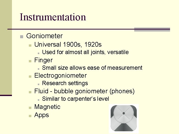
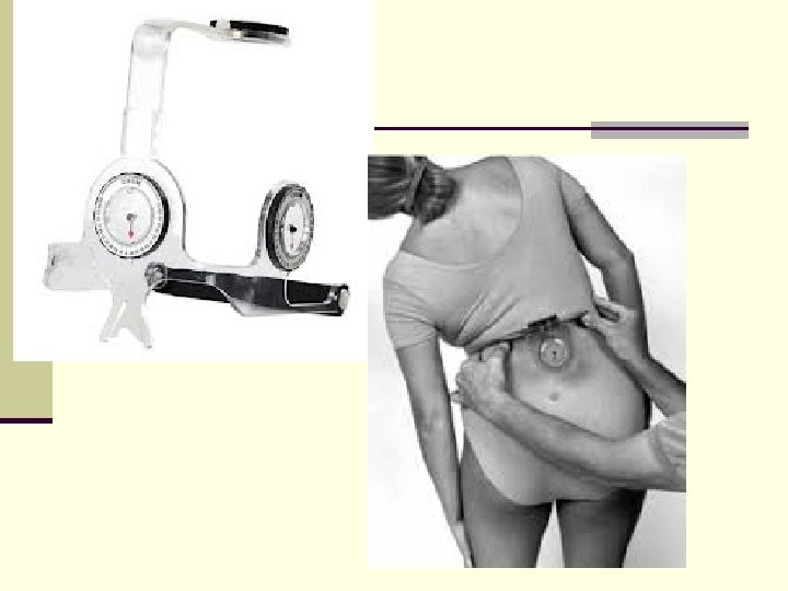
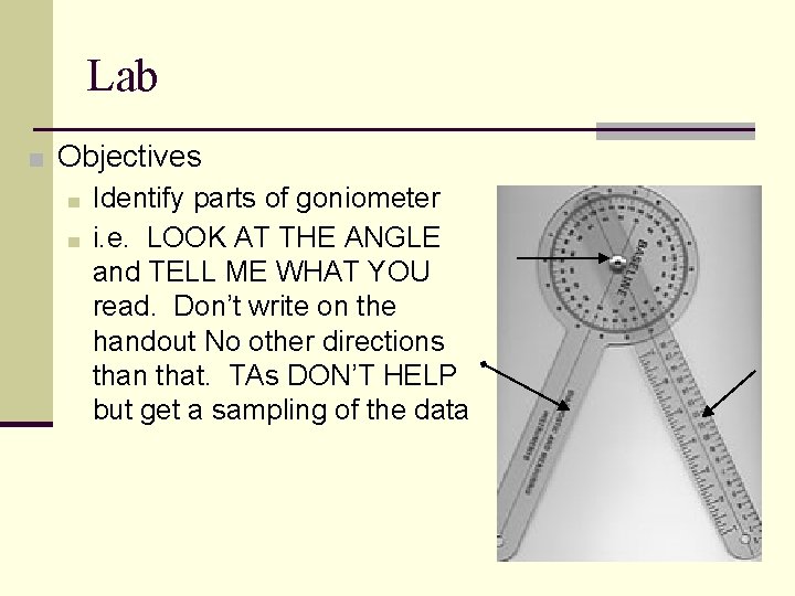
Instrumentation ■ Goniometer ■ Universal 1900 s, 1920 s ■ ■ Finger ■ ■ ■ Research settings Fluid - bubble goniometer (phones) ■ ■ Small size allows ease of measurement Electrogoniometer ■ ■ Used for almost all joints, versatile Similar to carpenter’s level Magnetic Apps
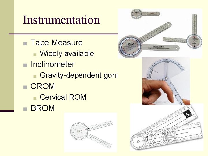
Instrumentation ■ Tape Measure ■ Widely available ■ Inclinometer ■ Gravity-dependent goni ■ CROM ■ Cervical ROM ■ BROM
Lab ■ Objectives ■ Identify parts of goniometer ■ i. e. LOOK AT THE ANGLE and TELL ME WHAT YOU read. Don’t write on the handout No other directions than that. TAs DON’T HELP but get a sampling of the data
Background Information ■ Things you need to know: ■ Anatomy ■ Normal values ■ Proper goniometric alignment ■ Testing positions ■ Where to stabilize ■ Joint structure and function
Procedures ■ Positioning ■ Start at zero position (usually related to anatomic position) ■ May affect the amount of tension on soft tissue structures around the joint ■ May affect stability of proximal segment ■ Stabilization ■ Proximal segment ■ Manual stabilization ■ Helps to isolate the motion
Procedures ■ Alignment ■ Stationary arm (SA) // to the proximal segment ■ Moving arm (MA) // to the distal segment ■ Fulcrum over the axis of rotation ■ Palpate bony landmarks for proper alignment ■ Recording ■ Side of the body, joint motion, starting position, ending position, position of pt. , type of motion. ■ Document the total ROM of the joint
Psychometric Properties of Goniometry ■ Reliability ■ Indicates degrees to which successive measurements yield the same result ■ Validity ■ Indicates the degrees to which an instrument measures what it is purported to measure.
Findings ■ ROM is a measure to test for a(n) ______: (Pathology? Impairment? Special test? Posture? Diagnosis? ) ■ Terms often used: ■ Mobility: refers to the ability of a joint slide/glide/translate. This is not a motion a person can consciously create in isolation. It is a function of joint parameters (tissue extensibility, jt. shape/congruency etc. ) ■ ■ Hypermobility: excessive mobility at the joint Hypomobility: a lack of mobility at a joint
Documentation ■ Recording Results ■ Essential to have common documentation system so that values are understandable ■ Positional joint ROM method: ▪ 0 -140 degrees elbow ROM ■ Documenting start and end position of motion trying to measure ▪ 0 -10 degrees elbow extension ▪ 0 -140 degrees elbow flexion ▪ 10 -140 degrees elbow flexion (what does that mean? )
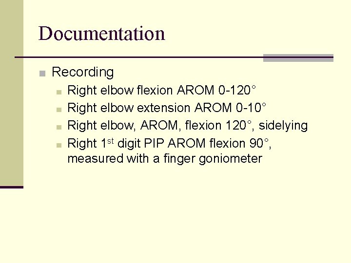
Documentation ■ Recording ■ Right elbow flexion AROM 0 -120° ■ Right elbow extension AROM 0 -10° ■ Right elbow, AROM, flexion 120°, sidelying ■ Right 1 st digit PIP AROM flexion 90°, measured with a finger goniometer
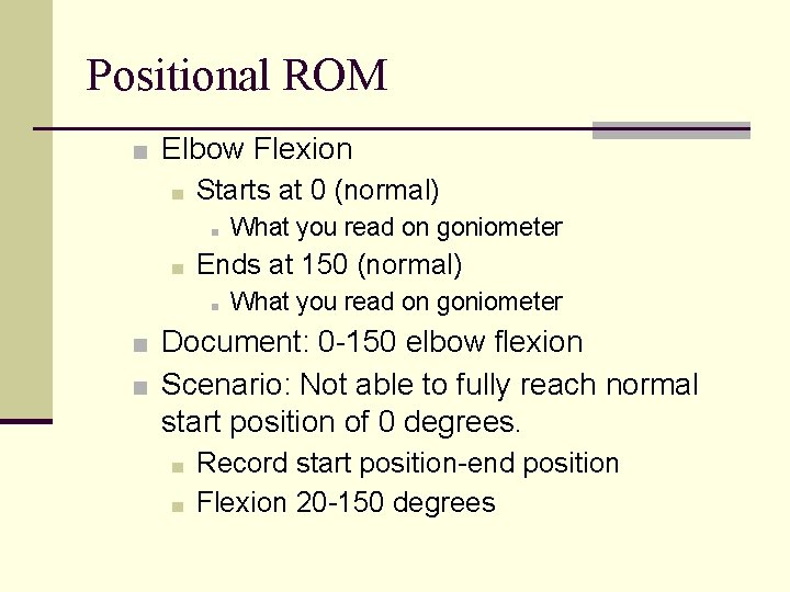
Positional ROM ■ Elbow Flexion ■ Starts at 0 (normal) ■ ■ What you read on goniometer Ends at 150 (normal) ■ What you read on goniometer ■ Document: 0 -150 elbow flexion ■ Scenario: Not able to fully reach normal start position of 0 degrees. ■ ■ Record start position-end position Flexion 20 -150 degrees
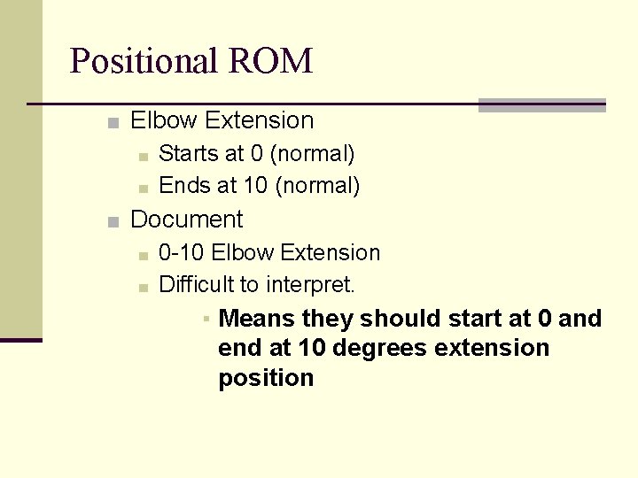
Positional ROM ■ Elbow Extension ■ Starts at 0 (normal) ■ Ends at 10 (normal) ■ Document ■ 0 -10 Elbow Extension ■ Difficult to interpret. ▪ Means they should start at 0 and end at 10 degrees extension position
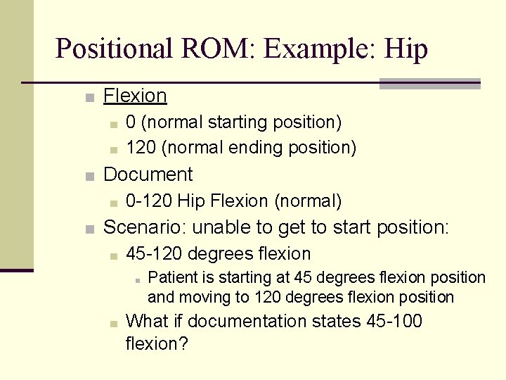
Positional ROM: Example: Hip ■ Flexion ■ 0 (normal starting position) ■ 120 (normal ending position) ■ Document ■ 0 -120 Hip Flexion (normal) ■ Scenario: unable to get to start position: ■ 45 -120 degrees flexion ■ ■ Patient is starting at 45 degrees flexion position and moving to 120 degrees flexion position What if documentation states 45 -100 flexion?
Positional ROM: Example: Hip ■ Extension ■ 0 (normal starting position) ■ 20 (normal ending position) ■ Document: ■ 0 -20 Hip Extension (normal) ■ Unable to get to start position: ■ 5 -20 degrees Extension ■ Do you expect normal hip flexion ROM? ■ Unable to get to normal start position and normal end position ■ 5 -10 degrees Extension
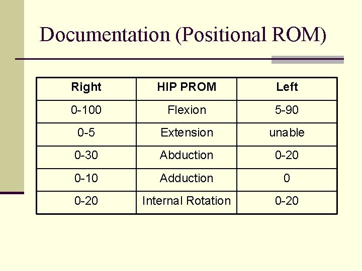
Documentation (Positional ROM) Right HIP PROM Left 0 -100 Flexion 5 -90 0 -5 Extension unable 0 -30 Abduction 0 -20 0 -10 Adduction 0 0 -20 Internal Rotation 0 -20
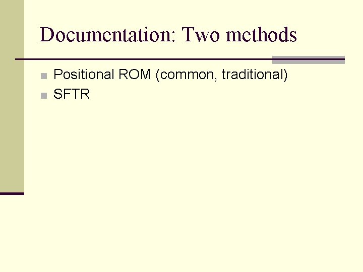
Documentation: Two methods ■ Positional ROM (common, traditional) ■ SFTR
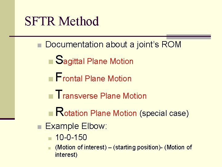
SFTR Method ■ Documentation about a joint’s ROM ■ Sagittal Plane Motion ■ Frontal Plane Motion ■ Transverse Plane Motion ■ Rotation Plane Motion (special case) ■ Example Elbow: ■ 10 -0 -150 ■ (Motion of interest) – (starting position)- (Motion of interest)
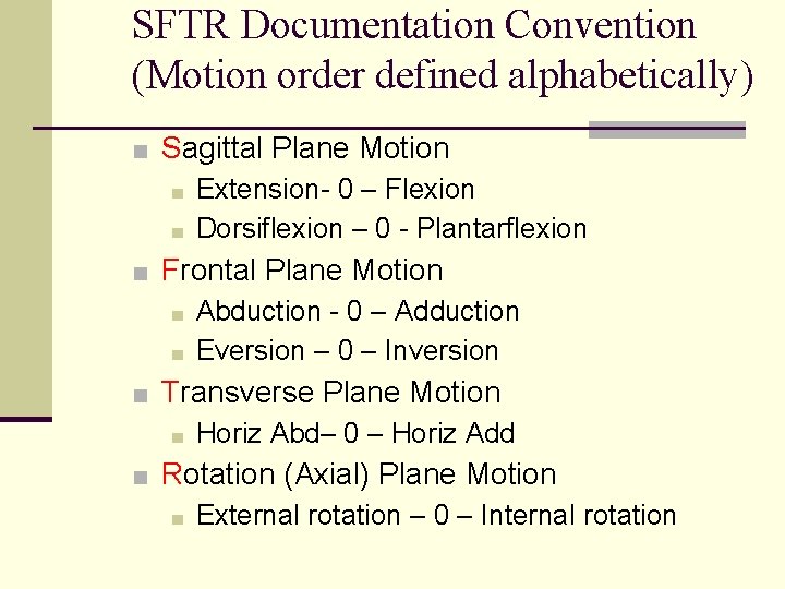
SFTR Documentation Convention (Motion order defined alphabetically) ■ Sagittal Plane Motion ■ Extension- 0 – Flexion ■ Dorsiflexion – 0 - Plantarflexion ■ Frontal Plane Motion ■ Abduction - 0 – Adduction ■ Eversion – 0 – Inversion ■ Transverse Plane Motion ■ Horiz Abd– 0 – Horiz Add ■ Rotation (Axial) Plane Motion ■ External rotation – 0 – Internal rotation
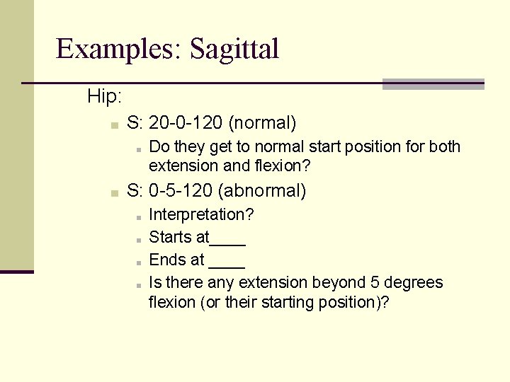
Examples: Sagittal Hip: ■ S: 20 -0 -120 (normal) ■ ■ Do they get to normal start position for both extension and flexion? S: 0 -5 -120 (abnormal) ■ ■ Interpretation? Starts at____ Ends at ____ Is there any extension beyond 5 degrees flexion (or their starting position)?
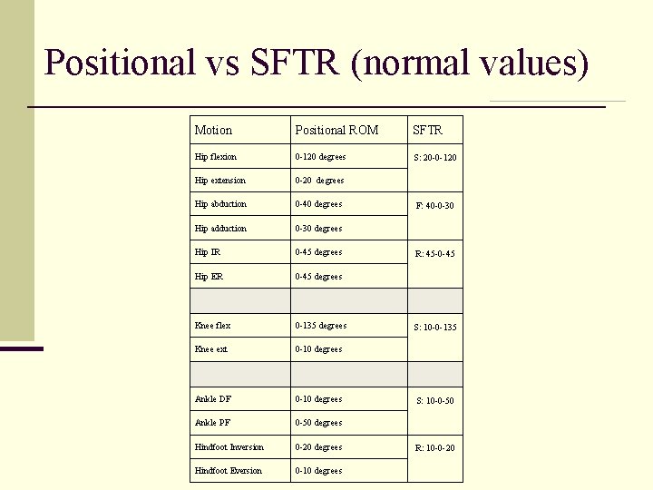
Positional vs SFTR (normal values) Motion Positional ROM SFTR Hip flexion 0 -120 degrees S: 20 -0 -120 Hip extension 0 -20 degrees Hip abduction 0 -40 degrees Hip adduction 0 -30 degrees Hip IR 0 -45 degrees Hip ER 0 -45 degrees Knee flex 0 -135 degrees Knee ext 0 -10 degrees Ankle DF 0 -10 degrees Ankle PF 0 -50 degrees Hindfoot Inversion 0 -20 degrees Hindfoot Eversion 0 -10 degrees F: 40 -0 -30 R: 45 -0 -45 S: 10 -0 -135 S: 10 -0 -50 R: 10 -0 -20
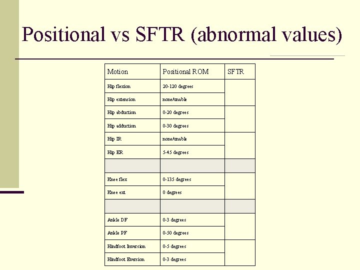
Positional vs SFTR (abnormal values) Motion Positional ROM Hip flexion 20 -120 degrees Hip extension none/unable Hip abduction 0 -20 degrees Hip adduction 0 -30 degrees Hip IR none/unable Hip ER 5 -45 degrees Knee flex 0 -135 degrees Knee ext 0 degrees Ankle DF 0 -3 degrees Ankle PF 0 -50 degrees Hindfoot Inversion 0 -5 degrees Hindfoot Eversion 0 -3 degrees SFTR
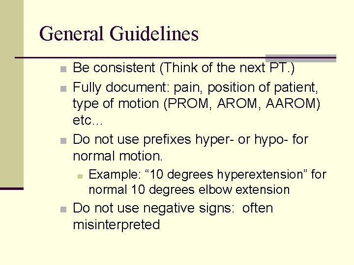
General Guidelines ■ Be consistent (Think of the next PT. ) ■ Fully document: pain, position of patient, type of motion (PROM, AAROM) etc… ■ Do not use prefixes hyper- or hypo- for normal motion. ■ Example: “ 10 degrees hyperextension” for normal 10 degrees elbow extension ■ Do not use negative signs: often misinterpreted
ROM Limitations ■ Factors affecting ROM ■ Age ■ under 2 and older adults ■ Sex ■ may vary ■ Active movement ■ ability/willingness to move ■ coordination ■ muscle strength ■ muscle hypertrophy, obesity ■ joint motion, edema
ROM Limitations ■ Factors Affecting ROM ■ Passive motion ■ ■ ■ integrity of the articular surfaces joint capsule extensibility ligamentous tissue muscular tissue end feels Occupation or culture
Real World Procedures: See lab packet 1. Position patient as per guidelines 2. Perform PROM to assess end feel and 3. 4. 5. 6. 7. 8. approximate available ROM Place patient at start position per lab packet Palpate landmarks Stabilize proximally at start position with SA Move into ROM of interest Re-align goniometer if needed Take measurement (with goniometer facing out)
References ■ Norkin, Cynthia C. & White, Joyce D. Measurement of Joint Motion: A Guide to Goniometry. , 5 th Ed. F. A. Davis, Philadelphia 2016. ■ Reese Berryman, Nancy & Bandy, William D. Joint Range of Motion and Muscle Length Testing. W. B. Saunders, Philadelphia, 2002. ■ (SFTR Method)